. 2001 May 15;29(10):2154-62.
doi: 10.1093/nar/29.10.2154.

CREB-H: a novel mammalian transcription factor belonging to the CREB/ATF family and functioning via the box-B element with a liver-specific expression

Affiliations

CREB-H: a novel mammalian transcription factor belonging to the CREB/ATF family and functioning via the box-B element with a liver-specific expression

Y Omori et al. Nucleic Acids Res. .

Abstract

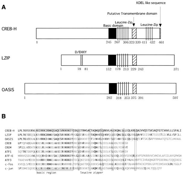
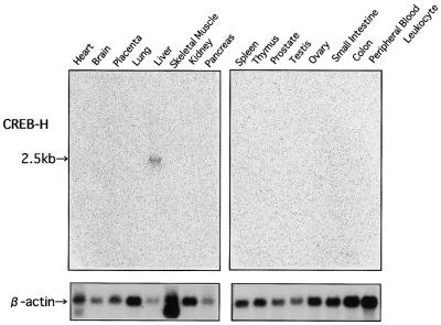
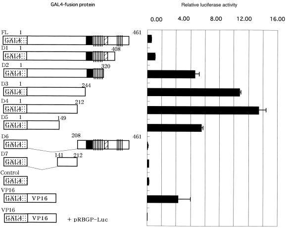
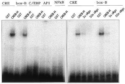
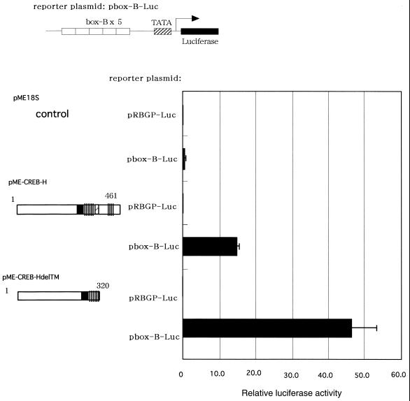
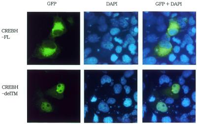
The expression of liver-specific genes is regulated by unequivocally allocated transcription factors via proper responsible elements within their promoters. We identified a novel transcription factor, CREB-H, and found that its expression was restricted in the liver among 16 human tissues tested. A region of CREB-H exhibited significant homology to the basic leucine zipper (b-Zip) domain of members of the CREB/ATF family: mammalian LZIP and Drosophila BBF-2 that binds to box-B, a Drosophila enhancer modulating the fat-body-specific gene expression. CREB-H contained a hydrophobic region representing a putative transmembrane domain, like LZIP. Constructing a variety of CREB-H fusion proteins with the GAL4 DNA-binding domain disclosed that CREB-H functioned as a transcriptional activator and its N-terminal 149 amino acids accounted for the activation ability. Gel mobility sift assays revealed that CREB-H did not bind to the C/EBP, AP-1 and NF-kappaB elements but specifically bound to CRE and the box-B element. Luciferase reporter assays demonstrated that like BBF-2, CREB-H activated transcription via the box-B element and that a deletion of the putative transmembrane domain increased the activation of reporter expression significantly. Furthermore, a fusion protein of GFP and full-length CREB-H was localized in reticular structures surrounding the nucleus, whereas a fusion protein of GFP and a deletion mutant lacking the putative transmembrane domain was mainly in the nucleus. These findings suggest that CREB-H plays an important role in transcriptional regulation of genes specifically expressed in the liver, and that the putative transmembrane domain may be associated with modulation of its function as the transcriptional activator.


Figures

Figure 1
Figure 1
(A) Schematic diagrams of CREB-H, LZIP and OASIS. The b-Zip, D/EHXY, putative transmembrane domain, KDEL-like sequence and leucine-zipper domains and their amino acid positions are indicated. (B) A multiple alignment of the b-Zip domain among CREB-H and other members of the CREB/ATF family. The basic and leucine zipper domains are indicated. Invariant residues are marked by bold letters.
Figure 2
Figure 2
(A) The genomic structure of CREB-H. CREB-H consisted of 10 exons distributing within a 19.4 kb region of the genome (cosmid R33590, accession no. AC005620; 40351 bp) mapped to human chromosome 19p13.3. (B) The mRNA start sites of CREB-H. The mRNA start site of two clones for full-length CREB-H were indicated. The nucleotide numbers in the cosmid R33590 sequence are shown.
Figure 3
Figure 3
The expression profile of the CREB-H mRNA in multiple human tissues. Each lane contained ∼2 µg of poly(A)+ RNA. A probe for CREB-H was a 708 bp fragment of the 5′-portion of the CREB-H cDNA outside the b-Zip domain. A 2.5 kb band corresponding to the CREB-H mRNA is marked. The blot hybridized with a β-actin probe is shown in the lower panel.
Figure 4
Figure 4
Mapping of the transcriptional activation domain in CREB-H. The GAL4-DNA binding domain and various deletion mutants of CREB-H were fused, and expression constructs of various GAL4–CREB-H fusion proteins were transfected with a reporter plasmid, pGAL-Luc, into COS7 cells. The luciferase activity was quantified 24 h after transfection. Relative luciferase activities are shown as mean values with standard errors of three independent experiments.
Figure 5
Figure 5
DNA-binding ability of CREB-H analyzed by gel-shift assays. A fusion protein of GST and a CREB-H mutant lacking the putative transmembrane domain was produced in bacteria and purified by glutathione–Sepharose 4B. Oligonucleotides representing CRE, C/EBP, AP1, NF-κB and box-B elements were annealed and labeled with [γ-32P]ATP and incubated with the GST–CREB-H fusion protein. Purified non-fused GST was used in a control lane. Competitive assays for CRE and the box-B element were performed with 5- and 50-fold excesses of unlabeled oligonucleotides for CRE and the box-B element, respectively.
Figure 6
Figure 6
CREB-H activates transcription of luciferase through the box-B element. The luciferase-reporter construct was transfected into COS7 cells with a CREB-H-expressing plasmid pME-CREB-H, or pME-CREB-HdelTM as well as a control vector plasmid, pME18S. The constructs expressing CREB-H are shown schematically on the left. The luciferase activity was quantified 24 h after transfection. Relative luciferase activities are shown as mean values with standard errors of three independent experiments.
Figure 7
Figure 7
Subcellular localization of fusion proteins of GFP and CREB-H. The full-length (CREBH-FL) or a deletion mutant of the transmembrane domain (CREBH-delTM) of CREB-H was fused to GFP and transfected to COS7 cells. The cells were stained by DAPI for visualization of the nucleus 24 h after transfection.

Similar articles

Cited by

References

    1. Lassar A.B., Buskin,J.N., Lockshon,D., Davis,R.L., Apone,S., Hauschka,S.D. and Weintraub,H. (1989) MyoD is a sequence-specific DNA binding protein requiring a region of myc homology to bind to the muscle creatine kinase enhancer. Cell, 58, 823–831. - PubMed
    1. Donoghue M., Ernst,H., Wentworth,B., Nadal-Ginard,B. and Rosenthal,N. (1988) A muscle-specific enhancer is located at the 3′ end of the myosin light-chain 1/3 gene locus. Genes Dev., 2, 1779–1790. - PubMed
    1. Christensen T.H., Prentice,H., Gahlmann,R. and Kedes,L. (1993) Regulation of the human cardiac/slow-twitch troponin C gene by multiple, cooperative, cell-type-specific, and MyoD-responsive elements. Mol. Cell. Biol., 13, 6752–6765. - PMC - PubMed
    1. Courtois G., Baumhueter,S. and Crabtree,G.R. (1988) Purified hepatocyte nuclear factor 1 interacts with a family of hepatocyte-specific promoters. Proc. Natl Acad. Sci. USA, 85, 7937–7941. - PMC - PubMed
    1. Landschulz W.H., Johnson,P.F. and McKnight,S.L. (1988) The leucine zipper: a hypothetical structure common to a new class of DNA binding proteins. Science, 240, 1759–1764. - PubMed

Publication types

MeSH terms

Associated data