. 2001 May 22;98(11):6074-9.
doi: 10.1073/pnas.111114298. Epub 2001 May 15.

Stimulation of phosphatidylinositol 3-kinase by fibroblast growth factor receptors is mediated by coordinated recruitment of multiple docking proteins

Affiliations

Stimulation of phosphatidylinositol 3-kinase by fibroblast growth factor receptors is mediated by coordinated recruitment of multiple docking proteins

S H Ong et al. Proc Natl Acad Sci U S A. .

Abstract

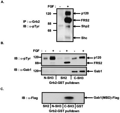
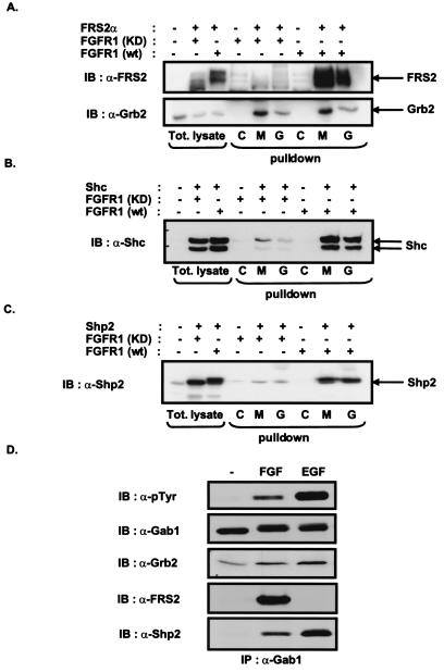
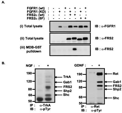
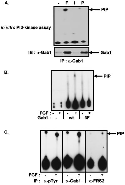
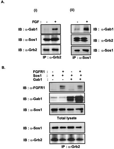
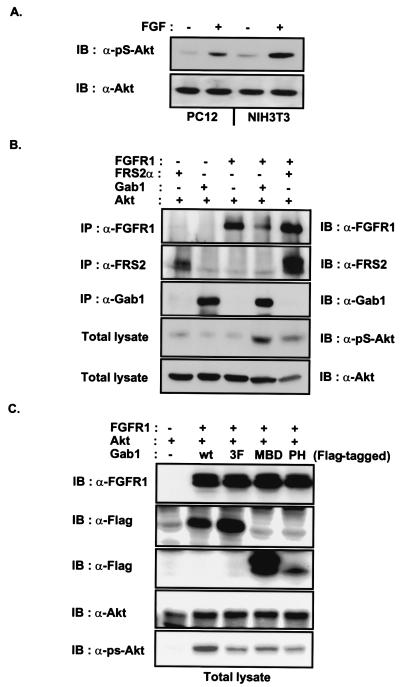
The docking protein FRS2 is a major downstream effector that links fibroblast growth factor (FGF) and nerve growth factor receptors with the Ras/mitogen-activated protein kinase signaling cascade. In this report, we demonstrate that FRS2 also plays a pivotal role in FGF-induced recruitment and activation of phosphatidylinositol 3-kinase (PI3-kinase). We demonstrate that tyrosine phosphorylation of FRS2alpha leads to Grb2-mediated complex formation with the docking protein Gab1 and its tyrosine phosphorylation, resulting in the recruitment and activation of PI3-kinase. Furthermore, Grb2 bound to tyrosine-phosphorylated FRS2 through its SH2 domain interacts primarily via its carboxyl-terminal SH3 domain with a proline-rich region in Gab1 and via its amino-terminal SH3 domain with the nucleotide exchange factor Sos1. Assembly of FRS2alpha:Grb2:Gab1 complex induced by FGF stimulation results in activation of PI3-kinase and downstream effector proteins such as the S/T kinase Akt, whose cellular localization and activity are regulated by products of PI3-kinase. These experiments reveal a unique mechanism for generation of signal diversity by growth factor-induced coordinated assembly of a multidocking protein complex that can activate the Ras/mitogen-activated protein kinase cascade to induce cell proliferation and differentiation, and PI3-kinase to activate a mediator of a cell survival pathway.


Figures

Figure 1
Figure 1
Complex formation between Grb2 and Gab1 in FGF-stimulated cells. (A) Quiescent Swiss 3T3 cells were unstimulated or stimulated with FGF1 and heparin (100 ng/ml and 5 μg/ml, respectively) for 10 min. Lysates were immunoprecipitated with anti-Grb2 antibodies and followed by SDS–PAGE and immunoblotting with anti-pTyr antibodies. (B) Swiss 3T3 cells were unstimulated or stimulated with FGF1 and heparin (100 ng/ml and 5 μg/ml, respectively) for 10 min. Cell lysates were incubated with immobilized GST fusion proteins of the N-terminal SH3, SH2, or C-terminal SH3 domains of Grb2. Bound proteins were eluted and resolved by SDS–PAGE and then followed by immunoblotting with anti-pTyr (Upper) or anti-Gab1 antibodies (Lower). (C) Gab1 binds to the C-terminal SH3 domain of Grb2 through the MBD. 293 cells were transfected with the expression vector for Flag-tagged MBD of Gab1. The lysates were incubated with immobilized GST fusion proteins of the SH2, N-SH3, or C-SH3 domains of Grb2. Bound proteins were eluted and resolved by SDS–PAGE and followed by immunoblotting with anti-Flag antibodies.
Figure 2
Figure 2
Gab1 and the MBD of Gab1 form a complex with FRS2α, Shc, or Shp2 in cells expressing activated FGF receptor. 293 cells were transfected with expression vectors for kinase-inactive FGFR1 (KD) mutant, wt FGFR1, and FRS2α, Shc, or Shp2 (AC) as indicated. Cell lysates were incubated with immobilized GST fusion proteins of full-length Gab1 (G) or the MBD of Gab1 (M). GST alone was used as a control (C). Bound proteins were eluted and analyzed by SDS–PAGE and immunoblotting with FRS2, Grb2, Shc, Shp2 antibodies (AC). (D) Quiescent PC12 cells were unstimulated or stimulated with FGF1 and heparin (100 ng/ml and 5 μg/ml, respectively) or EGF (50 ng/ml) for 10 min. The lysates were immunoprecipitated with anti-Gab1 antibodies, resolved by SDS–PAGE, and followed by immunoblotting with anti-pTyr, Gab1, Grb2, FRS2, or Shp2 antibodies.
Figure 3
Figure 3
Gab1 and the MBD of Gab1 form a complex with tyrosine-phosphorylated FRS2α. (A) 293 cells were transfected with expression vectors for wt FGFR1 or kinase-inactive FGFR1 (KD) mutant and wt FRS2α or an FRS2α mutant in which six tyrosine phosphorylation sites were replaced by phenylalanines (6F) as indicated. Equivalent amounts of the total cell lysates were resolved and immunoblotted with anti-FGFR1 (i) or anti-FRS2 antibodies (ii). Alternatively, cell lysates were incubated with immobilized GST fusion proteins of the MBD of Gab1 (iii). Bound proteins were eluted and resolved by SDS–PAGE and then followed by immunoblotting with anti-FRS2 antibodies. (B) FRS2 and Gab1 form a complex with tyrosine-phosphorylated NGF or GDNF receptors. Quiescent PC12 cells were unstimulated or stimulated with NGF (100 ng/ml, 10 min). Lysates were immunoprecipitated with anti-TrkA antibodies (Left). Similarly, lysates from unstimulated or GDNF-stimulated (50 ng/ml, 10 min) SH-SY5Y neuroblastoma cells were immunoprecipitated with anti-Ret antibodies (Right). In both cases the immunoprecipitates were resolved by SDS–PAGE and followed by immunoblotting with anti-pTyr antibodies.
Figure 4
Figure 4
FGF induces PI3-kinase activity associated with Gab1. (A) Quiescent L6-myoblasts stably expressing FGFR1 were unstimulated or stimulated with FGF1 and heparin (100 ng/ml and 5 μg/ml, respectively), insulin (100 ng/ml), or PDGF (100 ng/ml) for 10 min. The lysates were immunoprecipitated with anti-Gab1 antibodies. One-half of the immunocomplex was subjected to an in vitro PI3-kinase assay (Upper), and the second half was resolved by SDS–PAGE and immunoblotted with anti-pTyr antibodies (Lower). (B) COS-1 cells were transfected with expression vectors for Gab1-Flag (wt) or for 3F mutant of Gab1-Flag, (3F). Lysates from unstimulated or FGF1 and heparin- (100 ng/ml and 5 μg/ml, respectively, 10 min) stimulated cells were immunoprecipitated with anti-Flag antibodies, and the immunocomplexes were assayed for associated PI3-kinase activity. (C) Quiescent PC12 cells were unstimulated or stimulated with FGF1 and heparin (100 ng/ml and 5 μg/ml, respectively). The lysates were immunoprecipitated with anti-pTyr, anti-Gab1, or anti-FRS2 antibodies and the immunocomplexes assayed for associated PI3-kinase activity.
Figure 5
Figure 5
Gab1 and Sos1 associate with Grb2 in a same complex. (A) Quiescent NIH 3T3 cells were unstimulated or stimulated with FGF1 and heparin (100 ng/ml and 5 μg/ml, respectively) for 5 min. The lysates were immunoprecipitated with Grb2 (i) or Sos1 (ii) antibodies and followed by SDS–PAGE and immunoblotting with Gab1, Sos1, or Grb2 antibodies. (B) The 293 cells were transfected with expression vectors for FGFR1, Sos1, and Gab1 as indicated. Equivalent amounts of total cell lysates were resolved by SDS–PAGE and immunoblotted with anti-FGFR1, Gab1, or Sos1 antibodies. Alternatively, the lysates were immunoprecipitated with anti-Grb2 and followed by SDS–PAGE and immunoblotting with anti-Sos1 or Grb2 antibodies.
Figure 6
Figure 6
FRS2 and Gab1 potentiate FGF-induced activation of Akt. (A) Quiescent PC12 or NIH 3T3 cells were unstimulated or stimulated with FGF1 and heparin (100 ng/ml and 5 μg/ml, respectively) for 5 min. Equivalent amounts of total cell lysates were resolved by SDS–PAGE and then followed by immunoblotting with anti-pS-Akt or anti-Akt antibodies. (B) The 293 cells were transfected with expression vectors for FGFR1, FRS2α, Gab1 and Akt as indicated. The expression of FGFR1, FRS2α, and Gab1 was verified by immunoprecipitation with antibodies specific to these proteins and followed by SDS–PAGE and immunoblotting with the same antibodies. Equivalent amounts of total cell lysates were resolved by SDS–PAGE and immunoblotted with anti-pS-Akt or anti-Akt antibodies. (C) The MBD of Gab1 inhibits Gab1-potentiated FGFR-induced activation of Akt. 293 cells were transfected with the expression vectors for FGFR1, Akt and Gab1-Flag, Gab1(3F)-Flag, Gab1-MBD-Flag, or Gab1-PH-Flag as indicated. Equivalent amounts of total cell lysate were resolved by SDS–PAGE and followed by immunoblotting with anti-FGFR1, anti-Flag, anti-Akt, or anti-pS-Akt antibodies.

References

    1. Barde Y A. Neuron. 1989;2:1525–1534. - PubMed
    1. Goldfarb M. Cytokine Growth Factor Rev. 1996;7:311–325. - PubMed
    1. Kouhara H, Hadari Y R, Spivak-Kroizman T, Schilling J, Bar-Sagi D, Lax I, Schlessinger J. Cell. 1997;89:693–702. - PubMed
    1. Ong S H, Guy G R, Hadari Y R, Laks S, Gotoh N, Schlessinger J, Lax I. Mol Cell Biol. 2000;20:979–989. - PMC - PubMed
    1. Dhalluin C, Yan S K, Plotnikova O, Lee W K, Zeng L, Kuti M, Mujtaba S, Goldfarb P M, Zhou M M. Mol Cell. 2000;6:921–929. - PMC - PubMed

Publication types

MeSH terms

Substances