. 2001 May 22;98(11):6215-20.
doi: 10.1073/pnas.111139298. Epub 2001 May 15.

Biomechanics of the movable pretarsal adhesive organ in ants and bees

Affiliations

Biomechanics of the movable pretarsal adhesive organ in ants and bees

W Federle et al. Proc Natl Acad Sci U S A. .

Abstract

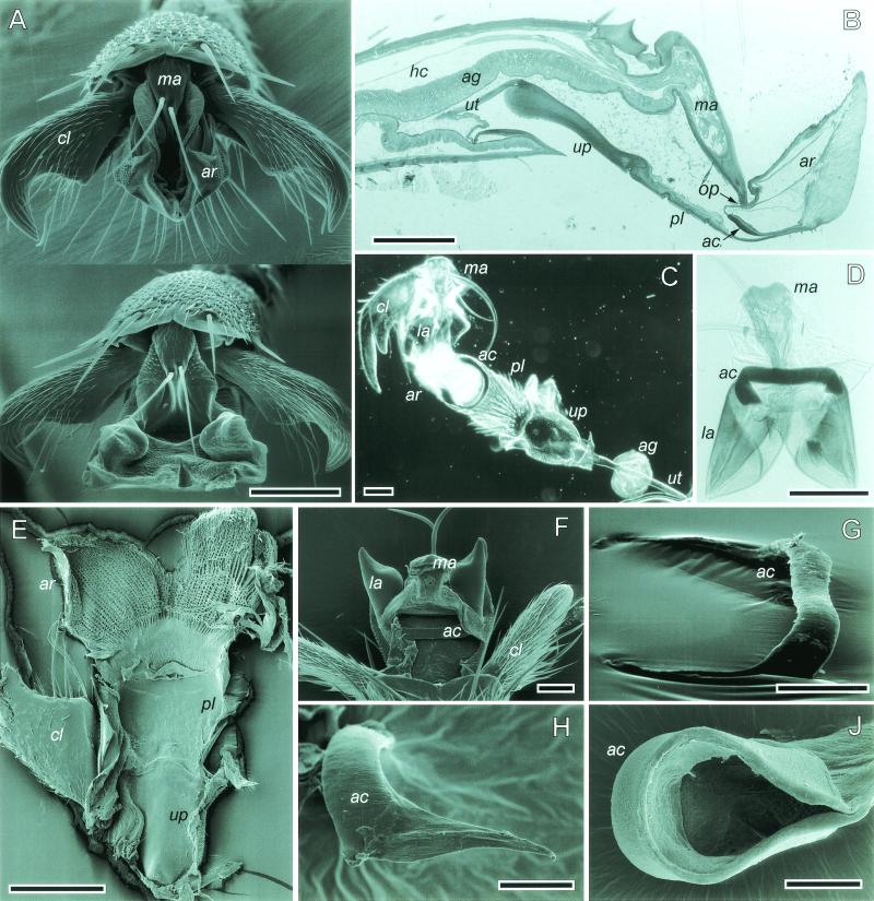
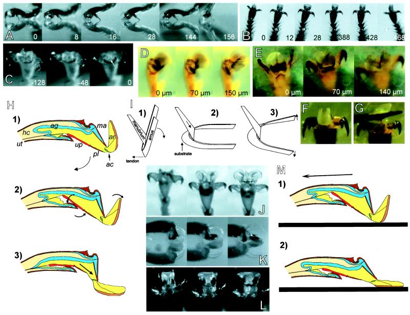
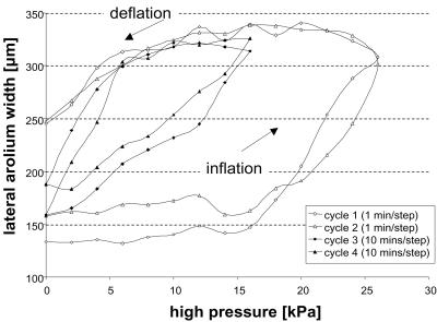
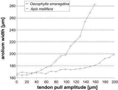
Hymenoptera attach to smooth surfaces with a flexible pad, the arolium, between the claws. Here we investigate its movement in Asian weaver ants (Oecophylla smaragdina) and honeybees (Apis mellifera). When ants run upside down on a smooth surface, the arolium is unfolded and folded back with each step. Its extension is strictly coupled with the retraction of the claws. Experimental pull on the claw-flexor tendon revealed that the claw-flexor muscle not only retracts the claws, but also moves the arolium. The elicited arolium movement comprises (i) about a 90 degrees rotation (extension) mediated by the interaction of the two rigid pretarsal sclerites arcus and manubrium and (ii) a lateral expansion and increase in volume. In severed legs of O. smaragdina ants, an increase in hemolymph pressure of 15 kPa was sufficient to inflate the arolium to its full size. Apart from being actively extended, an arolium in contact also can unfold passively when the leg is subject to a pull toward the body. We propose a combined mechanical-hydraulic model for arolium movement: (i) the arolium is engaged by the action of the unguitractor, which mechanically extends the arolium; (ii) compression of the arolium gland reservoir pumps liquid into the arolium; (iii) arolia partly in contact with the surface are unfolded passively when the legs are pulled toward the body; and (iv) the arolium deflates and moves back to its default position by elastic recoil of the cuticle.


Figures

Figure 1
Figure 1
(A) O. smaragdina arolium in the retracted (Top) and the extended phases (Bottom). (B) Sagittal section of a honeybee hind-leg pretarsus. (C and D) Whole mounts of pretarsi in A. mellifera and O. smaragdina, respectively. (E) O. smaragdina, inner view of ventral arolium cuticle. (F) O. smaragdina, dorsal view into opened arolium. (G and H) O. smaragdina, dissected arcus, lateral views. (H) Nonsclerotized arcus arm. (I) A. mellifera, dissected arcus with attached arolium cuticle. ac, arcus; ag, arolium gland; ar, arolium; cl, claw; hc, hemocoel; la, lateral arolium walls; ma, manubrium; pl, planta; up, unguitractor plate; ut, unguitractor tendon; op, opening. [Bars = 100 μm (AE), 50 μm (F, G, and I), and 20 μm (H).]
Figure 2
Figure 2
(AC) High-speed recordings of steps taken upside down on a glass plate (numbers indicate time in milliseconds). (A) A. mellifera, lateral view. (B) A. mellifera, ventral view. (C) O. smaragdina, arolium deflation before detachment (arrow indicates point of detachment). (D and E) O. smaragdina, experimental pull on the unguitractor tendon; numbers indicate the amplitude of the tendon pull (“0” is defined as the tendon-pull amplitude where the first pretarsus movement was visible). (D) Pretarsus lateral view. (E) Ventral view, arolium surface focused. (F) A. mellifera, arolium at maximal pull of the tendon (200 μm). (G) Same as F, arolium spread laterally by application of upward pressure to the planta with an insect pin. (H) Model of arolium extension caused by the contraction of the claw-flexor muscle. For abbreviations see legend for Fig. 1. (I) Model of the interaction between the two arolium sclerites, arcus and manubrium. (JL) Passive extension of arolium in contact with a glass surface. (J) O. smaragdina, pull of severed leg in the direction toward the body. (K and L) Pull of legs of freshly killed A. mellifera toward the body, lateral and frontal view of arolium, respectively. (M) Model of passive arolium extension caused by substratum contact and horizontal pull of the leg.
Figure 3
Figure 3
Experimental arolium inflation and deflation by applied pressure in O. smaragdina.
Figure 4
Figure 4
Lateral arolium extension caused by experimental pull on the unguitractor tendon in O. smaragdina and A. mellifera.

References

    1. Eisner T, Aneshansley D J. Proc Natl Acad Sci USA. 2000;97:6568–6573. - PMC - PubMed
    1. Federle W, Rohrseitz K, Hölldobler B. J Exp Biol. 2000;203:505–512. - PubMed
    1. Snodgrass R E. Anatomy of the Honey Bee. Ithaca, NY: Cornell Univ. Press; 1956.
    1. Heming B S. Can J Zool. 1971;49:91–108. - PubMed
    1. Walker G, Yue A B, Ratcliffe J. J Zool. 1985;205:297–307.

Publication types

LinkOut - more resources