. 2001 Jun 1;21(11):3705-14.
doi: 10.1523/JNEUROSCI.21-11-03705.2001.

Time-dependent reversal of long-term potentiation by low-frequency stimulation at the hippocampal mossy fiber-CA3 synapses

Affiliations

Time-dependent reversal of long-term potentiation by low-frequency stimulation at the hippocampal mossy fiber-CA3 synapses

Y L Chen et al. J Neurosci. .

Abstract

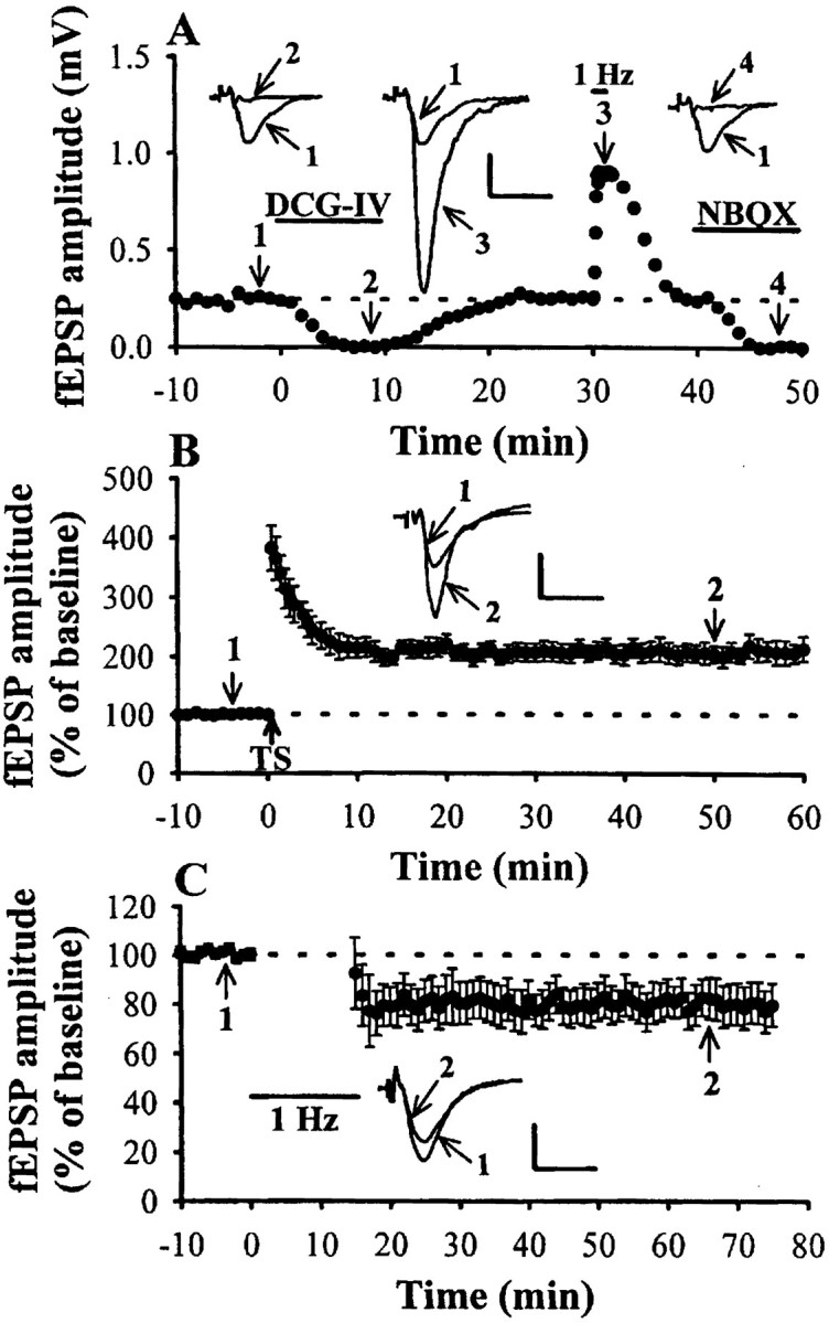
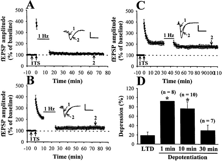
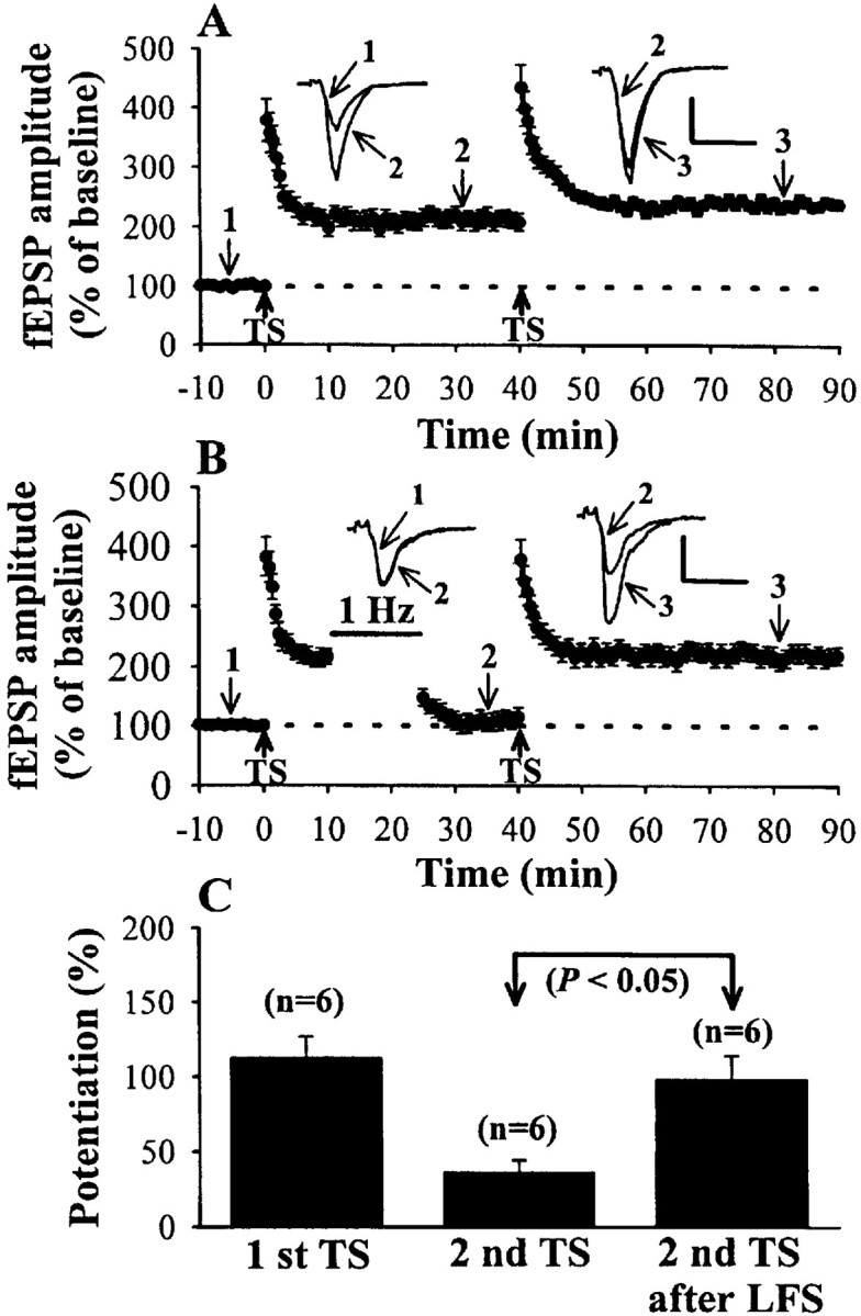
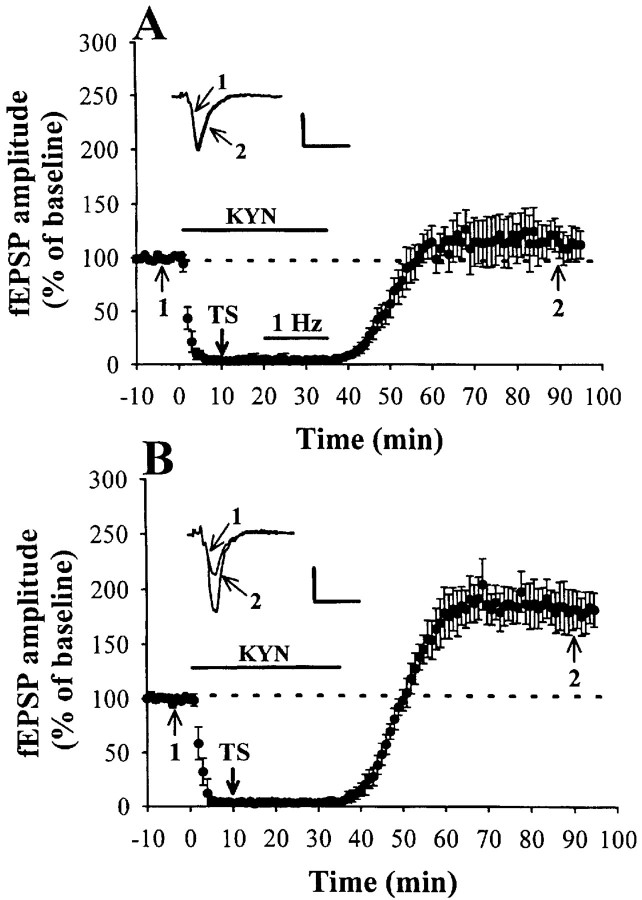
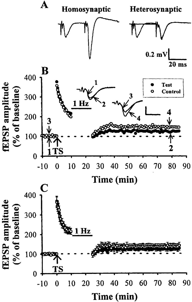
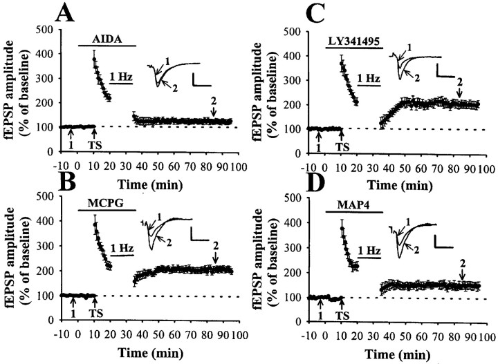
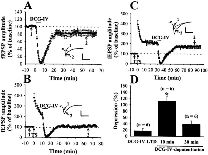
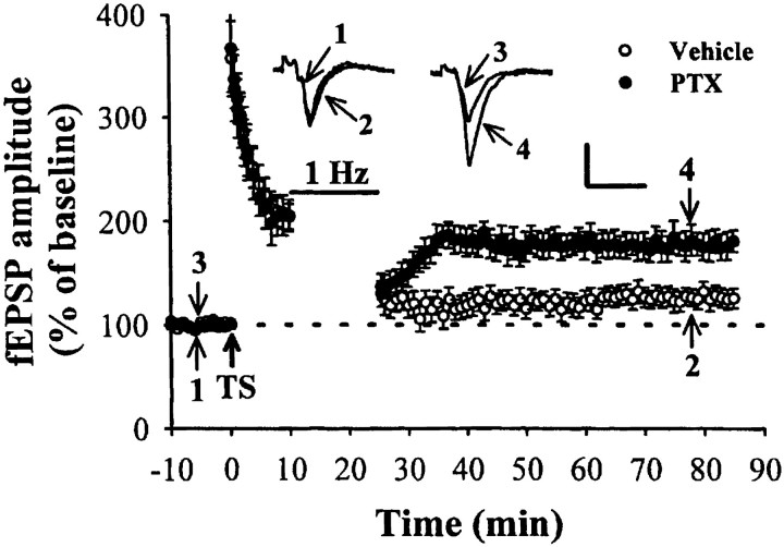
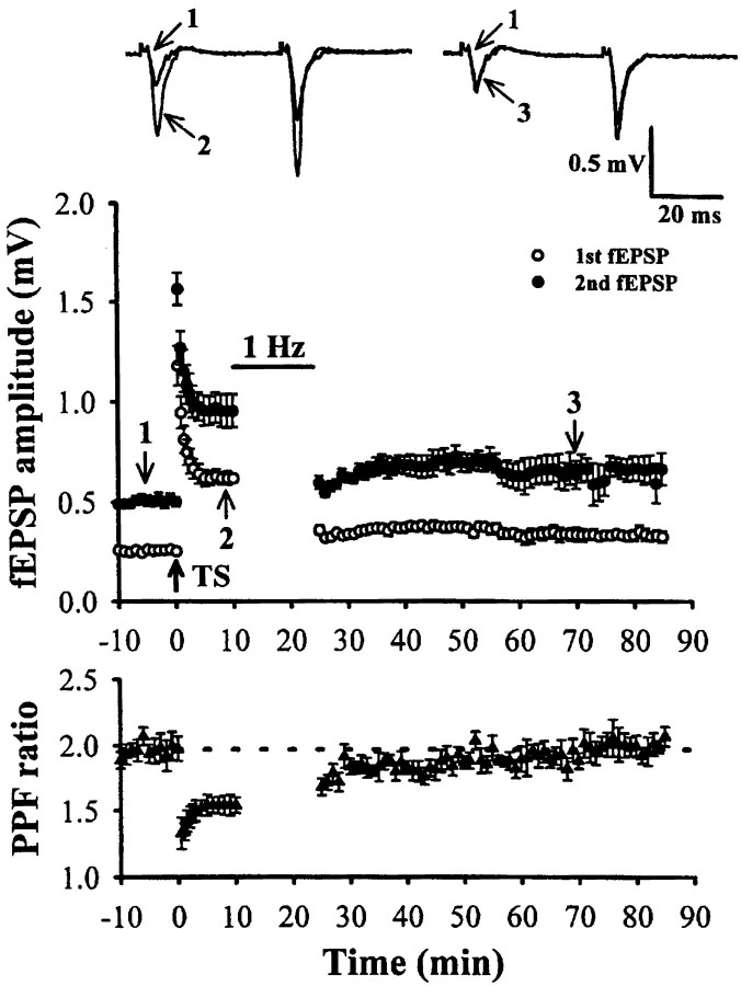
Using mouse hippocampal slices, we studied the induction of depotentiation of long-term potentiation (LTP) at the mossy fiber synapses onto CA3 pyramidal neurons. A long train of low-frequency (1 Hz/900 pulses) stimulation (LFS) induced a long-term depression of baseline synaptic transmission or depotentiation of previously established LTP, which was reversible and was independent of NMDA receptor activation. This LFS-induced depotentiation was observed when the stimulus was delivered 1 or 10 min after LTP induction. However, when LFS was applied at 30 min after induction, significantly less depotentiation was found. The induction of depotentiation on one input was associated with a heterosynaptic reverse of the LTP induced previously on a separate pathway. In addition, this LFS-induced depotentiation appeared to be mediated by the activation of group 2 metabotropic glutamate receptors (mGluRs), because it was mimicked by the bath-applied group 2 agonist (2S,2'R,3'R)-2-(2', 3'-dicarboxycyclopropyl) glycine and was specifically inhibited by the group 2 antagonists (S)-alpha-methyl-4-carboxyphenylglycine and (alphaS)-alpha-amino-alpha-(1S,2S)-2-carboxycyclopropyl-9H-xanthine-9-propanic acid. Moreover, the induction of depotentiation was entirely normal when synaptic transmission is blocked by glutamate receptor antagonist kynurenic acid and was associated with a reversal of paired-pulse facilitation attenuation during LTP expression. Pretreatment of the hippocampal slices with G(i/o)-protein inhibitor pertussis toxin (PTX) prevented the LFS-induced depotentiation. These results suggest that the activation of presynaptic group 2 mGluRs and in turn triggering a PTX-sensitive G(i/o)-protein-coupled signaling cascade may contribute to the LFS-induced depotentiation at the mossy fiber-CA3 synapses.


Figures

Fig. 1.
Fig. 1.
The induction of mossy fiber LTP and LTD.A, An example of pharmacological and physiological characterization of mossy fiber synaptic responses. To stimulate mossy fiber inputs, a bipolar stimulating electrode was placed in the stratum granulosum of dentate gyrus, and extracellular fEPSPs were recorded from the stratum lucidum of the CA3 region. The selective group 2 mGluR agonist DCG-IV was used to confirm that the responses were mediated by mossy fibers. Application of DCG-IV (0.5 μm) in the perfusion medium quickly and completely suppressed mossy fiber-evoked fEPSPs. The mossy fiber synapse also exhibits a greater frequency facilitation behavior. Increasing the stimulus frequency from 0.033 to 1 Hz caused pronounced frequency facilitation. This facilitation was completely reversed upon returning to the control stimulus frequency. At the end of the experiment, NBQX (20 μm) was applied.B, The time course and magnitude of mossy fiber LTP. High-frequency TS to the mossy fibers resulted in a large post-tetanic potentiation lasting for several minutes after TS, followed by stable LTP expression (n = 12).C, Mossy fiber LTD induced by LFS. Summary of nine experiments showing that LFS at 1 Hz for 15 min elicits LTD. The superimposed fEPSP in the inset of each graph illustrates respective recordings from example experiments taken at the time indicated by the numbers. All experiments were done in the presence of the NMDA receptor antagonist d-APV (50 μm). Upward arrows indicate application of TS. Horizontal bars indicate the period of the delivery of LFS or pharmacological agents as indicated. The horizontal dashed lines indicate the average value of the normalized amplitude during the control period. Calibration: 0.2 mV, 10 msec.
Fig. 2.
Fig. 2.
Time-dependent reversal of LTP by LFS.A–C, Summary of experiments in which LTP was induced at the mossy fiber–CA3 synapses. LFS (1 Hz, 15 min) was applied at various times after LTP induction: A, 1 min delay (n = 8); B, 10 min delay (n = 10); or C, 30 min delay (n = 7). D, Histogram comparing effects of LFS applied to naive synapses (LTD) or applied 1, 10, or 30 min after LTP induction. The data of LTD are taken from Figure1C. The magnitude of LTD was calculated at 40 min after the end of LFS at naive synapses. The magnitude of 1 min delay depotentiation was calculated by comparing the synaptic responses at 40 min after the end of LFS from the experiments illustrated in A with the magnitude of LTP at 50 min from the experiments illustrated in Figure 1B. Because both sets of data have variance, it is not possible to calculate an SE of this depotentiation value. Statistical analysis using ANOVA indicates that this value is significantly different from LTP at naive synapses (p < 0.05). The magnitude of 10 or 30 min delay depotentiation was calculated by comparing the synaptic responses at 40 min after the end of LFS from the experiments illustrated in B orC with the individual baseline magnitude just before each LFS application. Horizontal bars indicate the period of the delivery of LFS or pharmacological agents as indicated.Asterisks represent the significant difference from LTD group (p < 0.05). Calibration: 0.2 mV, 10 msec.
Fig. 3.
Fig. 3.
The depressed synapses can be potentiated.A, Summary of six experiments in which TS was applied twice with a 40 min interval. Note that the first TS nearly saturates the LTP; little additional potentiation is caused by a second TS.B, Summary of six experiments identical to those shown in A with the exception that LFS (1 Hz, 15 min) was delivered 10 min after the first TS. In this case, subsequent TS 40 min later was able to reverse the synaptic depression caused by LFS.C, Summary data in which the magnitude of the potentiation measured 40 min after each TS was calculated relative to the baseline period before each TS applied. In control experiments, as illustrated in A, we found that the first TS produced a potentiation of 112.3 ± 14.5% above baseline, whereas the second TS produced an additional increase of only 34.5 ± 8.7%. In the experiments shown in B, in which the second TS followed LFS, it caused a potentiation of 98.1 ± 16.3% above baseline (p < 0.05). Calibration: 0.2 mV, 10 msec.
Fig. 4.
Fig. 4.
Postsynaptic ionotropic glutamate receptor activation is not required for the induction of LFS-induced depotentiation. A, The time course of experiments in which mossy fiber depotentiation was induced in the absence of excitatory synaptic transmission. After complete blockade of fEPSPs with kynurenic acid (KYN; 20 mm), high-frequency TS was applied, and LFS was given 10 min after TS. Washout of kynurenic acid was started at the end of LFS. Note that, after washout of kynurenic acid, the fEPSPs recovered to near-baseline level (n = 6). B, Summary of six experiments identical to those shown in A, with the exception that LFS was not delivered after TS. In this case, the synaptic responses exhibited LTP after washout of kynurenic acid.Horizontal bars indicate the period of the delivery of LFS or kynurenic acid. Calibration: 0.2 mV, 10 msec.
Fig. 5.
Fig. 5.
Heterosynaptic reversal of LTP by LFS.A, An example showing that two stimulatory electrodes were used to activate two independent groups of afferents. Field EPSPs were evoked by paired stimulations applied at 30 msec intervals to the first and/or second afferents. Paired-pulse facilitation was present when stimuli were applied twice to the same afferent (homosynaptic facilitation) but not when the stimuli were applied to different afferents (no heterosynaptic facilitation). B, Example of an experiment showing that LTP was first induced on two afferents by coactivation, followed by LFS (1 Hz, 15 min) applied to only one afferent (test pathway). This resulted in a homosynaptic reversal of LTP at the test pathway but also in a heterosynaptic reversal of LTP induced previously at the other control pathway. The superimposed fEPSP in the inset illustrates respective recordings from example experiments taken at the time indicated bynumbers. Horizontal bars denote the period of the delivery of LFS. Calibration: 0.5 mV, 10 msec.C, Summary of data from six experiments performed as inB.
Fig. 6.
Fig. 6.
Application of group 2 mGluR antagonists selectively prevents the LFS-induced depotentiation. A, Summary of six experiments in which the group 1 mGluR antagonist AIDA (250 μm) was applied 10 min before and left until the end of LFS. AIDA does not affect the LFS-induced depotentiation.B, Summary of six experiments in which LFS-induced depotentiation was inhibited by the nonselective group 2 mGluR antagonist MCPG (250 μm). C, Pooled data from six experiments in which application of the selective group 2 mGluR antagonist LY341495 (3 μm), before TS and left until the end of LFS, results in an inhibition of the induction of LFS-induced depotentiation. D, Summary of eight experiments in which LFS-induced depotentiation was partially but significantly inhibited by the group 3 mGluR antagonist MAP4 (100 μm). Note that only group 2 mGluR antagonists could completely block the LFS-induced depotentiation. Horizontal bars indicate the period of the delivery of LFS or pharmacological agents as indicated. Calibration: 0.5 mV, 10 msec.
Fig. 7.
Fig. 7.
Time-dependent reversal of LTP can be induced by the bath-applied group 2 mGluR agonist DCG-IV.A, Application of DCG-IV (5 μm) for 5 min in the bath medium caused LTD that lasted over 60 min after washout of agonist (n = 6). B,C, DCG-IV (5 μm) was applied at various times after LTP induction: B, 10 min after (n = 8); or C, 30 min after (n = 6). D, Histogram compares the effect of DCG-IV applied to naive synapses (DCG-IV-LTD) or applied 10 or 30 min after LTP induction. The magnitude of DCG-IV LTD was calculated at 40 min after washout of DCG-IV at naive synapses. The magnitude of 10 or 30 min delay DCG-IV-induced depotentiation was calculated by comparing the synaptic responses at 40 min after washout of DCG-IV from the experiments illustrated in B orC with the individual baseline magnitude just before DCG-IV application. Note that DCG-IV erased potentiation when delivered 10 min after the TS but was without effect when applied 30 min after. Calibration: 0.5 mV, 10 msec.
Fig. 8.
Fig. 8.
PTX pretreatment blocks the induction of LFS-induced depotentiation. Mossy fiber fEPSP amplitude plotted as a function of time for each evoked potential observed during the course of experiments examining depotentiation induction by LFS in control vehicle- (n = 5) or PTX- (5 μg/ml) pretreated slices (n = 5). LTP could be successfully induced by high-frequency TS in all tested control vehicle- and PTX-pretreated slices, whereas the induction of LFS-induced depotentiation is impaired in PTX-pretreated slices. Calibration: 0.5 mV, 10 msec.
Fig. 9.
Fig. 9.
LFS-induced depotentiation reverses the reduction of PPF during LTP expression. TS-induced LTP is accompanied by attenuation of PPF. When LFS was delivered 10 min after, LTP induction significantly reversed PPF attenuation close to baseline values. Effects of TS and LFS on the PPF ratio calculated from the amplitude of the second of two fEPSPs divided by the first fEPSP amplitude to paired-pulse stimulation at an interstimulus interval of 40 msec. The superimposed fEPSPs in the top panel illustrate respective recordings from example experiments taken at the time indicated by numbers.

Similar articles

Cited by

References

    1. Amaral DG, Witter MP. The three-dimensional organization of the hippocampal formation: a review of anatomical data. Neuroscience. 1989;31:571–591. - PubMed
    1. Anwyl R. Metabotropic glutamate receptors: electrophysiological properties and role in plasticity. Brain Res Rev. 1999;29:83–120. - PubMed
    1. Arai A, Kessler M, Lynch G. The effects of adenosine on the development of long-term potentiation. Neurosci Lett. 1990a;119:41–44. - PubMed
    1. Arai A, Larson J, Lynch G. Anoxia reveals a vulnerable period in the development of long-term potentiation. Brain Res. 1990b;511:353–357. - PubMed
    1. Barrionuevo G, Schottler F, Lynch G. The effects of repetitive low frequency stimulation on control and “potentiated” responses in the hippocampus. Life Sci. 1980;27:2385–2391. - PubMed

Publication types

MeSH terms

Substances

LinkOut - more resources