. 2001 Jun 1;21(11):3771-9.
doi: 10.1523/JNEUROSCI.21-11-03771.2001.

Metabotropic glutamate receptor subtypes 1 and 5 are activators of extracellular signal-regulated kinase signaling required for inflammatory pain in mice

Affiliations

Metabotropic glutamate receptor subtypes 1 and 5 are activators of extracellular signal-regulated kinase signaling required for inflammatory pain in mice

F Karim et al. J Neurosci. .

Abstract

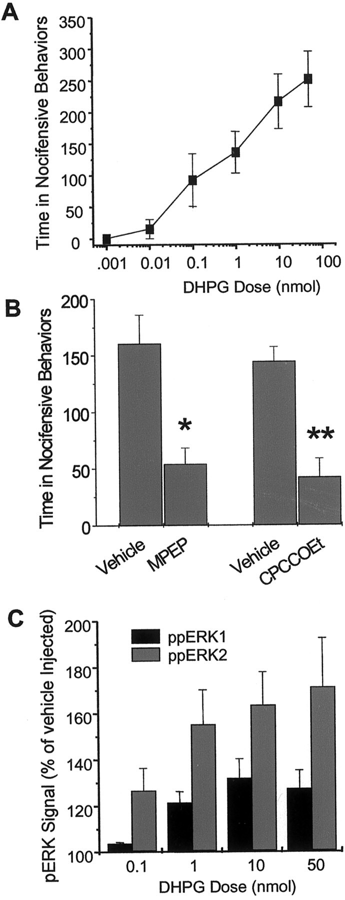
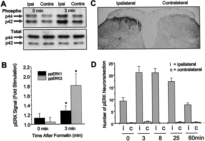
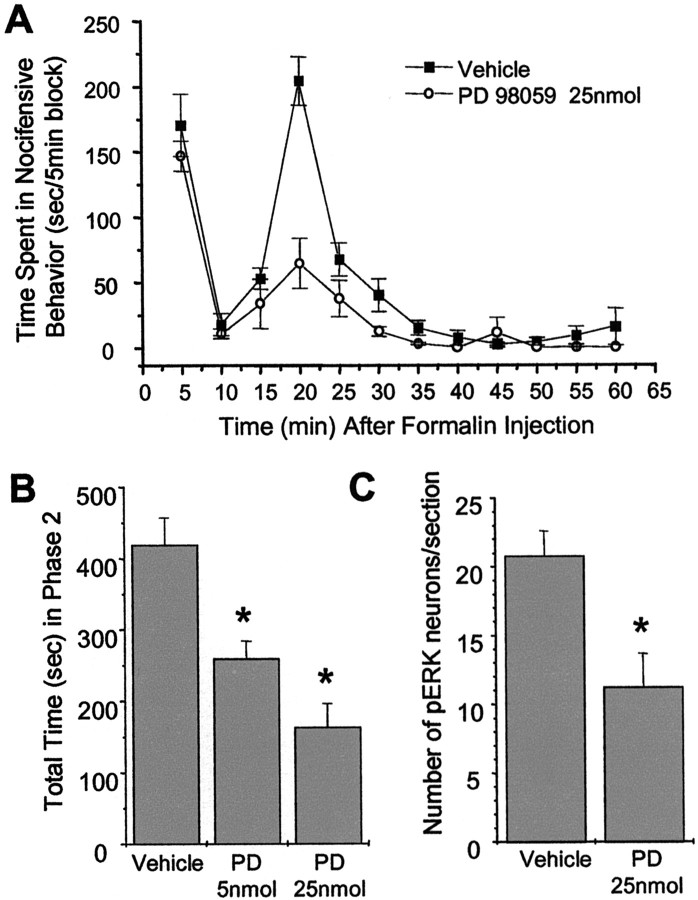
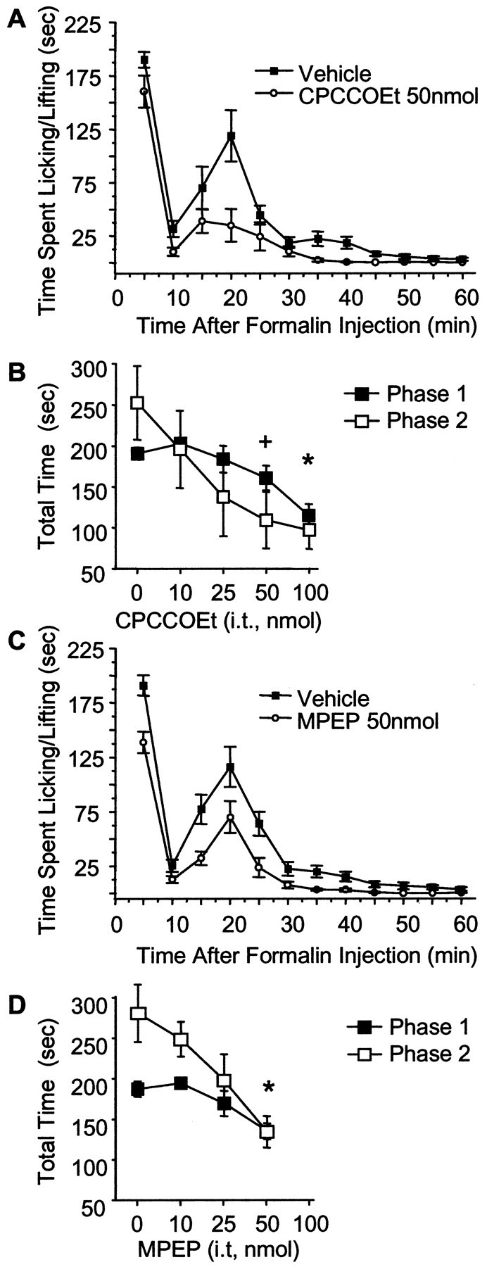
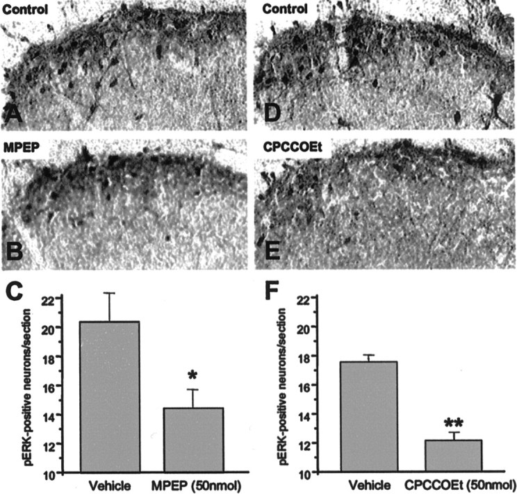
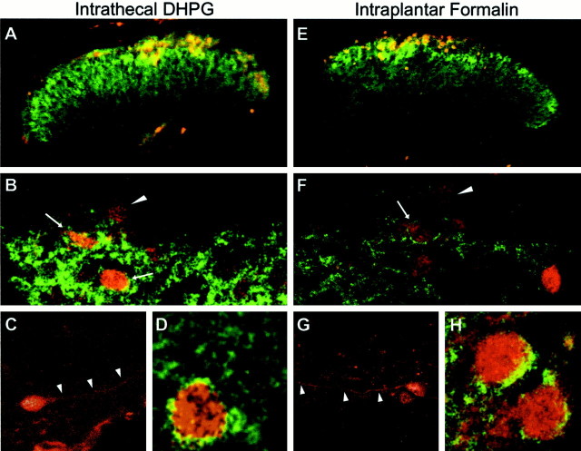
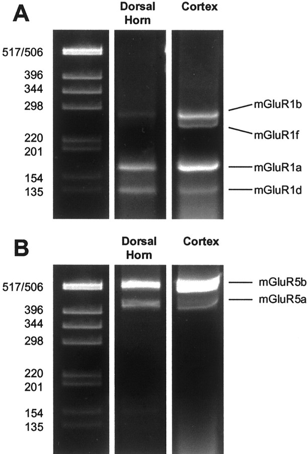
Metabotropic glutamate receptors are expressed abundantly in the spinal cord and have been shown to play important roles in the modulation of nociceptive transmission and plasticity. Most previous studies have focused on the group I metabotropic glutamate receptors (mGluR1 and mGluR5) and activation of phospholipase C signaling by these receptors in modulating nociception. Recently, it was shown that the extracellular signal-regulated kinases (ERKs)/mitogen-activated protein kinases are activated in spinal cord dorsal horn neurons in response to stimulation of nociceptors and that ERK signaling is involved in nociceptive plasticity. In the present studies, we sought to test the hypothesis that group I mGluRs modulate nociceptive transmission or plasticity via modulation of ERK signaling in dorsal horn neurons. We show that activation of mGluR1 and mGluR5 leads to activation of ERK1 and ERK2 in the spinal cord. Furthermore, we find that inflammation-evoked ERK activation, which is required for nociceptive plasticity, is downstream of mGluR1 and mGluR5. Finally, we show colocalization of group I mGluRs with activated ERK in dorsal horn neurons. These results show that mGluR1 and mGluR5 are activated in dorsal horn neurons in response to peripheral inflammation and that activation of these group I mGluRs leads to activation of ERK1 and ERK2, resulting in enhanced pain sensitivity.


Figures

Fig. 1.
Fig. 1.
The group 1 mGluR agonist DHPG increases nociceptive behavior and activates ERK1/2 in mouse spinal cord dorsal horn. A, Total time (in seconds) spent in spontaneous nocifensive behaviors after intrathecal injection of (RS)-DHPG. Mice were given a single intrathecal injection of various doses of (RS)-DHPG, and the time spent in nocifensive behaviors was recorded for 5 min.Points represent the mean ± SEM;n = 4–5 animals per dose. B, Effect of the mGluR5 antagonist MPEP and the mGluR1 antagonist CPCCOEt on DHPG-induced spontaneous nocifensive behavior. Mice were pretreated intrathecally with 50 nmol of either antagonist 15 min before intrathecal (RS)-DHPG (1 nmol), and the time spent in nocifensive behaviors was recorded for 5 min. Pointsrepresent the mean ± SEM; n = 4 animals per dose. *p < 0.05; **p < 0.01.C, Immunoblot analysis of phosphorylated ERK1 and ERK2 bands in mouse spinal cord homogenates from mice injected intrathecally with (RS)-DHPG. Points represent the mean ± SEM densities of phospho-ERK1 and phospho-ERK2 bands normalized to total ERK for each sample from four separate experiments. In C, all points are significant atp < 0.05 compared with vehicle-injected controls, with the exception of the p44 signal at the 0.1 nmol dose.
Fig. 2.
Fig. 2.
Subcutaneous injection of formalin in the hindpaw activates ERK1/2 ipsilaterally in the spinal cord.A, Representative immunoblot of mouse spinal cord homogenates using a phospho-ERK1/2 antibody (top) or total ERK1/2 antibody (bottom). Ipsilateral and contralateral lumbar spinal cord samples were taken at various time points after injection of 2% formalin. The arrows show the position of the 44 kDa (ERK1) and 42 kDa (ERK2) ERK isoforms.B, Quantitation of ERK activation after injection of formalin (2–5%). The phospho-ERK (pERK) bands were densitized and normalized to total ERK immunoblotted from the same samples using an anti-total ERK1/2 antibody and are expressed as fold stimulation of phospho-ERK on the ipsilateral side compared with the contralateral side. n = 7. *p < 0.05 compared with the contralateral side.C, Immunocytochemistry showing ipsilateral activation of ERKs 8 min after formalin injection. D, Time course of the number of dorsal horn neurons positive for phosphorylated ERK after formalin treatment. Data represent the mean ± SEM of 20 sections taken from two animals. Vehicle-injected animals showed no significant phospho-ERK staining (data not shown).
Fig. 3.
Fig. 3.
The MEK inhibitor PD98059 attenuates the second phase of formalin-induced nociceptive behavior and decreases ERK activation. A, Effect of a 20 min pretreatment with a single intrathecal injection of PD98059 (25 nmol) in the mouse formalin test. B, Effect of 5 and 25 nmol doses of PD98059 in the second phase of the formalin test (15–30 min). Each barrepresents the mean ± SEM; n = 5–7. *p < 0.05. C, Effect of PD98059 on activation of spinal ERK after formalin injection determined by cell counts of phospho-ERK-positive neurons. n = 3 each. *p < 0.05.
Fig. 4.
Fig. 4.
Intrathecal MPEP and CPCCOEt attenuate formalin-induced nociceptive behavior. A,C, Time course graphs of 15 min pretreatment with 50 nmol of CPCCOEt and 50 nmol of MPEP. B,D, Dose–response curves of CPCCOEt (B) and MPEP (D) on the first phase (the 5 min time point; filled squares) and the second phase (sum of 15–30 min points; open squares). Points represent the mean ± SEM;n = 5 - 11 per dose.+p < 0.05 for phase 2 only; *p < 0.05 for phase 1 and phase 2.
Fig. 5.
Fig. 5.
Intrathecal MPEP and CPCCOEt attenuate formalin-induced ERK activation in the spinal cord dorsal horn. Representative spinal cord lumbar sections immunostained with phospho-ERK antibody after a unilateral injection of formalin subcutaneously into the right hindpaw. Mice were pretreated with either vehicle (A) (100 mm HEPES, pH 7.4) or MPEP (B) (50 nmol). D andE show staining of spinal cords from vehicle- and CPCCOEt-pretreated mice. Quantitation of the total number of phospho-ERK-positive neurons in the dorsal horn of the lumbar spinal cord after pretreatment with either vehicle or MPEP (C) or vehicle or CPCCOEt (F) before subcutaneous formalin injection.Bars represent the mean ± SEM;n = 3–6 for each treatment. *p< 0.05; **p < 0.01.
Fig. 6.
Fig. 6.
Colocalization of phospho-ERK and mGluR5 in mouse dorsal horn after intrathecal DHPG (10 nmol) (A–D) or intraplantar formalin injection (E–H). A, E, Fluorescence images showing the distribution of mGluR5 (green) and phospho-ERK (red) immunoreactivity in the lumbar spinal cord dorsal horn 5 min after DHPG (A) or 8 min after 2% subcutaneous injection of formalin in the right hindpaw (E).B and F show higher-power examples of confocal images showing the distribution of mGluR5 (green) in relation to phospho-ERK (red) in the dorsal horn. Note that some phospho-ERK cells also have apparent membrane labeling for mGluR5 (arrows), whereas other phospho-ERK-positive cells contain no detectable mGluR5 (arrowheads).C and G show higher magnifications of phospho-ERK staining of dorsal horn neurons. Note the labeling of dendritic processes (arrowheads), which are typically seen when cells are observed using a confocal microscope.D and H show higher magnification of additional example neurons with apparent membrane labeling for mGluR5 and somatic phospho-ERK. These images are representative of similar results obtained from three separate animals.
Fig. 7.
Fig. 7.
Expression of various splice variants of mGluR1 and mGluR5 in mouse spinal cord dorsal horn. RT-PCR amplification of the alternatively spliced forms of mGluR1 (A) and mGluR5 (B) from total RNA prepared from dorsal horn of the mouse spinal cord and mouse cortex. The position of predicted band sizes for mGluR1a, mGluR1b, mGluR1d, mGluR1f, mGluR5a, and mGluR5b are indicated. Molecular markers are shown in base pairs.

References

    1. Alvarez FJ, Villalba RM, Carr PA, Grandes P, Somohano PM. Differential distribution of metabotropic glutamate receptors 1a, 1b, and 5 in the rat spinal cord. J Comp Neurol. 2000;422:464–487. - PubMed
    1. Bordi F, Ugolini A. Involvement of mGluR(5) on acute nociceptive transmission. Brain Res. 2000;871:223–233. - PubMed
    1. Boxall SJ, Thompson SW, Dray A, Dickenson AH, Urban L. Metabotropic glutamate receptor activation contributes to nociceptive reflex activity in the rat spinal cord in vitro. Neuroscience. 1996;74:13–20. - PubMed
    1. Budai D, Larson AA. The involvement of metabotropic glutamate receptors in sensory transmission in dorsal horn of the rat spinal cord. Neuroscience. 1998;83:571–580. - PubMed
    1. Coderre TJ, Melzack R. The contribution of excitatory amino acids to central sensitization and persistent nociception after formalin-induced tissue injury. J Neurosci. 1992;12:3665–3670. - PMC - PubMed

Publication types

MeSH terms

Substances