. 2001 Jun 1;21(11):3830-8.
doi: 10.1523/JNEUROSCI.21-11-03830.2001.

A common exocytotic mechanism mediates axonal and dendritic outgrowth

Affiliations

A common exocytotic mechanism mediates axonal and dendritic outgrowth

S Martinez-Arca et al. J Neurosci. .

Abstract

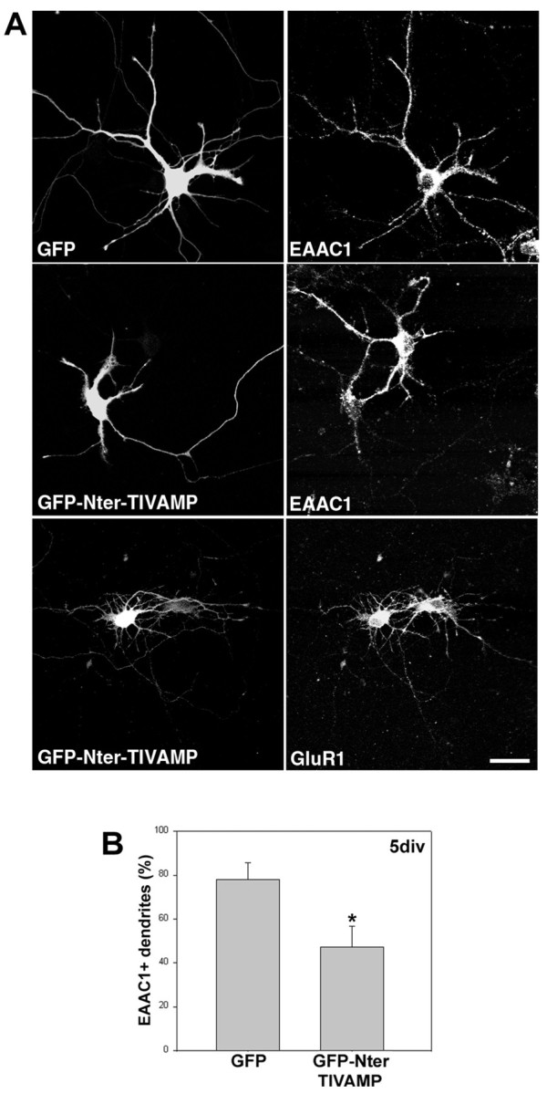
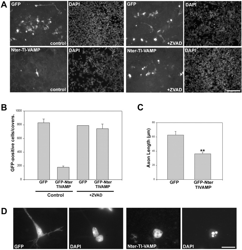
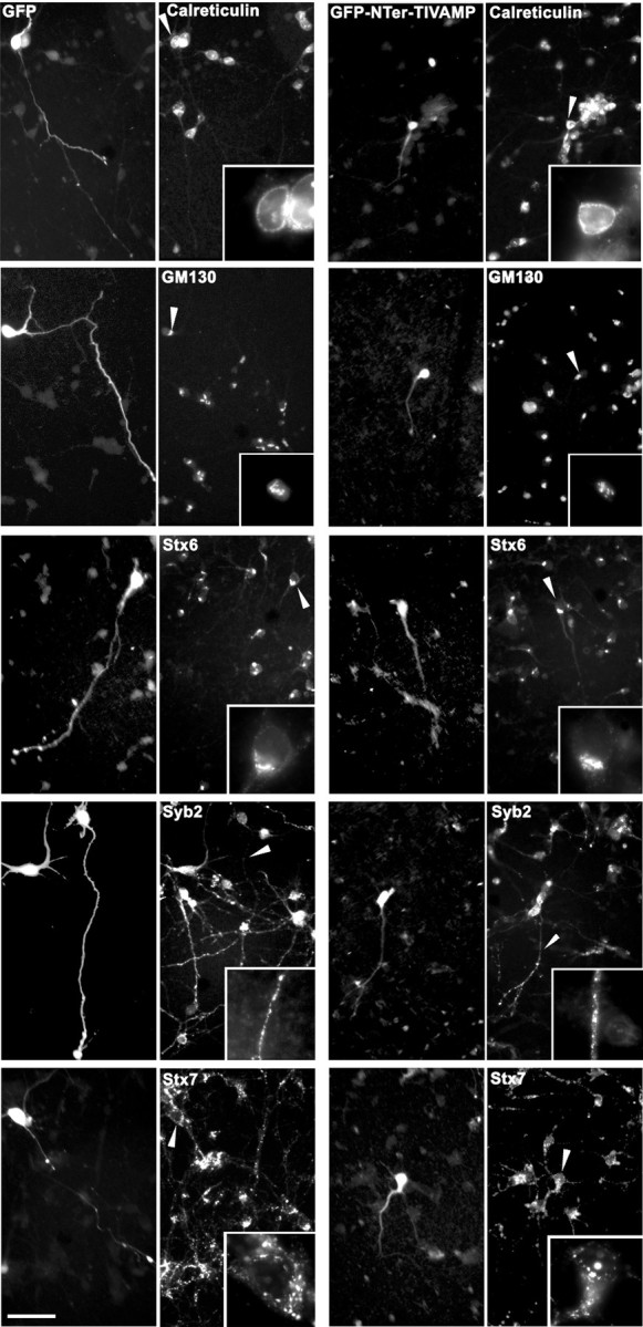
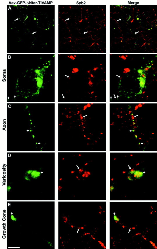
Outgrowth of the dendrites and the axon is the basis of the establishment of the neuronal shape, and it requires addition of new membrane to both growing processes. It is not yet clear whether one or two exocytotic pathways are responsible for the respective outgrowth of axons and dendrites. We have previously shown that tetanus neurotoxin-insensitive vesicle-associated membrane protein (TI-VAMP) defines a novel network of tubulovesicular structures present both at the leading edge of elongating dendrites and axons of immature hippocampal neurons developing in primary culture and that TI-VAMP is an essential protein for neurite outgrowth in PC12 cells. Here we show that the expression of the N-terminal domain of TI-VAMP inhibits the outgrowth of both dendrites and axons in neurons in primary culture. This effect is more prominent at the earliest stages of the development of neurons in vitro. Expression of the N-terminal domain deleted form of TI-VAMP has the opposite effect. This constitutively active form of TI-VAMP localizes as the endogenous protein, particularly concentrating at the leading edge of growing axons. Our results suggest that a common exocytotic mechanism that relies on TI-VAMP mediates both axonal and dendritic outgrowth in developing neurons.


Figures

Fig. 1.
Fig. 1.
Expression of the N-terminal domain of TI-VAMP inhibits axonal and dendritic growth. A, Hippocampal neurons from E18 rats transfected with GFP or GFP-Nter-TI-VAMP 4 hr after plating and fixed 24 hr later. Scale bar, 32 μm.B, Cells transfected as in A after 1 or 4 div were recorded 24 hr later, and the dendritic length was measured.C, Cells transfected as in A after 1 or 4 div were recorded 24 hr after transfection, and the number of dendrites on each cell was counted. D, Cells were transfected as in A after 1 div with GFP or GFP-Nter-TI-VAMP, and the axonal length was measured 24 hr later; shown are the mean values (±SEM) of between 40 and 60 analyzed cells. **p < 0.006; *p < 0.06.
Fig. 2.
Fig. 2.
Expression of the N-terminal domain of TI-VAMP affects the distribution of EAAC1 but not GluR1 in hippocampal neurons.A, Four-day-old hippocampal neurons from E18 rats were transfected with GFP or GFP-Nter-TI-VAMP, and after 24 hr they were fixed and stained for the indicated proteins. Note the expected dendritic localization of EAAC1 in control neurons and in neurons transfected with GFP (right top panel and nontransfected cell in the right middle panel) compared with its general lower expression and specifically its absence from the dendrites in cells expressing Nter-TI-VAMP (transfected cell in theright middle panel). By contrast, the level of expression and the localization of GluR1 were not affected by expression of GFP-Nter-TI-VAMP (compare the two cells in theright bottom panel). Scale bar, 21 μm.B, Cells transfected as in A were stained 24 hr later for EAAC1; shown are the mean values (±SEM) of percentage of GFP- or Nter-TI-VAMP-positive dendrites labeled also for EAAC1.
Fig. 3.
Fig. 3.
Expression of the N-terminal domain of TI-VAMP induces apoptosis. A, Corticostriatal neurons from intact embryonic brains were electroporated with the indicated constructs and cultured for 24 hr in the absence (left panels) or presence (right panels) of the caspase inhibitor zVAD. Observe the increase in the number of transfected cells in zVAD-treated Nter-TI-VAMP-electroporated cells compared with nontreated cells. In the case of GFP-electroporated cells, there is no difference between zVAD-treated or nontreated cells. Scale bar, 100 μm. B, Quantification of the apoptotic effect of the N-terminal domain of TI-VAMP in cells treated as inA; shown are the mean values (±SEM) of the number of positive cells on each coverslip. C, Quantification of the effect in axonal length of the expression of the N-terminal domain of TI-VAMP in cells treated as in A. Shown are the mean values (±SEM) of a minimum of 40 cells. D, Neurons infected with Aav carrying GFP or Aav carrying GFP-Nter-TI-VAMP fixed 3 d after infection. A representative cell of each type is shown. Note that the cell expressing GFP displays neurites and a normal nucleus compared with a noninfected cell, whereas the cell expressing Nter-TI-VAMP is round, with no neurites and presents a typical apoptotic nucleus as seen with DAPI staining. Scale bar, 20 μm.
Fig. 4.
Fig. 4.
Cells expressing the N-terminal domain of TI-VAMP show normal secretory and endocytic pathways. Corticostriatal neurons from E16 rats were infected with Aav carrying GFP or Aav carrying GFP-Nter-TI-VAMP, fixed 1 d after infection, and double-labeled for GFP and the indicated markers of the secretory (calreticulin, GM130, syntaxin 6, synaptobrevin 2) and endocytic (syntaxin 7) pathways. At this time, some Nter-TI-VAMP-expressing cells are not yet apoptotic, although they already present shorter neurites compared with GFP-expressing cells. Note, however, that the different markers localized similarly in GFP- and GFP-Nter-TI-VAMP-expressing neurons. Insets show higher magnifications of the areas indicated by the arrowheads. GM130, Golgi matrix protein of 130 kDa; Stx6, syntaxin 6;Syb2, synaptobrevin 2; Stx7, syntaxin 7. Scale bar, 60 μm (15 μm in insets).
Fig. 5.
Fig. 5.
Expression of ΔNter-TI-VAMP activates axonal growth. A, Intact brains from E13 mice (top panels) or corticostriatal neurons from E16 rats (bottom panels) were electroporated or infected with the indicated Aavs, respectively. Cells in primary culture were fixed after 2 (electroporation) or 3 div (Aavs). Note the punctate distribution in the cell body and along the axon of both full-length GFP-TI-VAMP and GFP-ΔNter-TI-VAMP and the fact that GFP-ΔNter-TI-VAMP-expressing cells present longer axons than cells expressing GFP-TI-VAMP. Scale bar: 20 μm (top panels); 60 μm (bottom panels). B, Quantification of the effect in axonal growth of the expression of ΔNter-TI-VAMP in electroporated neurons. Neurons expressing GFP, GFP-TI-VAMP, or GFP-ΔNter-TI-VAMP were fixed after the indicated times, and the length of their axons was measured. In the top panels the mean values (±SEM) of percentage of axons longer than 50 or 100 μm are shown from three independent experiments; the bottom panels show two representative experiments. C, Quantification of the effect on axonal growth of the expression of ΔNter-TI-VAMP in Aav-infected neurons. Neurons expressing the indicated constructs were fixed after 3 or 6 div, and their axonal length was measured; each panel shows a representative experiment. **p < 0.001; *p < 0.005.
Fig. 6.
Fig. 6.
GFP-ΔNter-TI-VAMP does not colocalize with synaptobrevin 2. Rat embryonic neurons were infected with Aav carrying GFP-ΔNter-TI-VAMP. After 6 div, the cells were fixed and permeabilized, incubated with a polyclonal antibody anti-GFP and with a monoclonal antibody anti-synaptobrevin 2 (Syb2), and observed by confocal microscopy. Low magnification images are shown inA. In all the other panels high magnification images of a cell body (B), an axon (C), a varicosity (D), and a growth cone (E), respectively, are shown. GFP-ΔNter-TI-VAMP (small arrows) does not colocalize with endogenous synaptobrevin 2 (B–E, large arrows) in any of the different neuronal domains. A significant amount of GFP-ΔNter-TI-VAMP was detected at the leading edge of the growth cone, in a region devoid of synaptobrevin 2. Scale bar:A, 90 μm; B, C,E, 4.6 μm; D, 3 μm.

References

    1. Advani RJ, Bae HR, Bock JB, Chao DS, Doung YC, Prekeris R, Yoo JS, Scheller RH. Seven novel mammalian SNARE proteins localize to distinct membrane compartments. J Biol Chem. 1998;273:10317–10324. - PubMed
    1. Advani RJ, Yang B, Prekeris R, Lee KC, Klumperman J, Scheller RH. VAMP-7 mediates vesicular transport from endosomes to lysosomes. J Cell Biol. 1999;146:765–775. - PMC - PubMed
    1. Atwal JK, Massie B, Miller FD, Kaplan DR. The TrkB-Shc site signals neuronal survival and local axon growth via MEK and P13-kinase. Neuron. 2000;27:265–277. - PubMed
    1. Bock JB, Scheller RH. SNARE proteins mediate lipid bilayer fusion. Proc Natl Acad Sci USA. 1999;96:12227–12229. - PMC - PubMed
    1. Bock JB, Klumperman J, Davanger S, Scheller RH. Syntaxin 6 functions in trans-Golgi network vesicle trafficking. Mol Biol Cell. 1997;8:1261–1271. - PMC - PubMed

Publication types

MeSH terms

Grants and funding

LinkOut - more resources