. 2001 Jun 1;21(11):3986-4001.
doi: 10.1523/JNEUROSCI.21-11-03986.2001.

Temporary inactivation of the retrosplenial cortex causes a transient reorganization of spatial coding in the hippocampus

Affiliations

Temporary inactivation of the retrosplenial cortex causes a transient reorganization of spatial coding in the hippocampus

B G Cooper et al. J Neurosci. .

Abstract

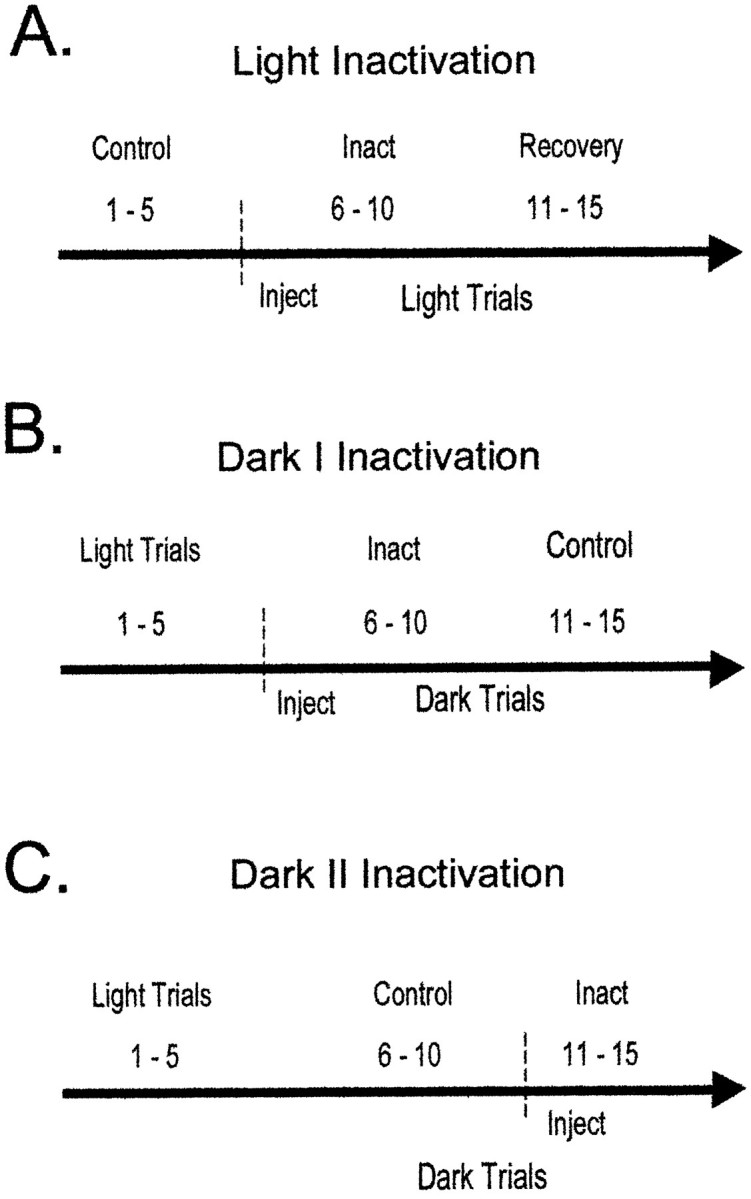
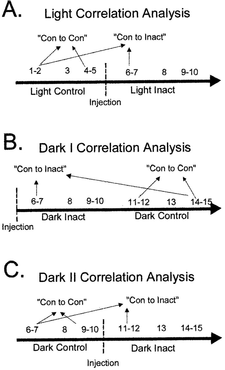
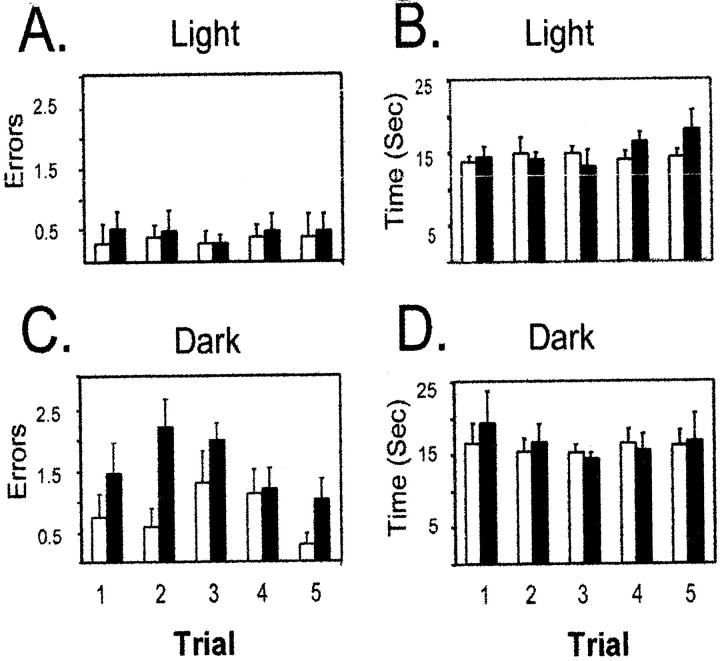
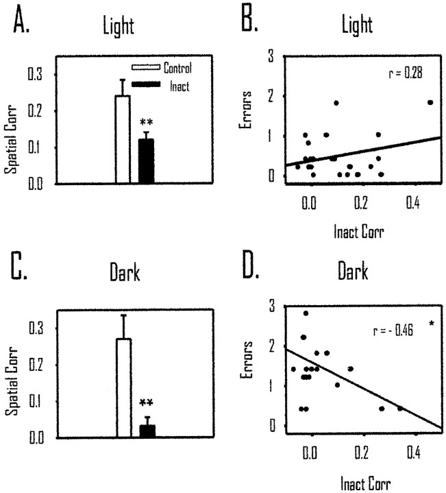
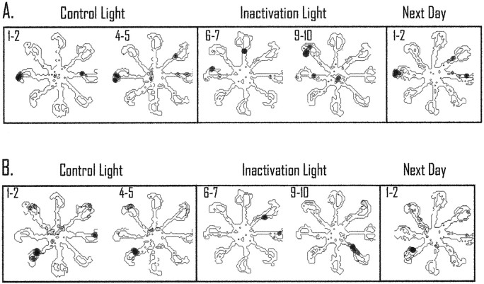
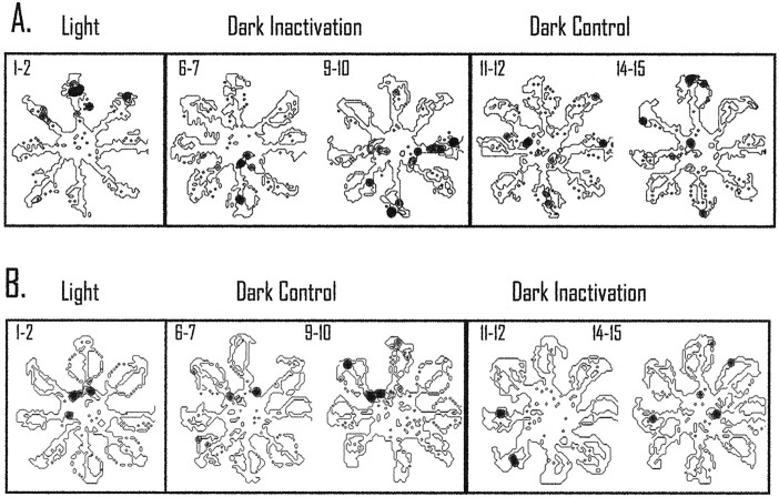
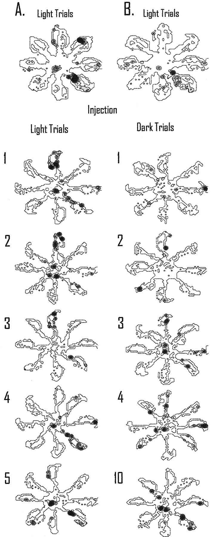
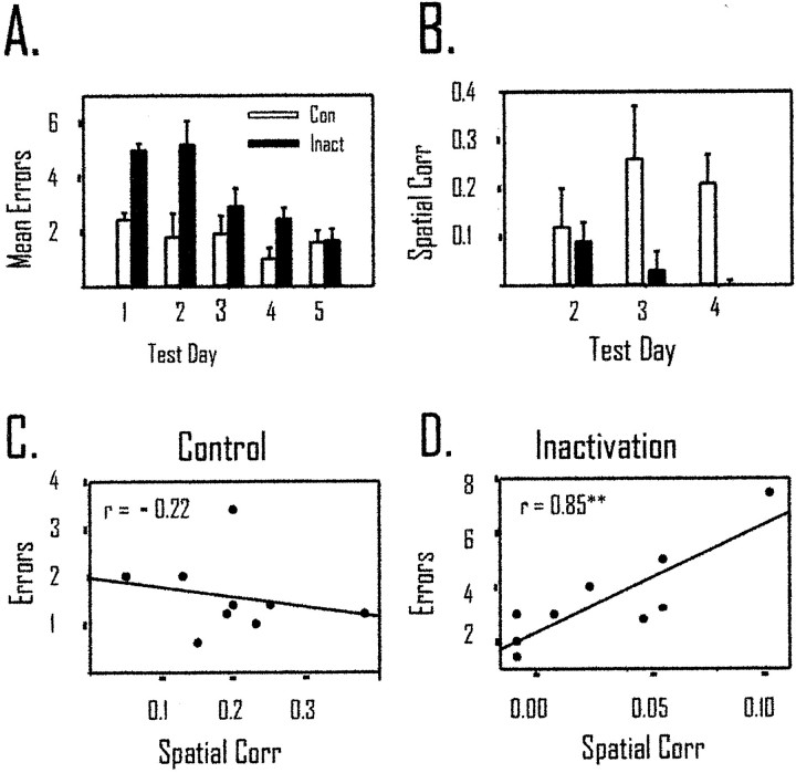
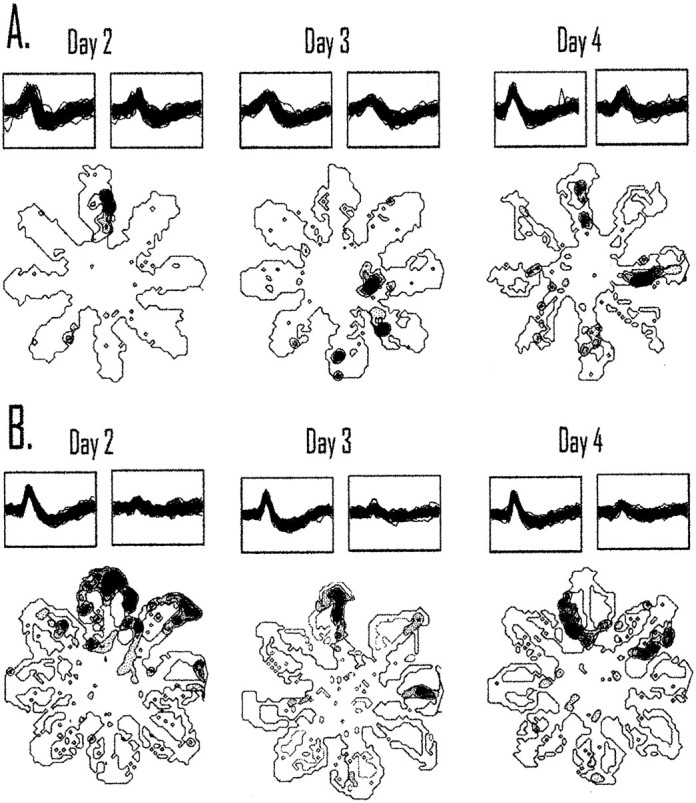
The ability to navigate accurately is dependent on the integration of visual and movement-related cues. Navigation based on metrics derived from movement is referred to as path integration. Recent theories of navigation have suggested that posterior cortical areas, the retrosplenial and posterior parietal cortex, are involved in path integration during navigation. In support of this hypothesis, we have found previously that temporary inactivation of retrosplenial cortex results in dark-selective impairments on the radial maze (Cooper and Mizumori, 1999). To understand further the role of the retrosplenial cortex in navigation, we combined temporary inactivation of retrosplenial cortex with recording of complex spike cells in the hippocampus. Thus, behavioral performance during spatial memory testing could be compared with place-field responses before, and during, inactivation of retrosplenial cortex. In the first experiment, behavioral results confirmed that inactivation of retrosplenial cortex only impairs radial maze performance in darkness when animals are at asymptote levels of performance. A second experiment revealed that retrosplenial cortex inactivation impaired spatial learning during initial light training. In both experiments, the normal location of hippocampal "place fields" was changed by temporary inactivation of retrosplenial cortex, whereas other electrophysiological properties of the cells were not affected. The changes in place coding occurred in the presence, and absence, of behavioral impairments. We suggest that the retrosplenial cortex provides mnemonic spatial information for updating location codes in the hippocampus, thereby facilitating accurate path integration. In this way, the retrosplenial cortex and hippocampus may be part of an interactive neural system that mediates navigation.


Figures

Fig. 1.
Fig. 1.
The injection procedure used for light inactivation and dark I and dark II inactivation. A dashed vertical line and Inject denote the time of injection; Inact indicates the trials used for the inactivation condition. A, In light inactivation, the first five trials serve as control trials for comparison with trials 6–10 that are inactivation trials. The final set of five trials was used to ensure that behavioral and electrophysiological changes returned toward control levels. B, In dark I inactivation, the injection occurred before the onset of dark trials.C, In dark II inactivation, the injection took place after the first five dark trials.
Fig. 2.
Fig. 2.
The trials used for the spatial correlation analysis during control and inactivation trials are displayed in the figure. The last five trials during light inactivation and the first five light trials for dark I and dark II inactivation are omitted from the spatial analysis; accordingly they are not included in the figure.A, For light inactivation, the firing rates on visited pixels during the first two trials of the control trials were correlated with the rates on visited pixels during the last two trials of the control condition (Con to Con). This provided the control stability assessment of place fields. To assess the effects of inactivation, the first two trials of the control trials were correlated with the first two inactivation trials (Con to Inact). B, The first two dark trials after inactivation were compared with the last two dark trials performed (Con to Inact) for the spatial correlation. Trials 11 and 12 were compared with trials 14 and 15 for the Con to Con spatial correlation. C, For dark II inactivation, the first two dark trials (6, 7) were compared with trials 9 and 10 for the Con to Con spatial correlation. The Con to Inact spatial correlation was derived from comparing trials 6 and 7 with the first two trials after inactivation (trials 11, 12).
Fig. 3.
Fig. 3.
Location of the tips and recording sites of the guide cannulas. A, Each filled circlecorresponds to a single guide cannula. Previous work and ink injections have shown that the spread of tetracaine is just slightly more than a 1 mm circumference around the injection site. Therefore, retrosplenial granular and agranular areas, cingulum bundle, and Oc2MM of posterior parietal cortex were likely affected by injections of tetracaine. B, Each filled circlecorresponds to two to eight cells recorded in that location. For electrode tracks that passed through the same area in different animals, a single filled circle is used to signify the recording site of multiple cells. The majority of cells (n = 43) were recorded from CA3 in the left hemisphere; a smaller number of cells (n = 15) were also recorded in CA1.
Fig. 4.
Fig. 4.
Temporary inactivation of retrosplenial cortex only impairs dark spatial memory performance. A, C, The average number of errors (±SEM) for control and inactivation trials is displayed. Inactivation of retrosplenial cortex did not change performance during light testing (A) but caused a significant impairment when animals were tested in darkness with retrosplenial cortex inactivated (C). B, D, The average time per choice on the maze was not affected by retrosplenial cortex inactivation during light (B) or dark (D) inactivation. Because the data obtained from dark I and dark II were similar, they were combined for the present analysis.
Fig. 5.
Fig. 5.
Temporary inactivation of retrosplenial cortex decreased after inactivation of retrosplenial cortex in light and dark but was only related to behavior during dark testing. A, The spatial correlation during con to con (Control) is significantly higher than that during con to inact (Inact; p < 0.01). This suggests that hippocampal place cells changed their preferred firing location during light inactivation. B, In light inactivation, there was no relationship between errors and the spatial correlation, suggesting that changes in place coding by hippocampal cells do not predict behavioral performance in the light.C, The spatial correlation in dark inactivation decreased significantly when retrosplenial cortex was inactivated (p < 0.01). D, There was a significant correlation between errors in darkness and changes in place coding by hippocampal place cells. *p < 0.05; **p < 0.01. Corr, Correlation.
Fig. 6.
Fig. 6.
Responses of two simultaneously recorded place cells during light inactivation. Pairs of trials are shown to illustrate the spatially selective activity that occurred during the con-to-con and con-to-inact spatial correlation. A, In this case the cell showed a preferred field on the western maze arm during trials 1 and 2 and during trials 4 and 5 (con-to-con trials). During retrosplenial cortex inactivation, the location of the field changed to firing on two arms and then began to fire on the northwestern maze arm in the subsequent trials. The preferred location for the cell did not return until the subsequent test day.B, This cell showed a similar consistent field during control light trials; during inactivation the field changed locations and then began to fire on the southeastern maze arm. Interestingly, the simultaneously recorded cells both rotated their preferred fields during inactivation but by different amounts. In both cases, the preferred field did not return until the next test day. All of the spatial plots omit cellular activity that is <20% of the maximum rate of the cell during the trials. The maximum rate is shown as dark areas, and shaded areas correspond to intermediate rates. This form of presentation is the same for Figures 7, 8, and 10. It should be noted that the small sample size for the spatial plots reduces the variability in the firing of the place cell (the animal only passes through the place field a total of four times, and if the field is directional the cell only has two opportunities to be active for a given plot). Accordingly, the firing-rate distribution is reduced substantially because of the small sample size.
Fig. 7.
Fig. 7.
Responses of two different place cells recorded in dark I and dark II inactivation conditions.A, This cell showed a preferred field on the north maze arm during the initial light trials, and the preferred location changed across trials during the inactivation condition. The field did not return to the original location until the last pair of control dark trials. B, The preferred field of this cell was on the northern portion of the center platform during light trials and control dark trials. During inactivation, the field shifted to firing on two maze arms and began to return during the last inactivation trials.
Fig. 8.
Fig. 8.
Recovery after inactivation requires more time in darkness than in light. The top spatial plots inA and B display five light trials preceding tetracaine injection. Individual trials after inactivation of retrosplenial cortex are displayed below the injection line; the number to the left of the spatial plot corresponds to the trial number after injection.A, After inactivation of retrosplenial cortex, the field shifted from the southeast maze arm to firing on the north arm of the maze and maintained that firing pattern until the fourth light trial. The field then maintained this location for the majority of the remaining light trials (only one more trial is displayed in the figure). B, For the dark I inactivation procedure, the cell did not show a consistent preferred firing location until the third dark trial after inactivation. For this trial and the majority of the remaining trials, the cell continued to fire on the north arm of the maze. This is the same location found to be the preferred firing pattern during light inactivation trials. The original preferred location for this cell did not return until the last dark trial (10 trials later). Thus, without visual information to update the place-coding system, the cellular correlate requires more trials to return to the original location compared with when visual information is available.
Fig. 9.
Fig. 9.
Inactivation of retrosplenial cortex impairs spatial learning and place-field stability. A, Spatial memory acquisition is impaired when tetracaine is infused into the retrosplenial cortex immediately before testing. The mean number of errors is significantly (p < 0.01) higher in the tetracaine group (Inact) compared with the control group (Con). There is a significant improvement across test days in both groups (p < 0.05). B, The spatial correlation also shows a significant difference between tetracaine and control groups (p < 0.05). Only test days 2–4 are displayed because those are the only days during which animals ran five trials and multiple hippocampal CS cells were recorded from the animals. Although there is a significant difference between groups in the spatial correlation, there was not a significant within-group effect of training. These data suggest that although behavior improves across trials, the spatial correlation does not. C, The spatial correlation did not relate to behavioral performance during acquisition in the animals receiving vehicle control injections [r(9) = −0.22; NS].D, In contrast to control animals, tetracaine injections into retrosplenial cortex resulted in a highly significant correlation between errors and place-field stability [r(9) = 0.85; p < 0.01]. This suggests that when place fields remain in the same location across trials after inactivation of retrosplenial cortex there is a greater likelihood for behavioral impairments (**p < 0.01).
Fig. 10.
Fig. 10.
Inactivation of retrosplenial cortex causes place fields to be less stable across days. A, The location of a place field recorded across 3 consecutive days of acquisition recorded from an animal undergoing retrosplenial cortex inactivation is shown. The field changes the preferred firing location across days from the north arm to the eastern edge of the center platform and to the east maze arm on days 2–4 of acquisition. B, Most of the cells recorded from an animal receiving vehicle control injections remained in the same location across days. The place field remains on the northern arm for 3 consecutive days of acquisition. Place fields appeared less stable after tetracaine injections than what was observed for animals receiving control injections. To illustrate recording stability, a set of 50 waveforms from each day is displayedabove each spatial plot.

Similar articles

Cited by

References

    1. Alyan S, McNaughton BL. Hippocampectomized rats are capable of homing by path integration. Behav Neurosci. 1999;113:19–31. - PubMed
    1. Barnes CA, Suster MS, Shen J, McNaughton BL. Multistability of cognitive maps in the hippocampus of old rats. Nature. 1997;388:272–275. - PubMed
    1. Blair HT, Sharp PE. Visual and vestibular influences on head-direction cells in the anterior thalamus of the rat. Behav Neurosci. 1996;110:643–660. - PubMed
    1. Brown MF, Bing MN. In the dark: spatial choice when access to spatial cues is restricted. Anim Learn Behav. 1997;25:21–30.
    1. Chen LL, Lin LH, Green EJ, Barnes CA, McNaughton BL. Head-direction cells in the rat posterior cortex. I. Anatomical distribution and behavioral modulation. Exp Brain Res. 1994a;101:8–23. - PubMed

Publication types

MeSH terms