. 2001 Jun;75(12):5448-56.
doi: 10.1128/JVI.75.12.5448-5456.2001.

Postentry restriction to human immunodeficiency virus-based vector transduction in human monocytes

Affiliations

Postentry restriction to human immunodeficiency virus-based vector transduction in human monocytes

S Neil et al. J Virol. 2001 Jun.

Abstract

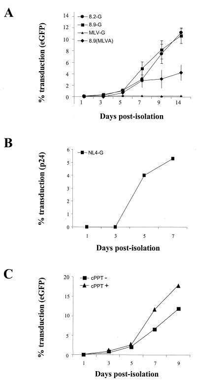
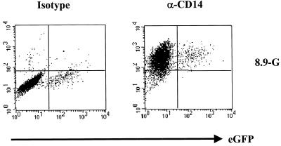
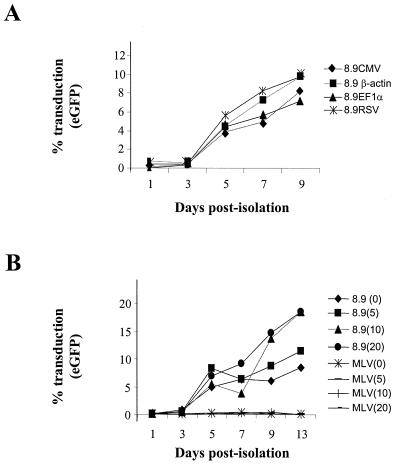
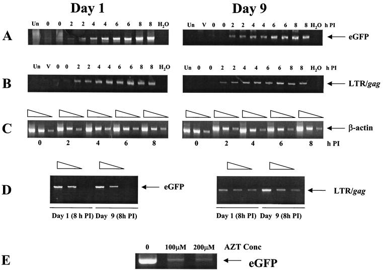
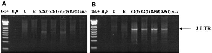
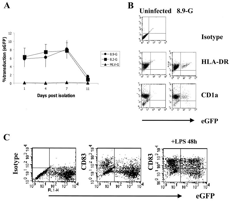
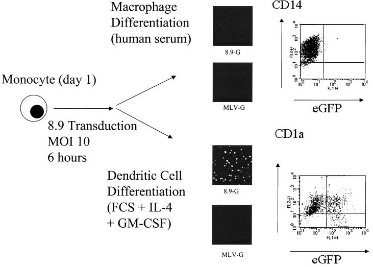
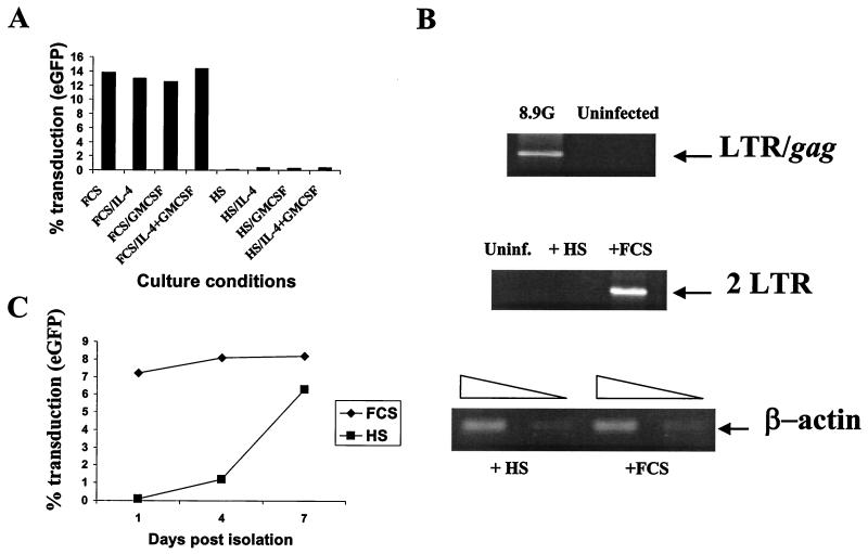
Cells of the monocyte lineage can be infected with human immunodeficiency virus type 1 (HIV-1) both during clinical infection and in vitro. The ability of HIV-1-based vectors to transduce human monocytes, monocyte-derived macrophages, and dendritic cells (DCs) was therefore examined, in order to develop an efficient protocol for antigen gene delivery to human antigen-presenting cells. Freshly isolated monocytes were refractory to HIV-1-based vector transduction but became transducible after in vitro differentiation to mature macrophages. This maturation-dependent transduction was independent of the HIV-1 accessory proteins Vif, Vpr, Vpu, and Nef in the packaging cells and of the central polypurine tract in the vector, and it was also observed with a vesicular stomatitis virus-pseudotyped HIV-1 provirus, defective only in envelope and Nef. The level and extent of reverse transcription of the HIV-1-based vector was similar after infection of immature monocytes and of mature macrophages. However, 2LTR vector circles could not be detected in monocytes, suggesting a block to vector nuclear entry in these cells. Transduction of freshly isolated monocytes exposed to HIV-1-based vector could be rescued by subsequent differentiation into DCs. This rescue was induced by fetal calf serum in the DC culture medium, which promoted vector nuclear entry.


Figures

FIG. 1
FIG. 1
Maturation dependence of HIV-1 infection in human MDMs. (A) HIV vector transduction. Adherent PBMCs were plated at 104 cells/well of a 96-well plate and infected with CMVΔR8.2(VSV-G) and CMVΔR8.9(VSV-G) packaged vectors (8.2-G and 8.9-G, respectively) or CMVΔR8.9(MLV-A) packaged vector [8.9(MLVA)] at an MOI of 10, as standardized on human 293T cells, on various days postisolation. Cells were cultured for 7 days posttransduction; percent cell transduction was then quantified by fluorescence-activated cell sorting. MLV vectors pseudotyped with VSV-G (MLV-G) were used as a control at the same MOI. Values are means ± standard errors of the means for three separate experiments. (B) HIV-1 infection. Titers of NL4-3 pseudotyped with VSV-G (NL4-G) were determined as for panel A, with the MOI standardized on 293T cells. Infection was scored by in situ p24 immunoassay. (C) Effect of cPPT on vector transduction (cPPT− or cPPT+). Titers of CMVΔR8.9(VSV-G) particles packaging pHR′-CMV-eGFP or HR′-cPPT-CMV-eGFP were determined as for panel A. The levels of p24 in both supernatants were equal (not shown), and infections were performed with an equivalent volume for which the cPPT− vector gave an MOI of 10 to standardize for virion input.
FIG. 2
FIG. 2
Identification of infected cells as macrophages Macrophages transduced with a CMVΔR8.9(VSV-G) packaged vector on day 7 postisolation were analyzed on day 14 for eGFP expression and stained with a PE-conjugated monoclonal antibody against the monocyte/macrophage marker CD14 or the equivalent PE-conjugated isotype control (y axis).
FIG. 3
FIG. 3
Maturation-dependent transduction is independent of transgene promoter and dNTP concentration. (A) MDMs were transduced as for Fig. 1A at an MOI of 10 with CMVΔR8.9(VSV-G) packaged vector with eGFP driven from either CMV, human β-actin, human EF1α, or RSV promoters. Cells were analyzed for eGFP expression 7 days posttransduction. (B) CMVΔR8.9(VSV-G) or MLV(VSV-G) vector stocks were treated for 1 h prior to infection with 0, 5, 10, or 20 μM concentrations of dNTPs as shown in parentheses and then used to infect MDMs at an MOI of 10 at various days postisolation. Cells were analyzed for eGFP expression 7 days posttransduction.
FIG. 4
FIG. 4
Analysis of reverse transcription in monocytes (day 1) or mature macrophages (day 9). Cells were harvested at 2-h intervals after exposure to CMVΔR8.9(VSV-G) packaged vector at an MOI of 10. PCR was then performed for intermediate reverse transcripts containing eGFP sequences (A) and second-strand-transfer products containing LTR/gag sequences (B), and serial twofold dilutions were used for PCR of cellular β-actin for a loading control (C). PCR was also performed on supernatants containing an equivalent amount of virus to check for DNA contamination in the viral preparations (lanes V). Serial twofold dilutions of samples from the the 8-h time points of days 1 and 9 were used for PCR to estimate relative amounts of eGFP and LTR/gag transcripts (D). The reverse transcriptase inhibitor zidovudine (AZT) was added to day 1 cells exposed to CMVΔR8.9(VSV-G) packaged vector at an MOI of 10. After 14 h, PCR was performed on cell lysates to detect eGFP transcripts (E). Un, uninfected cells; PI, postinfection.
FIG. 5
FIG. 5
Analysis of nuclear 2LTR circles in infected monocytes (day 1 [A]) and mature macrophages (day 9 [B]). Cells were exposed to CMVΔR8.2(VSV-G) or CMVΔR8.9(VSV-G) packaged vectors overnight at MOIs of 5 and 1 (8.2 and 8.9, respectively). MLV(VSV-G), unenveloped CMVΔR8.2 (E) vectors, and uninfected cell lysates (U) were used as controls. Cells were then washed, cultured for 48 h, and lysed. PCR with primers specific for 2LTR circles (36) was performed on an equivalent of 1.5 × 10⁵ cells. The 2LTR product of 680 bp is indicated.
FIG. 6
FIG. 6
Transduction of monocyte-derived DCs by HIV-based vectors. (A) Adherent monocytes were differentiated to DCs in RPMI medium containing 10% FCS, IL-4, and GM-CSF. Cells were exposed to CMVΔR8.2(VSV-G), CMVΔR8.9(VSV-G), or MLV(VSV-G) packaged vectors at an MOI of 10 for 12 h on various days after isolation as monocytes. Cells were the washed and cultured for 7 days and analyzed for eGFP expression by FACScan analysis. Results are means ± standard errors from three separate experiments. (B) Transduced cells were identified as DCs by expression of eGFP and staining with the Langerhans cell marker CD1a and HLA-DR (y axis). (C) Day 1 monocytes transduced with CMVΔR8.9(VSV-G) packaged HR′cPPTCMVeGFP vectors at at MOI of 10 were differentiated to DCs, cultured for a further 48 h with and without 50 ng of LPS/ml, and stained for surface expression of the DC activation marker CD83 (y axis). Cell populations shown were previously gated on HLA-DR+ CD1a+ DCs.
FIG. 7
FIG. 7
Monocyte differentiation to DCs rescues transduction. Adherent monocytes (106; day 1) were infected with CMVΔR8.9(VSV-G) or MLV(VSV-G) packaged vectors (8.9 and MLV, respectively) at an MOI of 10. At 6 h posttransduction, cells were washed and either plated in macrophage medium (RPMI medium plus 10% human serum) or DC differentiation medium (10% FCS plus IL-4 and GM-CSF). eGFP expression was assessed 7 days later by confocal microscopy of infected cells. Images were acquired on a Bio-Rad MRC 1064 confocal microscope at a magnification of ×200. eGFP expression in CMVΔR8.9-transduced cells was analyzed by fluorescence-activated cell sorting, and cells were phenotype stained for CD14 (macrophages) or CD1a (DCs).
FIG. 8
FIG. 8
FCS rescues transduction of monocytes by HIV-based vectors. (A) Monocytes exposed to CMVΔR8.9(VSV-G) packaged vectors at an MOI of 10 for 6 h were subsequently cultured in RPMI medium containing either FCS or human serum with IL-4, GM-CSF, or both cytokines. eGFP expression was analyzed 7 days later by fluorescence-activated cell sorting. (B) Effect of FCS on accumulation of 2LTR circles in monocytes. Day 1 monocytes were exposed to CMVΔR8.9(VSV-G) (8.9-G) packaged vectors at an MOI of 10 for 6 h in RPMI medium plus human serum. PCR was performed on a sample of these cells to detect second-strand-transfer reverse transcription products (LTR/gag). The monocytes were washed and replated in medium containing either human serum (HS) or FCS and cultured for a further 24 h. The cells were lysed, and PCR was performed to detect 2LTR circles and β-actin as a loading control. (C) DC culture in human serum renders these cells maturation dependent. DCs were cultured in either human serum or FCS and exposed to CMVΔR8.9(VSV-G) packaged vectors at an MOI of 10 on various days postisolation as monocytes, washed, and cultured for a further 7 days. eGFP expression was analyzed by fluorescence-activated cell sorting.

Similar articles

Cited by

References

    1. Arthos J, Rubbert A, Rabin R L, Cicala C, Machado E, Wildt K, Hanbach M, Steenbeke T D, Swofford R, Farber J M, Fauci A S. CCR5 signal transduction in macrophages by human immunodeficiency virus and simian immunodeficiency virus envelopes. J Virol. 2000;74:6418–6424. - PMC - PubMed
    1. Ayehunie S, Garcia-Zepeda E, Hoxie J, Horuk R, Kupper T, Luster A, Ruprecht R. Human immunodeficiency virus-1 entry into purified blood dendritic cells through CC and CXC chemokine coreceptors. Blood. 1997;90:1379–1386. - PubMed
    1. Banchereau J, Steinman R M. Dendritic cells and the control of immunity. Nature. 1998;392:245–252. - PubMed
    1. Blauvert A, Asada H, Saville H W, Klaus-Kovtun V, Altman D, Yarchoan R, Katz S. Productive infection of dendritic cells by HIV-1 and their ability to capture virus are mediated through separate pathways. J Clin Investig. 1997;100:2043–2053. - PMC - PubMed
    1. Briant L, Robert-Hebmann V, Acquaviva C, Pelchen-Matthews A, Marsh M, Devaux C. The protein tyrosine kinase p56lck is required for triggering NF-kappaB activation upon interaction of human immunodeficiency virus type 1 envelope glycoprotein gp120 with cell surface CD4. J Virol. 1998;72:6207–6214. - PMC - PubMed

Publication types