. 2001 Jun;75(12):5498-503.
doi: 10.1128/JVI.75.12.5498-5503.2001.

Inhibition of L-deleted foot-and-mouth disease virus replication by alpha/beta interferon involves double-stranded RNA-dependent protein kinase

Affiliations

Inhibition of L-deleted foot-and-mouth disease virus replication by alpha/beta interferon involves double-stranded RNA-dependent protein kinase

J Chinsangaram et al. J Virol. 2001 Jun.

Abstract

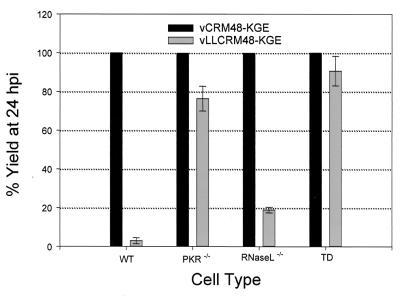
We previously demonstrated that the ability of foot-and-mouth disease virus (FMDV) to form plaques in cell culture is associated with the suppression of alpha/beta interferon (IFN-alpha/beta). In the present study, we used Escherichia coli-expressed porcine and bovine IFN-alpha or -beta individually to demonstrate that each was equally effective in inhibiting FMDV replication. The block in FMDV replication appeared to be at the level of protein translation, suggesting a role for double-stranded RNA-dependent protein kinase (PKR). In support of these findings, treatment of porcine and bovine cells with 2-aminopurine, an inhibitor of PKR, increased the yield of virus 8.8- and 11.2-fold, respectively, compared to that in untreated infected cells. In addition, results of FMDV infection in mouse embryonic fibroblast cells derived from gene knockout mice lacking the gene for RNase L(-/-) or PKR(-/-) or both indicated an important role for PKR in the inhibition of FMDV replication.


Figures

FIG. 1
FIG. 1
Expression of bovine and porcine IFN-α or -β in E. coli. Lysates of BL21(DE3) cells containing bovine and porcine pET-IFN-α or -β (bIFN-α or -β and pIFN-α or -β), respectively, were collected at 0, 1.5, or 3 h after IPTG induction, run on an SDS-PAGE (15% acrylamide) and stained with Coomassie blue. Arrows indicate induced IFN proteins. Lanes M, protein molecular mass markers in kilodaltons.
FIG. 2
FIG. 2
(A) Plaque formation ability of A12-LLV2 on PK or IBRS2 cells. Cells were infected with approximately 100 PFU of A12-LLV2, overlaid, and stained at 36 h postinoculation. (B) Induction of IFN-α or IFN-β mRNA in PK or IBRS2 cells. Cells were infected with A12-IC, A12-LLV2, or mock infected for 6 h and used in RT-PCR as described in Materials and Methods. Aliquots from RT reactions were used in three separate PCR assays with IFN-α, IFN-β, and β-actin primers. DNA from uninfected PK or IBRS2 cells was used in PCR assays as target controls. IFN-α, IFN-β, and β-actin RT-PCR products are 379, 452, and 890 bp, respectively. Lanes MW, 1-kb-ladder DNA molecular weight markers.
FIG. 3
FIG. 3
Plaque assay on IBRS2 cells. Cells were treated for 16 h with supernatant from mock-infected PK (A) or A12-LLV2-infected PK containing IFN-α/β (B) or with E. coli-expressed porcine IFN-α (C) or IFN-β (D) and then infected with approximately 100 PFU of A12-LLV2. Cells were overlaid and stained at 36 h postinoculation.
FIG. 4
FIG. 4
Synthesis of FMDV full-length RNA and protein. IBRS2 cells were treated for 16 h with supernatant from A12-LLV2-infected PK cells containing IFN-α/β (+) or mock-infected PK cells (−) and infected at an MOI of 10 with A12-IC virus. (A) RNA was harvested at 1, 2, and 3 hpi and run on a 1% formaldehyde agarose gel. RNA was transferred to a nylon membrane and hybridized to a peroxidase-labeled full-length FMDV probe, and the signal was detected using a chemiluminescent technique. The arrow indicates full-length FMDV RNA. (B) [35S]methionine-labeled proteins were harvested at 1.5, 2, 2.5, 3, 3.5, and 4 h postinoculation, immunoprecipitated using a polyclonal antibody against FMDV nonstructural protein 3D, and analyzed on an SDS-PAGE (15% acrylamide). Arrows indicate 3D and precursors. Lane c, A12-IC-infected IBRS2 lysate immunoprecipitated with 3D antiserum.
FIG. 5
FIG. 5
Growth of FMDV on EF cells. WT, RNase L−/−, PKR−/− or TD EF cells were infected with vCRM48-KGE or vLLCRM48-KGE virus at a low MOI. At 1 hpi, cells were treated with MES. Viruses were harvested at 1 and 24 hpi, titrated on BHK-21 cells, and the increase of titer over 24 h was determined as virus growth. The ratio of the growths of vLLCRM48-KGE and vCRM48-KGE in each cell type was reported as a percentage.

References

    1. Almeida M R, Rieder E, Chinsangaram J, Ward G, Beard C, Grubman M J, Mason P W. Construction and evaluation of an attenuated vaccine for foot-and-mouth disease: difficulty adapting the leader proteinase-deleted strategy to the serotype O1 virus. Virus Res. 1998;55:49–60. - PubMed
    1. Berinstein A, Roivainen M, Hovi T, Mason P W, Baxt B. Antibodies to the vitronectin receptor (integrin alpha V beta 3) inhibit binding and infection of foot-and-mouth disease virus to cultured cells. J Virol. 1995;69:2664–2666. - PMC - PubMed
    1. Brown C C, Piccone M E, Mason P W, McKenna T S, Grubman M J. Pathogenesis of wild-type and leaderless foot-and-mouth disease virus in cattle. J Virol. 1996;70:5638–5641. - PMC - PubMed
    1. Chinsangaram J, Akita G Y, Osburn B I. Detection of bovine group B rotaviruses in feces by polymerase chain reaction. J Vet Diagn Investig. 1994;6:302–307. - PubMed
    1. Chinsangaram J, Mason P W, Grubman M J. Protection of swine by live and inactivated vaccines prepared from a leader proteinase-deficient serotype A12 foot-and-mouth disease virus. Vaccine. 1998;16:1516–1522. - PMC - PubMed

MeSH terms

LinkOut - more resources