. 2001 Jun;75(12):5703-10.
doi: 10.1128/JVI.75.12.5703-5710.2001.

Conservation of the conformation and positive charges of hepatitis C virus E2 envelope glycoprotein hypervariable region 1 points to a role in cell attachment

Affiliations

Conservation of the conformation and positive charges of hepatitis C virus E2 envelope glycoprotein hypervariable region 1 points to a role in cell attachment

F Penin et al. J Virol. 2001 Jun.

Abstract

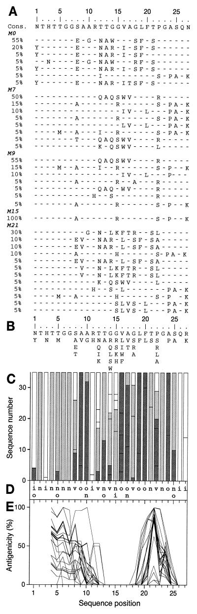
Chronic hepatitis C virus (HCV) infection is a major cause of liver disease. The HCV polyprotein contains a hypervariable region (HVR1) located at the N terminus of the second envelope glycoprotein E2. The strong variability of this 27-amino-acid region is due to its apparent tolerance of amino acid substitutions together with strong selection pressures exerted by anti-HCV immune responses. No specific function has so far been attributed to HVR1. However, its presence at the surface of the viral particle suggests that it might be involved in viral entry. This would imply that HVR1 is not randomly variable. We sequenced 460 HVR1 clones isolated at various times from six HCV-infected patients receiving alpha interferon therapy (which exerts strong pressure towards quasispecies genetic evolution) and analyzed their amino acid sequences together with those of 1,382 nonredundant HVR1 sequences collected from the EMBL database. We found that (i) despite strong amino acid sequence variability related to strong pressures towards change, the chemicophysical properties and conformation of HVR1 were highly conserved, and (ii) HVR1 is a globally basic stretch, with the basic residues located at specific sequence positions. This conservation of positively charged residues indicates that HVR1 is involved in interactions with negatively charged molecules such as lipids, proteins, or glycosaminoglycans (GAGs). As with many other viruses, possible interaction with GAGs probably plays a role in host cell recognition and attachment.


Figures

FIG. 1
FIG. 1
Analysis of HVR1 quasispecies sequences from patient E (genotype 1b) based on sequences of 20 clones per time point at five time points, i.e., 100 distinct clones. (A) Alignment of HVR1 quasispecies sequences at month zero (M0, before IFN-α treatment), M7 (1 month after the end of the first IFN-α course), M9 (beginning of the second IFN-α course), M15 (end of the second IFN-α course), and M21 (6 months after the end of the second IFN-α course). The frequency of each sequence in the quasispecies is given as a percentage in the left-hand column. Cons, the derived consensus amino acid sequence. Amino acids identical to those in the consensus sequence are represented by a hyphen. (B) Repertoire of patient E's HVR1 amino acids per position from the analysis of the 36 nonredundant sequences observed at the five time points. Amino acids are listed in decreasing order of observed frequency, from top to bottom. (C) Histogram showing the hydropathic character of the residues at each position in HVR1. The height of the box in each bar indicates the number of sequences with a given residue at a given position. The boxes are presented in order of decreasing hydrophobicity, from bottom to top, according to the hydrophobicity scale of Black and Mould (4). Each box is colored according to the hydrophobic character of the residue: dark gray for hydrophobic (F, I, W, Y, L, V, M, P, C, A), light gray for neutral (G, T, S), and white for hydrophilic (K, Q, N, H, E, D, R). (D) Consensus hydropathic pattern of HVR1 quasispecies deduced from the latter. o, hydrophobic residue; n, neutral residue; i, hydrophilic residue; v, variable residue. (E) Antigenicity profiles of HVR1 sequences calculated according to the method of Parker et al. (34) with a window of 7 amino acids. Therefore, the antigenicity of the first three HVR1 positions could not be estimated.
FIG. 2
FIG. 2
Analysis of 119 unambiguously genotyped HCV genotype 1b HVR1 sequences from the EMBL database. Residues observed at a given position in only one sequence were not taken into consideration. (A) Repertoire of genotype 1b HVR1 residues per position. Residues are listed in decreasing order of observed frequency, from top to bottom. Residues within the box correspond to those observed in more than 10% of the sequences. (B) Histogram showing the hydropathic character of residues at each position (see the legend to Fig. 1C). (C) Consensus hydropathic pattern of genotype 1b sequences (see the legend to Fig. 1D). (D) Antigenicity profiles of the 10 most distantly related sequences of genotype 1b sequences (see the legend to Fig. 1E). Selection of these 10 among 119 sequences was based on the HVR1 phylogenetic tree of genotype 1b.
FIG. 3
FIG. 3
Analysis of 1,382 HVR1 sequences from the EMBL database. Amino acids observed at a given position in fewer than five distinct sequences (<0.3%) were not taken into consideration. (A) Repertoire of HVR1 residues per position in the 1,382 unrelated EMBL HVR1 sequences. Amino acids are listed in decreasing order of observed frequency, from top to bottom. Residues within the box correspond to those observed in more than 10% of the sequences. (B) Histogram showing the hydropathic character of residues at each position in HVR1. The height of the box in each bar indicates the frequency of sequences with a given residue at a given position. (C) Consensus hydropathic pattern from the 1,382 HVR1 sequences. (D) Comparison of the antigenicity profiles of HVR1 sequences representative of the principal HCV subtypes of clades 1 to 6 (the EMBL accession number of each sequence is indicated in parentheses): 1a (M67463), 1b (D90208), 1c (D14853), 2a (D00944), 2b (D10988), 2c (D50409), 3a (D28917), 3b (D49374), 4a (Y11604), 5a (Y13184), 6a (Y12083), 10a (D63821; classified in clade 3), and 11a (D63822; classified in clade 6).
FIG. 4
FIG. 4
Frequency of acidic and basic residues and conserved basic patterns in the 1,382 HVR1 sequences from EMBL. The frequency of the residues at each position was calculated by dividing the number of observed acidic or basic residues by the total number of examined sequences (n = 1,382). (A) Frequency of acidic residues. (B) Frequency of basic residues. (C) Fourteen most common basic patterns among the 1,382 HVR1 sequences from EMBL. Basic residues (Arg, Lys, and His) are represented by a B, while nonbasic residues are represented by a hyphen. The observed frequency of each pattern is given in the left-hand column.
FIG. 5
FIG. 5
Summary of HVR1 structural analysis and predicted amino acid functions. The HVR1 consensus hydropathic pattern was established from the 1,382 HVR1 sequences from EMBL. o, n, i, and v, mean hydrophobic, neutral, hydrophilic, and variable positions, respectively; G is a fully conserved Gly residue. The black boxes indicate the main anchoring regions, and the gray box indicates a putative turn. The plus signs indicate positions often occupied by basic residues.

Similar articles

Cited by

References

    1. Afonso A M, Jiang J, Penin F, Tareau C, Samuel D, Petit M A, Bismuth H, Dussaix E, Feray C. Nonrandom distribution of hepatitis C virus quasispecies in plasma and peripheral blood mononuclear cell subsets. J Virol. 1999;73:9213–9221. - PMC - PubMed
    1. Agnello V, Abel G, Elfahal M, Knight G B, Zhang Q-X. Hepatitis C virus and other flaviviridae viruses enter cells via low density lipoprotein receptor. Proc Natl Acad Sci USA. 1999;96:12766–12771. - PMC - PubMed
    1. Alter H J. To C or not to C: these are the questions. Blood. 1995;85:1681–1695. - PubMed
    1. Black S D, Mould D R. Development of hydrophobicity parameters to analyze proteins which bear post- or cotranslational modifications. Anal Biochem. 1991;193:72–82. - PubMed
    1. Blanchet C, Combet C, Geourjon C, Deléage G. MPSA: integrated system for multiple protein sequence analysis with client/server capabilities. Bioinformatics. 2000;16:286–287. - PubMed

Publication types