. 2001 May 15;15(10):1182-7.
doi: 10.1101/gad.890501.

Unipolar membrane association of Dishevelled mediates Frizzled planar cell polarity signaling

Affiliations

Unipolar membrane association of Dishevelled mediates Frizzled planar cell polarity signaling

J D Axelrod. Genes Dev. .

Abstract

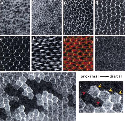
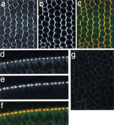
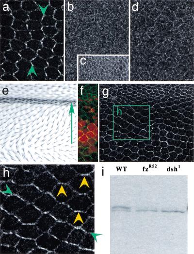
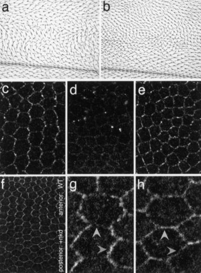
Drosophila epithelia acquire a planar cell polarity (PCP) orthogonal to their apical-basal axes. Frizzled (Fz) is the receptor for the PCP signal, and Dishevelled (Dsh) transduces the signal. Here, I demonstrate that unipolar relocalization of Dsh to the membrane is required to mediate PCP, but not Wingless (Wg) signaling. Dsh membrane localization reflects the activation of Fz/PCP signaling, revealing that the initially symmetric signal evolves to one that displays unipolar asymmetry, specifying the cells' ultimate polarity. This transition from symmetric to asymmetric Dsh localization requires Dsh function, and reflects an amplification process that generates a steep intracellular activity gradient necessary to determine PCP.


Figures

Figure 1
Figure 1
Subcellular localization of Dishevelled (Dsh). In panels c to j, proximal is left, distal is right. (a) Localization of Dsh::green fluorescent protein (GFP) in late third-instar subapical section. (b) Late third-instar apical section (including the dorsal-ventral boundary). A similar result was observed in fzR52 mutant wing discs (not shown, but see Fig. 3c). (c) Two-hours after-puparium-formation (apf) apical sections. (d) Thirty-hour apf apical sections. (e) Thirty-four-hour apf apical section. (e–g) Double label of 34-h apf wing with Dsh::GFP (e), phalloidin (actin, f), and overlay (g) (Dsh::GFP, green; phalloidin, red). In panels a to g, the Dsh::GFP transgene was expressed in a dshv26 null mutant background. The result was identical when expressed in a wild-type background (not shown). (h) Dsh1::GFP subcellular localization in a dsh1 mutant background at 30 h apf. The dsh1 allele carries a point mutation in the DEP domain, and is a strong allele for Frizzled (Fz)/planar cell polarity (PCP) signaling (Perrimon and Mahowald 1987; Axelrod et al. 1998). Similar results were obtained in dshv26 null and in wild-type backgrounds (not shown). (i–j) Clones of cells lacking the Dsh::GFP transgene in 30-h apf wings appear as holes in the GFP pattern, and were also marked with anti-βgal (not shown). All cells express endogenous dsh+, and thus are genetically wild type. Note that in cells abutting the clones, Dsh::GFP accumulates at the distal edges (yellow arrowheads), but not at the proximal edges (red arrowheads). Twinspots of the clones have two copies of Dsh::GFP and show enhanced fluorescence compared with heterozygous tissue. Panels a to h are of the same magnification.
Figure 2
Figure 2
Relationship of Dishevelled (Dsh)::green fluorescent protein (GFP) and Flamingo (Fmi) localization. (a–e) A 30-h after puparium formation (apf) pupal wing showing Dsh::GFP (a), Fmi (b), and the overlay of Dsh::GFP (green) and Fmi (red) (c). (d–f) Tangential section of a double-labeled wing taken through edge cells: Dsh::GFP (d), Fmi (e), overlay, as in c (f). Much of the Dsh colocalizes with Fmi, and some remains in the cytoplasm. (g) Dsh::GFP does not show significant association with the membrane in a 30-h apf fmi45/fmi59 mutant wing.
Figure 3
Figure 3
Frizzled (Fz) loss and gain of function alters Dishevelled (Dsh) localization. In a to f, proximal is left, distal is right. Dsh localization in 30-h after-puparium-formation (apf) wild type (a), fzR52 (b), and fzJ22 (d) mutant wings, and in a 2-h apf fzR52 mutant wing (c). (e) Ectopic Fz overexpressed in a gradient in the dpp domain produces hairs pointing down the Fz gradient. The arrow points from high toward low ectopic Fz expression. (f) In an equivalent region of a 30-h apf pupal wing in which dpp-GAL4 drives both UAS-fz and UAS-lacZ (red), the ectopic Fz gradient is visualized (anti-βgal, red) along with a reorganized Dsh::green fluorescent protein (GFP) pattern (green). (g) A 30-h pupal wing from a region equivalent to that in e, showing only the reorganized Dsh::GFP localization. (h) Enlarged view of the region marked by the box in g. Note that Dsh::GFP accumulates in a zigzag pattern that is perpendicular to the pattern of hairs in this region (green arrowheads; compare with a) and includes anterior–posterior boundaries (yellow arrowheads). (i) A Western blot of wild-type (WT), fzR52, and dsh1 pupal wings, probed with anti-Dsh. The slower migrating isoform seen in WT corresponds to a previously identified hyperphosphorylated form (Yanagawa et al. 1995) and is severely reduced in the mutants. Equal loading between lanes was assured by Ponceau staining before hybridization (not shown).
Figure 4
Figure 4
Loss of Dishevelled (Dsh) membrane localization or asymmetry blocks polarity signaling. In all panels, proximal is left, distal is right. Dominant negative planar cell polarity phenotypes result from expression of Dsh(ΔbPDZ) (a) or Dsh(ΔDEP+) (b) in the patched expression domain. (c) Anterior (internal control) region of a 30-h after-puparium-formation wing in which patched-GAL4 drives expression of Dsh(ΔbPDZ), showing wild-type Dsh::green fluorescent protein (GFP) localization. (d) In the patched expression domain of the same wing, Dsh::GFP membrane association is lost. (e) Dsh::GFP pattern in the patched expression domain of a wing expressing Dsh(ΔDEP+). Note that the enrichment of Dsh::GFP at proximal–distal boundaries is diminished, and the pattern is more nearly symmetric. (f) Dsh::GFP at the anterior–posterior compartment boundary of a wing overexpressing Naked cuticle (Nkd) in the posterior, engrailed domain. High magnification of a portion of the anterior, wild-type domain (g) and the posterior domain overexpressing Nkd (h). Note the wild-type, zigzag pattern in the anterior, and the nearly complete loss of asymmetry in the posterior. In the posterior, Dsh is no longer enriched at the proximal–distal boundaries (compare darker arrowheads) and no longer suppressed at the anterior–posterior boundaries (compare lighter arrowheads).

References

    1. Adler PN, Charlton J, Jones KH, Liu J. The cold-sensitive period for frizzled in the development of wing hair polarity ends prior to the start of hair morphogenesis. Mech Dev. 1994a;46:101–107. - PubMed
    1. Adler PN, Charlton J, Park WJ. The Drosophila tissue polarity gene inturned functions prior to wing hair morphogenesis in the regulation of hair polarity and number. Genetics. 1994b;137:829–836. - PMC - PubMed
    1. Adler PN, Krasnow RE, Liu J. Tissue polarity points from cells that have higher Frizzled levels towards cells that have lower Frizzled levels. Curr Biol. 1997;7:940–949. - PubMed
    1. Axelrod JD, Miller JR, Shulman JM, Moon RT, Perrimon N. Differential recruitment of Dishevelled provides signaling specificity in the Planar Cell Polarity and Wingless signaling pathways. Genes & Dev. 1998;12:2610–2622. - PMC - PubMed
    1. Bhat KM. frizzled and frizzled 2 play a partially redundant role in wingless signaling and have similar requirements to wingless in neurogenesis. Cell. 1998;95:1027–1036. - PubMed

Publication types

MeSH terms