

Concurrent occurrence of gastric adenocarcinoma and duodenal neuroendocrine cell carcinoma: a composite tumour or collision tumours ?
- PMID: 11358908
- PMCID: PMC1728320
- DOI: 10.1136/gut.48.6.853
Concurrent occurrence of gastric adenocarcinoma and duodenal neuroendocrine cell carcinoma: a composite tumour or collision tumours ?
Abstract
Background: Neuroendocrine cell (NEC) carcinoma is occasionally accompanied by adenocarcinoma but the relationship between these two morphologically distinct tumours is unclear. Two hypotheses have arisen regarding the mechanism for the association of adenocarcinoma and NEC carcinoma. One is that both are derived from a common multipotential epithelial stem cell. The second hypothesis is that adenocarcinoma and NEC carcinoma arise from a multipotential epithelial stem cell and a primitive NEC, respectively.
Aims: To elucidate the relationship between the two histologically distinct tumours, adenocarcinoma of the stomach and NEC carcinoma of the duodenum.
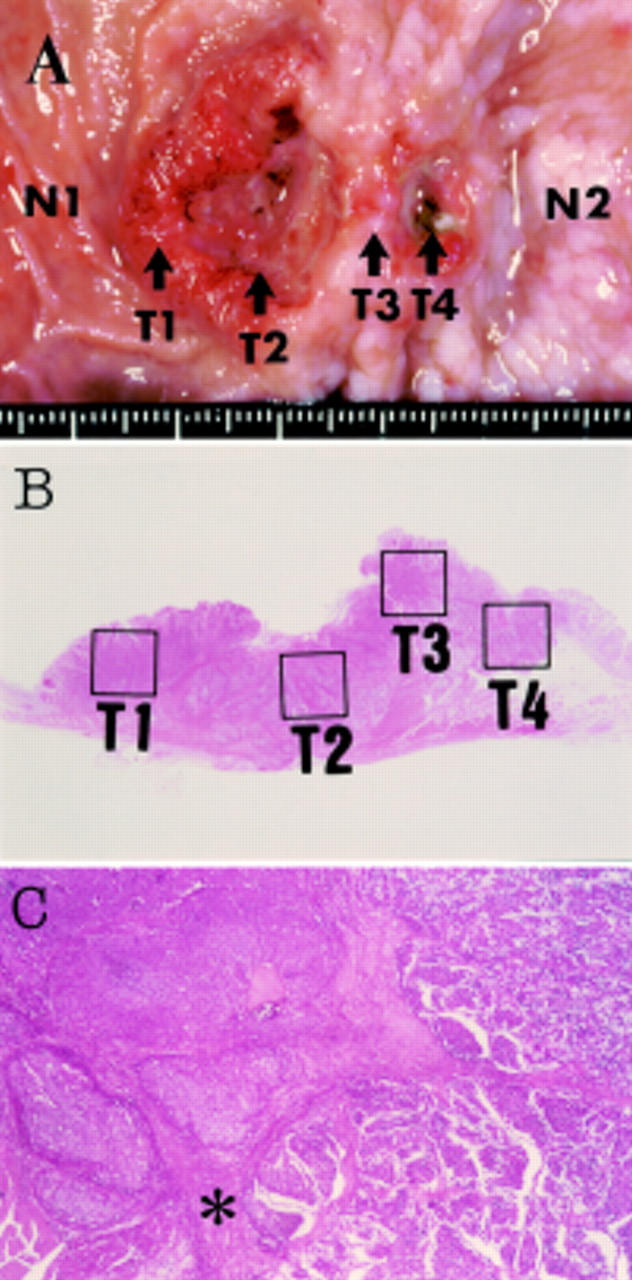
Patient/methods: We present a case in which the tumour extended across the pyloric ring, the gastric portion of which revealed adenocarcinoma while the duodenal portion showed argyrophil NEC carcinoma. The two histologically distinct lesions of the tumour were examined by immunohistochemistry and genetic analysis of p53.
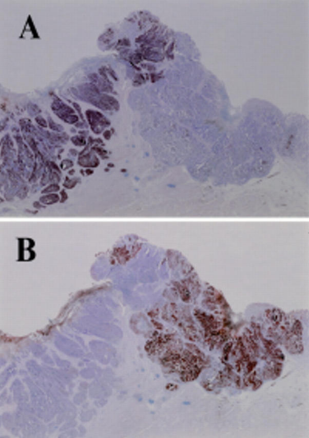
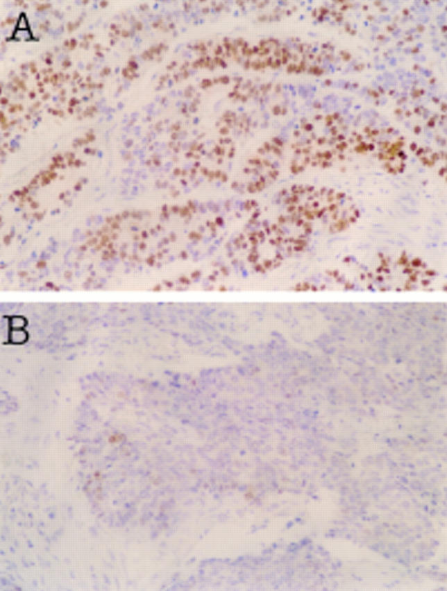
Results: The gastric region was negative for chromogranin A staining but positive for carcinoembryonic antigen (CEA) staining. In contrast, the duodenal region was positive for chromogranin A but negative for CEA. All tumour regions showed a point mutation in p53 gene at exon 7 (GGC (glycine)-->GTC (valine) at codon 245). The distal portion of the duodenal tumour showed an additional point mutation in p53 gene at exon 5 (GCC (alanine)-->GTC (valine) at codon 129).
Conclusions: The two histologically distinct tumours, adenocarcinoma of the stomach and NEC carcinoma of the duodenum, appear to be derived from a common epithelial cell.
Figures
Publication types
MeSH terms
LinkOut - more resources
Full Text Sources
Medical
Research Materials
Miscellaneous