

Quantitative analysis of inflammatory cells infiltrating the cystic fibrosis airway mucosa
- PMID: 11359444
- PMCID: PMC1906034
- DOI: 10.1046/j.1365-2249.2001.01456.x
Quantitative analysis of inflammatory cells infiltrating the cystic fibrosis airway mucosa
Abstract
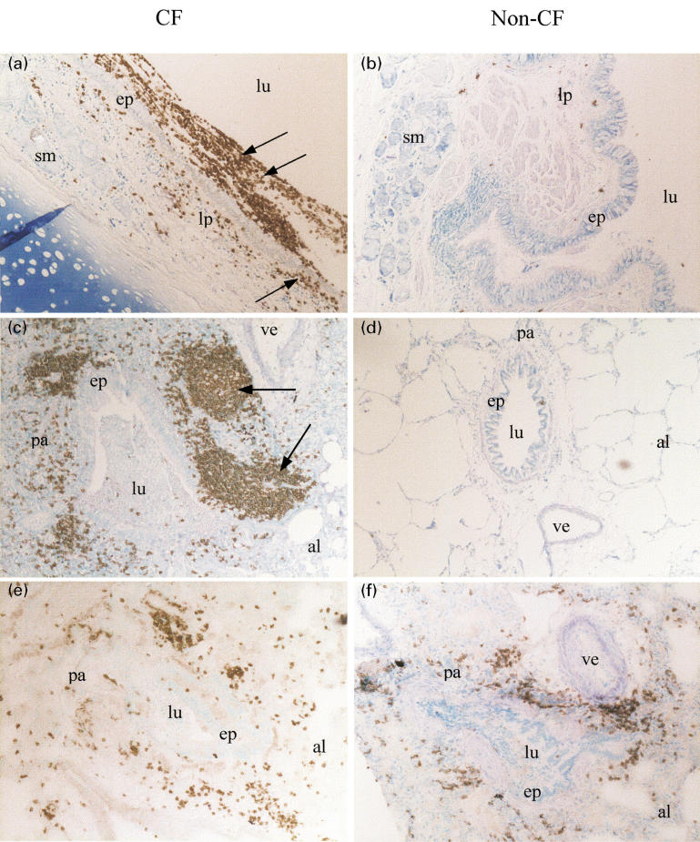
Airway inflammation represents a hallmark of the cystic fibrosis (CF) disease. However, the mucosal distribution of immune cells along the CF airways has not been clearly defined, particularly in intermediate bronchi and distal bronchioles. We analysed lung tissues collected at the time of transplantation from homozygous DeltaF508+/+CF patients versus non-CF donors. Using immunohistochemistry, the distribution of intercellular adhesion molecule-1 (ICAM-1), vascular cell adhesion molecule-1 (VCAM-1) and E-selectin, polymorphonuclear neutrophils (PMN), mast cells, CD3+ T cells, including the CD4+ and CD8+ subsets, CD20+ B cells, CD38+ plasma cells and CD68+ macrophages, was analysed at lobar, segmental and distal levels of the bronchial tree. Using image cytometry, the number of cells per mm2 was assessed in the depth of the bronchial wall. In CF airways, alterations mainly consisted in lesions of the surface epithelium. Numerous immune cells were heterogeneously distributed all along the bronchial tree and mainly located in the mucosa, beneath the surface epithelium. Compared to non-CF donors, the lymphoid aggregates formed by B cells were significantly larger all along the CF airways (P = 0.001). The number of T lymphocytes was higher at the CF distal level (P = 0.035), where we observed an intense tissue damage. PMN preferentially accumulated (P = 0.033) in the CF surface epithelium, which overexpressed ICAM-1 but not VCAM-1 and E-selectin. These results highlight the nature of the inflammatory infiltrate in the CF airway mucosa and emphasize a prominent implication of PMN, B and T lymphocytes in the CF disease.
Figures


References
-
- Boat TF, Welsh MJ, Beaudet AL. Cystic fibrosis. In: Scriver CR, Beaudet AL, Sly WL, Valle D, editors. The Metabolic Basis of Inherited Diseases. 6. New York: McGraw-Hill; 1989. pp. 2649–80.
-
- Quinton P. Chloride permeability in Cystic Fibrosis. Nature. 1983;301:421–2. - PubMed
-
- Accurso FJ. Early pulmonary disease in cystic fibrosis. Curr Opin Pulm Med. 1997;3:400–3. - PubMed
-
- Davis PB, Drumm M, Konstan MW. State of the art: cystic fibrosis. Am J Respir Crit Care Med. 1996;154:1229–56. - PubMed
Publication types
MeSH terms
Substances
LinkOut - more resources
Full Text Sources
Other Literature Sources
Medical
Research Materials
Miscellaneous
