

Sustained postinfarction myocardial oedema in humans visualised by magnetic resonance imaging
- PMID: 11359743
- PMCID: PMC1729755
- DOI: 10.1136/heart.85.6.639
Sustained postinfarction myocardial oedema in humans visualised by magnetic resonance imaging
Abstract
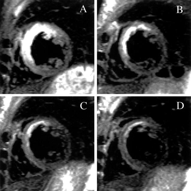
Objective: To demonstrate postinfarction myocardial oedema in humans with particular reference to the longitudinal course, using magnetic resonance imaging (MRI).
Design: Prospective observational study. Subjects were studied one week, one month, three months, six months, and one year after presenting with a myocardial infarct.
Setting: Cardiology and magnetic resonance departments in a Danish university hospital.
Patients: 10 patients (three women, seven men), mean (SEM) age 58.2 (3.20) years, with a first transmural myocardial infarct.
Main outcome measures: Location and duration of postinfarction myocardial oedema.
Results: All patients had signs of postinfarction myocardial oedema. The magnetic resonance images were evaluated by two blinded procedures, employing two MRI and two ECG observers: (1) MRI determined oedema location was compared with the ECG determined site of infarction and almost complete agreement was found; (2) the time course of postinfarction myocardial oedema was explored semiquantitatively, using an image ranking procedure. Myocardial oedema was greatest at the initial examination one week after the infarction, with a gradual decline during the following months (Spearman's rank correlation analysis: rho(observer 1) = 0.94 (p < 0.0001) and rho(observer 2) = 0.97 (p < 0.0001)). The median duration of oedema was six months.
Conclusions: Postinfarction myocardial oedema seems surprisingly long lasting. This observation is of potential clinical interest because the oedema may have prognostic significance.
Figures
Similar articles
-
Quantification of infarct size and myocardium at risk: evaluation of different techniques and its implications.Eur Heart J Cardiovasc Imaging. 2015 Jul;16(7):738-46. doi: 10.1093/ehjci/jev001. Epub 2015 Mar 2. Eur Heart J Cardiovasc Imaging. 2015. PMID: 25736308 Free PMC article.
-
Cardiac magnetic resonance evaluation of edema after ST-elevation acute myocardial infarction.Rev Esp Cardiol. 2009 Aug;62(8):858-66. doi: 10.1016/s1885-5857(09)72650-7. Rev Esp Cardiol. 2009. PMID: 19706241 English, Spanish.
-
Aborted myocardial infarction: a clinical-magnetic resonance correlation.Heart. 2005 Apr;91(4):e24. doi: 10.1136/hrt.2004.047183. Heart. 2005. PMID: 15772173 Free PMC article.
-
T2-weighted magnetic resonance imaging to assess myocardial oedema in ischaemic heart disease.Heart. 2009 Aug;95(16):1357-61. doi: 10.1136/hrt.2009.169961. Epub 2009 May 15. Heart. 2009. PMID: 19447836 Review.
-
Principles, current status and clinical implications of ischaemic heart disease assessment by cardiac magnetic resonance imaging.Intern Med J. 2012 Jan;42(1):7-17. doi: 10.1111/j.1445-5994.2011.02606.x. Intern Med J. 2012. PMID: 21999843 Review.
Cited by
-
Myocardium at risk: reasons and methods for measuring the extent.J Nucl Cardiol. 2013 Feb;20(1):23-6. doi: 10.1007/s12350-012-9659-x. J Nucl Cardiol. 2013. PMID: 23229648 No abstract available.
-
Myocardial area at risk and salvage measured by T2-weighted cardiovascular magnetic resonance: reproducibility and comparison of two T2-weighted protocols.J Cardiovasc Magn Reson. 2011 Sep 15;13(1):50. doi: 10.1186/1532-429X-13-50. J Cardiovasc Magn Reson. 2011. PMID: 21917186 Free PMC article.
-
Mechanisms regulating vascular and lymphatic regeneration in the heart after myocardial infarction.J Pathol. 2023 Aug;260(5):666-678. doi: 10.1002/path.6093. Epub 2023 Jun 5. J Pathol. 2023. PMID: 37272582 Free PMC article. Review.
-
Multiparametric CMR imaging of infarct remodeling in a percutaneous reperfused Yucatan mini-pig model.NMR Biomed. 2017 May;30(5):10.1002/nbm.3693. doi: 10.1002/nbm.3693. Epub 2017 Feb 6. NMR Biomed. 2017. PMID: 28164391 Free PMC article.
-
Thrombus aspiration during primary percutaneous coronary intervention is associated with reduced myocardial edema, hemorrhage, microvascular obstruction and left ventricular remodeling.J Cardiovasc Magn Reson. 2012 Mar 26;14(1):19. doi: 10.1186/1532-429X-14-19. J Cardiovasc Magn Reson. 2012. PMID: 22448853 Free PMC article.
References
Publication types
MeSH terms
LinkOut - more resources
Full Text Sources
Medical