. 2001 May;77(4):1058-66.
doi: 10.1046/j.1471-4159.2001.00304.x.

Sustained extracellular signal-regulated kinase activation by 6-hydroxydopamine: implications for Parkinson's disease

Affiliations

Sustained extracellular signal-regulated kinase activation by 6-hydroxydopamine: implications for Parkinson's disease

S M Kulich et al. J Neurochem. 2001 May.

Abstract

Although the toxin 6-hydroxydopamine (6-OHDA) is utilized extensively in animal models of Parkinson's disease, the underlying mechanism of its toxic effects on dopaminergic neurons is not completely understood. We examined the effects of 6-OHDA on the CNS-derived tyrosine hydroxylase expressing B65 cell line, with particular attention to the regulation of the extracellular signal-regulated protein kinases (ERK). 6-OHDA elicited a dose-dependent cytotoxicity in B65 cells. Toxic doses of 6-OHDA also elicited a biphasic pattern of ERK phosphorylation with a prominent sustained phase, a pattern that differed from that observed with hydrogen peroxide (H(2)O(2)) treatment. 6-OHDA-elicited ERK phosphorylation was blocked by PD98059, an inhibitor of the upstream mitogen activated protein kinase kinase (MEK) that phosphorylates and activates ERK. PD98059 also conferred protection against 6-OHDA cytotoxicity, but did not affect H(2)O(2) toxicity in B65 cells. These results suggest that ERK activation plays a direct mechanistic role in 6-OHDA toxicity, rather than representing a protective compensatory response, and raise the possibility that abnormal patterns of ERK activation may contribute to dopaminergic neuronal cell death.


Figures
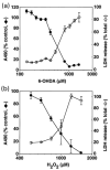
Fig. 1
Fig. 1
Cytotoxicity of 6-OHDA and H2O2 in B65 cells. B65 cells were exposed to (a) 6-OHDA or (b) H2O2 for 18–20 h. Cell injury was determined by measuring LDH activity released into the media (○) or by altered MTS metabolism (●). Values were normalized to total LDH activity or the A490 of vehicle treated control cells, respectively, as described in the Materials and methods section. Each value represents the mean of triplicate wells ± SE. Each experiment was performed independently at least three times. Representative plots are illustrated.
Fig. 2
Fig. 2
Dose–response study of ERK activation in B65 cells. Cells were treated with the indicated μM concentrations of 6-hydroxydopamine (6-OHDA), H2O2 (H2O2) or vehicle (0.05% ascorbate for 6-OHDA, water for H2O2) for 18 h. The cells were then lysed and equal amounts of protein (20 μg) were subjected to immunoblot analysis using an antibody specific for activated dually phosphorylated ERK (top, P-ERK). The blots were then stripped and reprobed with antibody against total ERK (bottom, T-ERK). Blots depicted are representative of eight independent experiments.
Fig. 3
Fig. 3
Kinetics of ERK activation in B65 cells in response to 6-OHDA and H2O2. (a) Cells treated with 500 μM 6-hydroxydopamine (6-OHDA) or 1 mM H2O2 (H2O2) for the indicated times (minutes) were lysed and equal amounts of protein (50 μg) were subjected to immunoblot analysis using antibody against the activated form of ERK (top, P-ERK). The blots were then stripped and reprobed with antibody against total ERK (bottom, T-ERK). Lanes designated with * were harvested following an overnight (16 h) incubation. Lanes designated with – were treated with the appropriate vehicle (0.05% ascorbate for 6-OHDA, water for H2O2). (b) Cells were treated with either 1000 or 500 μM 6-OHDA or 1 mM H2O2 for the indicated time (hours) then subjected to immunoblot analysis as in (a). (c) Immunoblots from cells treated with 1000 mM 6-OHDA were subjected to densitometric analysis, and the data for the ERK1 (top panel) and ERK2 (bottom panel) isoforms from a representative time-course is shown. Closed circles (●) represent the relative band intensity of phosphorylated ERK/total ERK elicited by 6-OHDA; open circles (○) represent the phosphorylated ERK/total ERK ratio observed in vehicle-treated cells. Cells treated with only fresh DH10 exhibited a pattern of ERK phosphorylation similar to that seen with the vehicle controls. (d) Immunoblots from cells treated with 500 μM 6-OHDA and 1 mM H2O2 were subjected to densitometric analysis, and the data for the ERK1 (top panel) and ERK2 (bottom panel) isoforms from a representative time-course is shown. Closed circles represent the relative band intensity elicited by 6-OHDA while open circles represent the relative band intensity elicited by H2O2. Data are expressed as the ratio of phosphorylated ERK to total ERK elicited by the oxidizing agents (pERK/tERK-OX) normalized to the ratio of phosphorylated ERK to total ERK elicited in vehicle treated cells (pERK/tERK-V). Data shown in panels A–D are representative compilations of 10 independent time-course experiments.
Fig. 4
Fig. 4
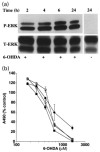
Pulse-chase studies comparing ERK phosphorylation and cytotoxicity. (a) Cells were pulsed with 1 mM 6-hydroxydopamine (6-OHDA) for the indicated times (hours) at which time 6-OHDA was removed. The cells were washed with DH10 and then incubated in fresh DH10 for a total incubation time of 24 h. Immunoblot analysis of cell lysates was performed as described above. Cells treated with vehicle did not exhibit increased phosphorylation at any time point (24 h time point shown as an example). Data shown are representative of four independent experiments. (b) Cells were pulsed with various concentrations of 6-OHDA for the indicated times (2 h: ●, 4 h: ○, 6 h: ■, 22 h: □), washed and placed in fresh DH10 for a total incubation time of 22 h. Cell injury was determined using the MTS viability assay. Each value represents the mean of triplicate wells ± SEM. The experiment was performed independently three times, and a representative plot is illustrated.
Fig. 5
Fig. 5
Effect of the MEK inhibitor PD98059 upon 6-OHDA mediated ERK phosphorylation. B65 cells treated for 20 h with 500 mM 6-hydroxydopamine (6-OHDA) in the presence (+) or absence (−) of 50 μM PD98059 were lysed and equal amounts of protein (50 μg) were subjected to immunoblot analysis using antibody against the activated form of ERK (top, P-ERK). The blots were then stripped and reprobed with antibody against total ERK (bottom, T-ERK).
Fig. 6
Fig. 6
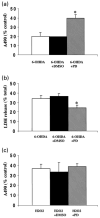
Effect of PD98059 on 6-OHDA-mediated cytotoxicity. Following overnight exposure of B65 cells to 655 mM 6-OHDA in the absence (white bar) or presence (hatched bar) of 50 mM PD98059, cytotoxicity was determined utilizing MTS (a) and LDH release (b) assays. Cells treated with 6-OHDA in the presence of 0.1% DMSO, the vehicle in which the inhibitor was dissolved (black bar), showed values similar to those lacking DMSO. PD98059 treatment had no effect upon the A490 or total LDH values of ascorbate-treated control wells against which the other treatment conditions were normalized (A490 values: 2.26 ± 0.06, untreated; 2.23 ± 0.08 PD98059 treated; total LDH values: 129 ± 27 IU/mL, untreated; 132 ± 23 IU/mL, PD 98059 treated). MTS data is a compilation of five independent experiments and LDH release data is a compilation of three independent experiments. Data are expressed as the mean ± SEM. Analysis by two-tailed Student's t-tests yielded p-values of <0.05 (*). (c) Following overnight exposure of B65 cells to 1 mM H2O2 in the absence (white bar) or presence (hatched bar) of 50 mM PD98059, cytotoxicity was determined utilizing a MTS assay. Cells treated with H2O2 in the presence of 0.1% DMSO (black bar), showed values similar to those lacking DMSO. MTS data is a compilation of five independent experiments, and is expressed as the mean ± SEM (p = 0.75, two-tailed Student's t-test).

Similar articles

Cited by

References

    1. Alessi DR, Cuenda A, Cohen P, Dudley DT, Saltiel AR. PD 098059 is a specific inhibitor of the activation of mitogen-activated protein kinase kinase in vitro and in vivo. J Biol Chem. 1995;270:27489–27494. - PubMed
    1. Ambani LM, Van Woert MH, Murphy S. Brain peroxidase and catalase in Parkinson disease. Arch Neurol. 1975;32:114–118. - PubMed
    1. Andrew R, Watson DG, Best SA, Midgley JM, Wenlong H, Petty RKH. The determination of hydroxydopamines and other trace amines in the urine of Parkinsonian patients and normal controls. Neurochem Res. 1993;18:1175–1177. - PubMed
    1. Asanuma M, Hirata H, Cadet JL. Attenuation of 6-hydroxydopamine-induced dopaminergic nigrostriatal lesions in superoxide dismutase transgenic mice. Neuroscience. 1998;85:907–917. - PubMed
    1. Beal MF. Aging, energy, and oxidative stress in neurodegenerative diseases. Ann Neurol. 1995;38:357–366. - PubMed

Publication types