. 2001 Jun;21(12):3964-73.
doi: 10.1128/MCB.21.12.3964-3973.2001.

NF-kappaB inducers upregulate cFLIP, a cycloheximide-sensitive inhibitor of death receptor signaling

Affiliations

NF-kappaB inducers upregulate cFLIP, a cycloheximide-sensitive inhibitor of death receptor signaling

S Kreuz et al. Mol Cell Biol. 2001 Jun.

Abstract

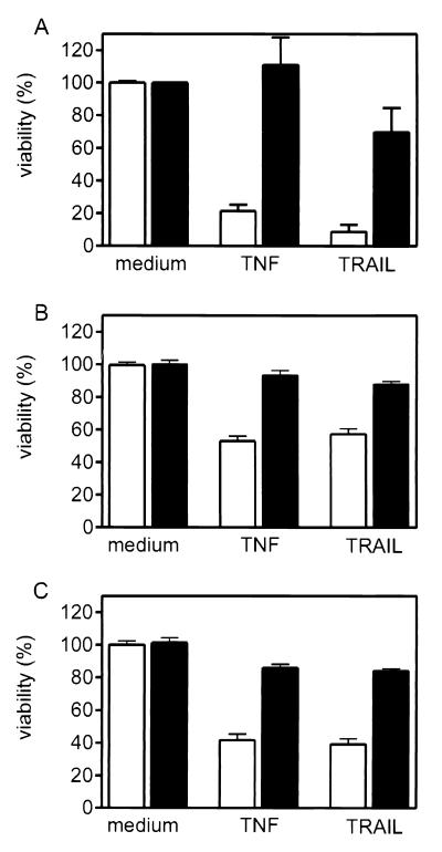
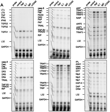
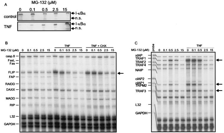
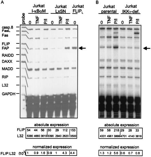
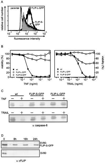
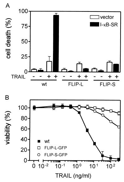
The caspase 8 homologue FLICE-inhibitory protein (cFLIP) is a potent negative regulator of death receptor-induced apoptosis. We found that cFLIP can be upregulated in some cell lines under critical involvement of the NF-kappaB pathway, but NF-kappaB activation was clearly not sufficient for cFLIP induction in all cell lines. Treatment of SV80 cells with the proteasome inhibitor N-benzoyloxycarbonyl (Z)-Leu-Leu-leucinal (MG-132) or geldanamycin, a drug interfering with tumor necrosis factor (TNF)-induced NF-kappaB activation, inhibited TNF-induced upregulation of cFLIP. Overexpression of a nondegradable IkappaBalpha mutant (IkappaBalpha-SR) or lack of IkappaB kinase gamma expression completely prevented phorbol myristate acetate-induced upregulation of cFLIP mRNA in Jurkat cells. These data point to an important role for NF-kappaB in the regulation of the cFLIP gene. SV80 cells normally show resistance to TNF-related apoptosis-inducing ligand (TRAIL) and TNF, as apoptosis can be induced only in the presence of low concentrations of cycloheximide (CHX). However, overexpression of IkappaBalpha-SR rendered SV80 cells sensitive to TRAIL-induced apoptosis in the absence of CHX, and cFLIP expression was able to reverse the proapoptotic effect of NF-kappaB inhibition. Western blot analysis further revealed that cFLIP, but not TRAF1, A20, and cIAP2, expression levels rapidly decrease upon CHX treatment. In conclusion, these data suggest a key role for cFLIP in the antiapoptotic response of NF-kappaB activation.


Figures

FIG. 1
FIG. 1
Impact of prestimulation with IL-1 (A), TNF (B), and the agonistic CD40-specific MAb G28.5 on TNF- and TRAIL-induced cytotoxicity. SV80-CD40 cells were cultivated in 96-well plates (15,000 cells/well) for 24 h and were then treated with IL-1 (10 ng/ml), TNF (10 ng/ml), and anti-CD40 MAb G28.5 (1 μg/ml) (A to C; solid bars) or remained untreated (A to C; empty bars). After 6 h the cells were washed twice with medium and challenged with TNF (50 ng/ml) or TRAIL-Flag (100 ng/ml) complexed with the anti-Flag MAb M2 (1 μg/ml) in the presence of 25 μg of CHX/ml for an additional 8 h. Cell viability was determined using the MTT assay.
FIG. 2
FIG. 2
(A) RNase protection assay analysis of steady-state levels of mRNA of various apoptosis-related genes in SV80-CD40 cells which were stimulated with TNF (10 ng/ml), IL-1 (10 ng/ml), the agonistic CD40-specific MAb G28.5 (αCD40) (1 μg/ml), or control MAb (1 μg/ml) or remained untreated. Whole RNAs were isolated after treatment, and 10 μg of each RNA was analyzed with the hCK-3, hApo-1c, hApo-2, hApo-5, hApo-3, hApo-3c, hApo-5b, and hApo-6 Multi-Probe template sets to detect the indicated mRNAs. No cytokine-induced changes were observed with the Multi-Probe template sets hApo-1c, containing caspase-specific probes, and hApo-2, containing templates of bcl-2 family members (data not shown). L32 and GAPDH specific probes were included in each template set as internal controls. (B) SV80-CD40 cells were treated for the indicated times with IL-1 (10 ng/ml) or TNF (10 ng/ml), and Daudi and Jurkat cells were treated for 6 h with the agonistic CD40-specific MAb G28.5 (αCD40) (1 μg/ml) or P/I (I+P). con., control. Total RNAs were isolated, and 10 μg of each RNA was analyzed using RNase protection assays with the hApo-3b Multi-Probe template set, which contains, among others, probes for cFLIP and Fas. The position of the FLIP-specific band is indicated with an arrow.
FIG. 2
FIG. 2
(A) RNase protection assay analysis of steady-state levels of mRNA of various apoptosis-related genes in SV80-CD40 cells which were stimulated with TNF (10 ng/ml), IL-1 (10 ng/ml), the agonistic CD40-specific MAb G28.5 (αCD40) (1 μg/ml), or control MAb (1 μg/ml) or remained untreated. Whole RNAs were isolated after treatment, and 10 μg of each RNA was analyzed with the hCK-3, hApo-1c, hApo-2, hApo-5, hApo-3, hApo-3c, hApo-5b, and hApo-6 Multi-Probe template sets to detect the indicated mRNAs. No cytokine-induced changes were observed with the Multi-Probe template sets hApo-1c, containing caspase-specific probes, and hApo-2, containing templates of bcl-2 family members (data not shown). L32 and GAPDH specific probes were included in each template set as internal controls. (B) SV80-CD40 cells were treated for the indicated times with IL-1 (10 ng/ml) or TNF (10 ng/ml), and Daudi and Jurkat cells were treated for 6 h with the agonistic CD40-specific MAb G28.5 (αCD40) (1 μg/ml) or P/I (I+P). con., control. Total RNAs were isolated, and 10 μg of each RNA was analyzed using RNase protection assays with the hApo-3b Multi-Probe template set, which contains, among others, probes for cFLIP and Fas. The position of the FLIP-specific band is indicated with an arrow.
FIG. 3
FIG. 3
The indicated cell lines were treated with TNF (10 ng/ml) for 6 h (+) or remained untreated (−). Total RNAs were isolated, and 10 μg of each RNA was analyzed as described above with the hApo-3b (A) and the hApo-5 (B) Multi-Probe template sets. The arrows indicate the positions of the bands specific for Fas, cFLIP, TRAF1, cIAP2, and cIAP1.
FIG. 4
FIG. 4
(A) SV80-CD40 cells were treated with the indicated concentrations of MG-132 or remained untreated. Cells were stimulated with TNF (10 ng/ml) for 20 min, and cellular IκB contents were compared by Western blotting. n.s., nonspecific. (B and C) SV80-CD40 cells were treated with the indicated concentrations of MG-132 for 7 h. In addition, the various groups were cotreated with TNF (10 ng/ml) or with TNF and CHX (25 μg/ml) for the last 6 h of MG-132 incubation. Total RNAs were isolated, and 10 μg of each RNA was analyzed using RNase protection assays with the hApo-3b (B) and the hApo-5 (C) Multi-Probe template sets. (D) SV80-CD40 cells were treated with the indicated concentrations of GA for 14 h. Cells were then challenged further with TNF (10 ng/ml) for 6 h or remained without additional treatment. Cell lysates were analyzed as described for panels B and C. Arrows indicate the positions of the bands specific for FLIP (B and D) and TRAF1, cIAP2, and cIAP1 (C).
FIG. 4
FIG. 4
(A) SV80-CD40 cells were treated with the indicated concentrations of MG-132 or remained untreated. Cells were stimulated with TNF (10 ng/ml) for 20 min, and cellular IκB contents were compared by Western blotting. n.s., nonspecific. (B and C) SV80-CD40 cells were treated with the indicated concentrations of MG-132 for 7 h. In addition, the various groups were cotreated with TNF (10 ng/ml) or with TNF and CHX (25 μg/ml) for the last 6 h of MG-132 incubation. Total RNAs were isolated, and 10 μg of each RNA was analyzed using RNase protection assays with the hApo-3b (B) and the hApo-5 (C) Multi-Probe template sets. (D) SV80-CD40 cells were treated with the indicated concentrations of GA for 14 h. Cells were then challenged further with TNF (10 ng/ml) for 6 h or remained without additional treatment. Cell lysates were analyzed as described for panels B and C. Arrows indicate the positions of the bands specific for FLIP (B and D) and TRAF1, cIAP2, and cIAP1 (C).
FIG. 5
FIG. 5
(A and B) Jurkat-I-κBαM cells and the corresponding parental cell line transfected with empty vector (Jurkat-LxSN) (A) or IKKγ-deficient Jurkat cells together with the parental control cell line (B) were treated with TNF (10 ng/ml) or P/I for 6 h or remained untreated (0). Ten micrograms of total RNA of each sample was analyzed using RNase protection assay analysis with the hApo-5 Multi-Probe template set. FLIPL, Jurkat clone stably transfected with cFLIP-L. Absolute expression and normalized expression are given in arbitrary units.
FIG. 6
FIG. 6
(A) Western blot analysis of cFLIP, caspase 8, TRAF1, and A20 expression levels in TNF- and IL-1-treated SV80-CD40 cells. SV80-CD40 cells (3 × 10⁶) were incubated for 0, 7, and 11 h with TNF (10 ng/ml) or IL-1 (10 ng/ml). To determine the CHX sensitivity of the investigated proteins, cells were further treated for 11 h with TNF or IL-1 during the last 4 h that CHX (25 μg/ml) was added. Expression of cFLIP (FLIPS), caspase 8, TRAF1, cIAP1, cIAP2, and A20 was analyzed by Western blotting. Values on the left are in kilodaltons. (B) SV80 cells were pretreated with the indicated concentrations of CHX for 2 h and were subsequently challenged with TNF (10 ng/ml) for 6 h. Cells were analyzed with the hApo-3b Multi-Probe template set as described for Fig. 2.
FIG. 7
FIG. 7
(A) FACS analysis of SV80 transfectants stably expressing FLIP-L–GFP or FLIP-S–GFP and mock-transfected SV80 cells. (B) Cells described for panel A were cultivated in 96-well plates (15,000 cells/well) for 24 h and were then treated with the indicated concentrations of TNF or TRAIL-Flag complexed with the anti-Flag MAb M2 (1 μg/ml) in the presence of 50 μg of CHX/ml for an additional 16 h. Cell viability was determined using the MTT assay. wt, wild type. (C) Cells described for panel A were challenged with TNF (10 ng/ml) and with cross-linked TRAIL-Flag in the presence of 50 μg of CHX/ml or remained untreated. Cells were lysed, and proteins were then separated by SDS-PAGE and transferred to nitrocellulose. The presence of the nonprocessed caspase 8 isoforms p53 and p55 was determined by Western blot analyses. wt, wild type; −, absence of TNF or TRAIL; +, presence of TNF or TRAIL. (D) SV80 and SV80 FLIP-S–GFP cells were incubated for the indicated times with CHX (25 μg/ml). Proteins (70 μg per lane) were then separated by SDS-PAGE and transferred to nitrocellulose, and the expression of endogenous cFLIP in SV80 cells and of cFLIP-S–GFP in the transfectants was detected on the same blot with the anti-FLIP MAb N19 and an alkaline-conjugated secondary antibody. (E) RNase protection assay analysis of various members of the TRAF and IAP protein families in SV80, SV80 FLIP-S–GFP, and SV80 FLIP-L–GFP. Cells were treated with the indicated combinations of TNF (20 ng/ml), agonistic anti-TRAIL-R2 antisera (αTR2) (1 μg/ml), z-VAD-fmk (Z) (20 μM), and CHX (C) (25 μg/ml) for 6 h. 0, untreated. Total RNAs were isolated after treatment, and 10 μg of each RNA was analyzed with the hApo-5 Multi-Probe template set to detect the indicated mRNAs. Absolute expression and normalized expression are given in arbitrary units. Relative expression levels were calculated as described in Materials and Methods. Arrows indicate the positions of the bands specific for TRAF1 and cIAP2.
FIG. 7
FIG. 7
(A) FACS analysis of SV80 transfectants stably expressing FLIP-L–GFP or FLIP-S–GFP and mock-transfected SV80 cells. (B) Cells described for panel A were cultivated in 96-well plates (15,000 cells/well) for 24 h and were then treated with the indicated concentrations of TNF or TRAIL-Flag complexed with the anti-Flag MAb M2 (1 μg/ml) in the presence of 50 μg of CHX/ml for an additional 16 h. Cell viability was determined using the MTT assay. wt, wild type. (C) Cells described for panel A were challenged with TNF (10 ng/ml) and with cross-linked TRAIL-Flag in the presence of 50 μg of CHX/ml or remained untreated. Cells were lysed, and proteins were then separated by SDS-PAGE and transferred to nitrocellulose. The presence of the nonprocessed caspase 8 isoforms p53 and p55 was determined by Western blot analyses. wt, wild type; −, absence of TNF or TRAIL; +, presence of TNF or TRAIL. (D) SV80 and SV80 FLIP-S–GFP cells were incubated for the indicated times with CHX (25 μg/ml). Proteins (70 μg per lane) were then separated by SDS-PAGE and transferred to nitrocellulose, and the expression of endogenous cFLIP in SV80 cells and of cFLIP-S–GFP in the transfectants was detected on the same blot with the anti-FLIP MAb N19 and an alkaline-conjugated secondary antibody. (E) RNase protection assay analysis of various members of the TRAF and IAP protein families in SV80, SV80 FLIP-S–GFP, and SV80 FLIP-L–GFP. Cells were treated with the indicated combinations of TNF (20 ng/ml), agonistic anti-TRAIL-R2 antisera (αTR2) (1 μg/ml), z-VAD-fmk (Z) (20 μM), and CHX (C) (25 μg/ml) for 6 h. 0, untreated. Total RNAs were isolated after treatment, and 10 μg of each RNA was analyzed with the hApo-5 Multi-Probe template set to detect the indicated mRNAs. Absolute expression and normalized expression are given in arbitrary units. Relative expression levels were calculated as described in Materials and Methods. Arrows indicate the positions of the bands specific for TRAF1 and cIAP2.
FIG. 8
FIG. 8
(A) Transient expression of IκB-SR renders SV80 cells, but not FLIP-L- or FLIP-S-expressing cells, sensitive to TRAIL in the absence of CHX. SV80, SV80 FLIP-L, and SV80 FLIP-S cells were transfected with pEGFP along with empty vector or pIκB-SR encoding a nondegradable form of IκB and were split. After 1 day of recovery they were challenged with TRAIL-Flag complexed with the anti-Flag MAb M2 (1 μg/ml) for an additional 16 h or remained untreated. Finally GFP-positive cells were analyzed for the percentage of cells with morphological features of apoptosis. −, absence of TRAIL; +, presence of TRAIL. (B) Cells were cultivated in 96-well plates (15,000 cells/well) for 24 h and were then treated 1 h with MG-132 (10 μM). Subsequently, the indicated concentrations of cross-linked TRAIL-Flag were added for an additional 16 h. Cell viability was determined using the MTT assay. wt, wild type.

Similar articles

Cited by

References

    1. Barkett M, Xue D, Horvitz H R, Gilmore T D. Phosphorylation of IkappaB-alpha inhibits its cleavage by caspase CPP32 in vitro. J Biol Chem. 1997;272:29419–29422. - PubMed
    1. Beauparlant P, Kwon H, Clarke M, Lin R, Sonenberg N, Wainberg M, Hiscott J. Transdominant mutants of IκBα block Tat-tumor necrosis factor synergistic activation of human immunodeficiency virus type 1 gene expression and virus multiplication. J Virol. 1996;70:5777–5785. - PMC - PubMed
    1. Beg A A, Sha W C, Bronson R T, Ghosh S, Baltimore D. Embryonic lethality and liver degeneration in mice lacking the RelA component of NF-kappa B. Nature. 1995;376:167–170. - PubMed
    1. Beg A A, Baltimore D. An essential role for NF-kappaB in preventing TNF-alpha-induced cell death. Science. 1996;274:782–784. - PubMed
    1. Chan F K-M, Lenardo M J. A crucial role for p80 TNF-R2 in amplifying p60 TNF-R1 apoptosis signals in T lymphocytes. Eur J Immunol. 2000;30:652–660. - PubMed

Publication types

MeSH terms

Substances