. 2001 Jun;21(12):4016-31.
doi: 10.1128/MCB.21.12.4016-4031.2001.

Epidermal growth factor-induced tumor cell invasion and metastasis initiated by dephosphorylation and downregulation of focal adhesion kinase

Affiliations

Epidermal growth factor-induced tumor cell invasion and metastasis initiated by dephosphorylation and downregulation of focal adhesion kinase

Z Lu et al. Mol Cell Biol. 2001 Jun.

Abstract

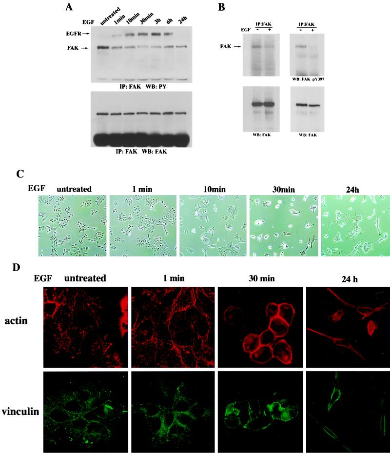
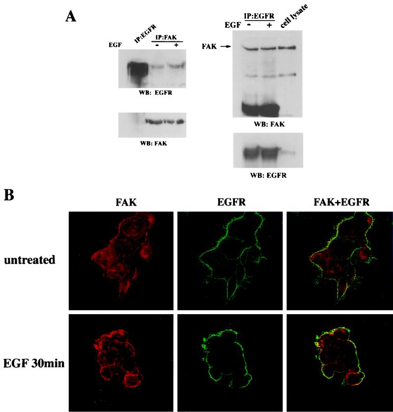
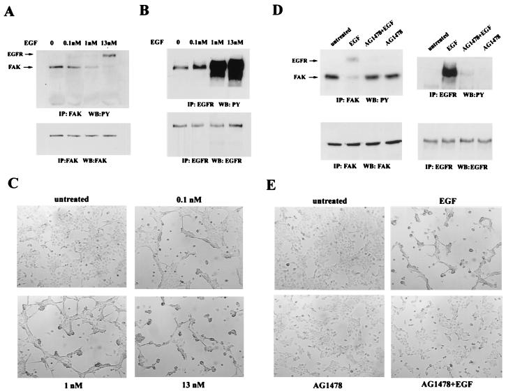
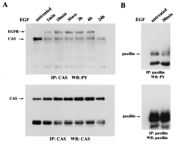
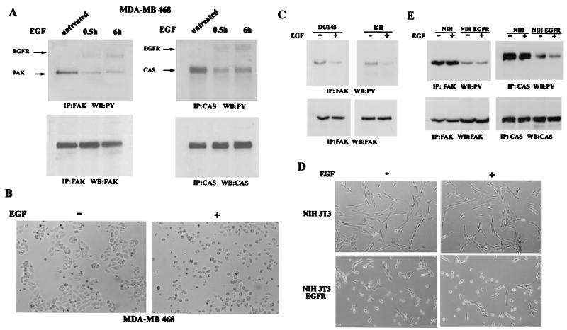
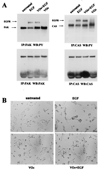
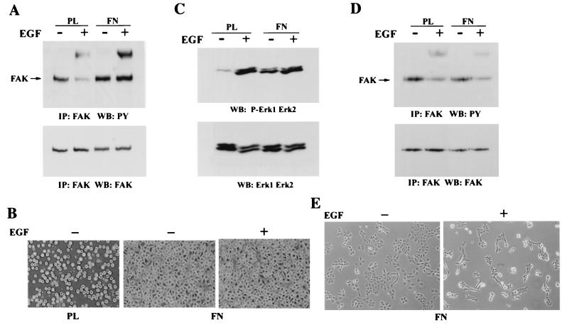
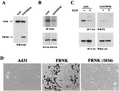
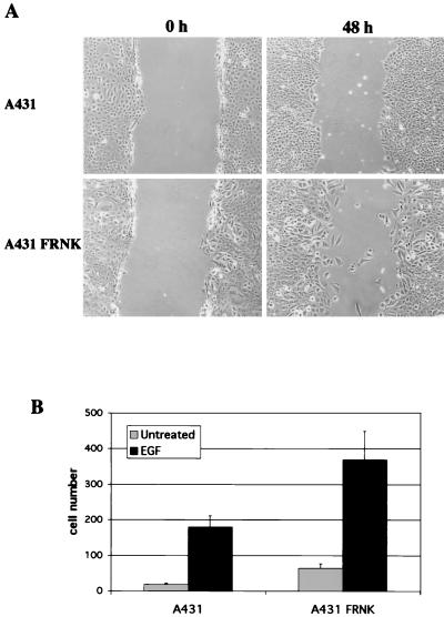
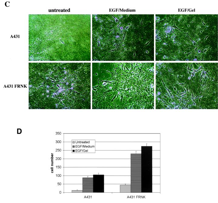
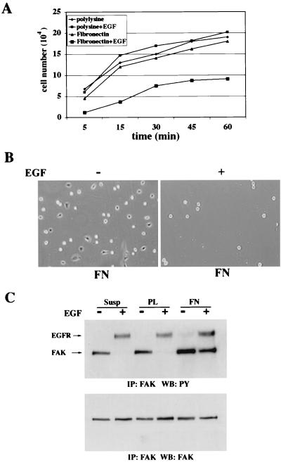
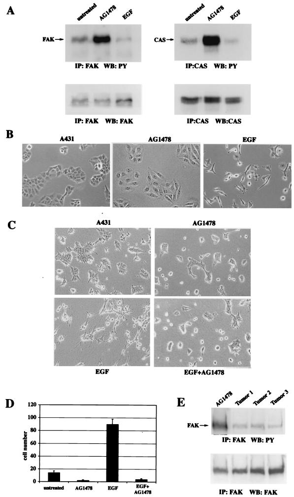
Upregulated epidermal growth factor (EGF) receptor (EGFR) expression and EGFR-induced signaling have been correlated with progression to invasion and metastasis in a wide variety of carcinomas, but the mechanism behind this is not well understood. We show here that, in various human carcinoma cells that overexpress EGFR, EGF treatment induced rapid tyrosine dephosphorylation of focal adhesion kinase (FAK) associated with downregulation of its kinase activity. The downregulation of FAK activity was both required and sufficient for EGF-induced refractile morphological changes, detachment of cells from the extracellular matrix, and increased tumor cell motility, invasion, and metastasis. Tumor cells with downregulated FAK activity became less adherent to the extracellular matrix. However, once cells started reattaching, FAK activity was restored by activated integrin signaling. Moreover, this process of readhesion and spreading could not be abrogated by further EGF stimulation. Interruption of transforming growth factor alpha-EGFR autocrine regulation with an EGFR tyrosine kinase inhibitor led to a substantial increase in FAK tyrosine phosphorylation and inhibition of tumor cell invasion in vitro. Consistent with this, FAK tyrosine phosphorylation was reduced in cells from tumors growing in transplanted, athymic, nude mice, which have an intact autocrine regulation of the EGFR. We suggest that the dynamic regulation of FAK activity, initiated by EGF-induced downregulation of FAK leading to cell detachment and increased motility and invasion, followed by integrin-dependent reactivation during readhesion, plays a role in EGF-associated tumor invasion and metastasis.


Figures

FIG. 1
FIG. 1
EGF induces FAK dephosphorylation and inactivation prior to cell morphology changes and detachment. (A) A431 cells were treated with EGF (100 ng/ml) for the indicated times. FAK was immunoprecipitated (IP) with polyclonal FAK antiserum followed by Western blot (WB) analysis with antiphosphotyrosine (PY) antibody (upper panel) and then reprobing with polyclonal FAK antiserum (lower panel). (B) A431 cells were treated with EGF (100 ng/ml) for 30 min. FAK was immunoprecipitated with polyclonal FAK antiserum and then analyzed by in vitro kinase assay as described in Materials and Methods (upper left panel). FAK levels were assessed by immunoblotting analysis using polyclonal FAK antiserum (lower left panel). Similarly, immunoprecipitated FAK was immunoblotted with anti-phospho-FAK Tyr-397 antibodies (upper right panel), and then reprobed with polyclonal FAK antiserum (lower right panel). (C) A431 cells treated with EGF (100 ng/ml) for the indicated times were examined using a digital camera mounted on a microscope with 100× magnification or stained for actin with TRITC-labeled phalloidin or for focal adhesion with antivinculin antibody (D).
FIG. 2
FIG. 2
FAK associates and colocalizes with EGFR. (A) A431 cells were treated with EGF (100 ng/ml) for 30 min. Immunoprecipitation (IP) was then carried out with anti-FAK antiserum, followed by immunoblotting (WB) with anti-EGFR monoclonal antibody (upper left panel). FAK protein levels were determined by immunoblot analysis using a FAK antiserum (lower left panel). Reciprocal immunoprecipitation with anti-EGFR monoclonal antibody was followed by immunoblotting with polyclonal FAK antiserum (upper right panel), and then reprobing with anti-EGFR monoclonal antibody (lower right panel). (B) Deconvolution microscopy for FAK (red), EGFR (green), and colocalized FAK and EGFR (yellow) in A431 cells that were either left untreated or treated with EGF for 30 min.
FIG. 3
FIG. 3
FAK dephosphorylation, refractile morphological changes, and detachment of A431 cells are dependent on EGFR activation. A431 cells were treated with different doses of EGF for 30 min. The tyrosine phosphorylation levels of FAK (A) and EGFR (B) and the morphology of the cells (C) were examined as described in the legend to Fig. 1. (D) A431 cells were treated with EGF (100 ng/ml) or AG1478 (300 nM) or pretreated with AG1478 (300 nM) for 30 min before EGF treatment. The tyrosine phosphorylation levels of FAK (upper left panel) and EGFR (upper right panel) were determined by blotting with antiphosphotyrosine (PY) monoclonal antibody following immunoprecipitation with either anti-FAK or anti-EGFR antibodies. The protein levels of FAK (lower left panel) and EGFR (lower right panel) in the immunoprecipitates were determined by immunoblotting for the indicated protein. (E) A431 cells were treated with EGF (100 ng/ml) or AG1478 (300 nM) or pretreated with AG1478 (300 nM) for 30 min before EGF treatment. The morphology of A431 cells was examined as described in the legend to Fig. 1.
FIG. 4
FIG. 4
p130cas and paxillin are also dephosphorylated upon EGF treatment. (A) A431 cells were treated with EGF (100 ng/ml) for the indicated times. p130cas was immunoprecipitated (IP) with polyclonal p130cas antiserum, followed by immunoblot (WB) analysis with antiphosphotyrosine antibody (upper panel). Immunoblots were then reprobed with p130cas antiserum (lower panel). (B) A431 cells were treated with EGF (100 ng/ml) for 30 min. Immunoprecipitation was carried out with antipaxillin monoclonal antibody, followed by immunoblotting with antiphosphotyrosine antibody (upper panel). Immunoblots were then reprobed with antipaxillin monoclonal antibody.
FIG. 5
FIG. 5
EGF-induced inactivation of FAK and refractile morphological changes are general phenotypes of human tumor cells that overexpress EGFR. (A) MDA-MB468 breast cancer cells were treated with EGF (100 ng/ml) for 30 min or 6 h. The tyrosine phosphorylation levels of FAK and p130cas as well as the morphology of the cells, which were either left untreated or treated with EGF for 6 h (B), were examined as described in the legend to Fig. 1. DU145 prostate cancer cells and KB oral carcinoma cells (C) and NIH 3T3 cells and NIH 3T3 EGFR cells (D and E) were either left untreated or treated with EGF (100 ng/ml) for 30 min. The morphology of the cells and tyrosine phosphorylation levels of FAK and p130cas were examined as described in the legend to Fig. 1.
FIG. 6
FIG. 6
EGF-induced phenotype changes of A431 cells is PTP dependent. A431 cells were treated with EGF (100 ng/ml) or pervanadate (50 μM) or pretreated with pervanadate (50 μM) for 30 min before EGF treatment. The tyrosine phosphorylation levels of FAK (A) and cell morphology (B) were examined as described in the legend to Fig. 1.
FIG. 7
FIG. 7
Activation of FAK by integrin engagement blocks EGF-induced FAK dephosphorylation and also prevents morphological changes and cell detachment from the ECM. A431 cells were trypsinized and kept suspended in DMEM with 0.1% BSA for 40 min before plating onto either fibronectin (FN)- or poly-l-lysine (PL)-coated plates for 40 min (A and B) or 12 h (D and E) prior to 20 min of EGF (100 ng/ml) treatment. The tyrosine phosphorylation levels of FAK and cell morphology were examined as described in the legend to Fig. 1. (C) The activation of ERK1 and ERK2 was determined by Western blot analysis using anti-phospho-ERK1 and -ERK2 monoclonal antibodies (upper panel). The blot was reprobed with anti-ERK1 and -ERK2 antisera (lower panel).
FIG. 8
FIG. 8
Inhibition of FAK by expression of pp41/43FRNK results in refractile morphological changes and cell detachment. (A) The level of pp41/43FRNK overexpression in A431 cells was determined by immunoblotting (WB) with anti-FAK polyclonal antiserum recognizing the C terminus of FAK. (B) FAK was immunoprecipitated (IP) with polyclonal FAK antiserum and used to perform the in vitro kinase assay as described in Materials and Methods. (C) The tyrosine phosphorylation levels of p130cas without or with EGF (100 ng/ml) treatment and the morphology of A431 cells, A431 cells expressing FRNK and A431 cells expressing the point mutant L1034S-FRNK (D) were examined as described in the legend to Fig. 1.
FIG. 9
FIG. 9
EGF-induced downregulation of FAK activity promotes tumor cell motility and invasion. (A) A431 cells or A431 cells expressing FRNK were plated at 70% confluence in DMEM with 10% serum. At 24 h after seeding, the cell monolayers were wounded by scraping with a 200-μl plastic micropipette tip, washed, and then refed with complete DMEM. After 48 h, cells were fixed with 4% paraformaldehyde and photographed as described in the legend to Fig. 1. (B) Six thousand A431 cells or A431 cells expressing pp41/43FRNK in DMEM with 10% serum were seeded in the 6.5-mm upper chamber of transwells containing a polycarbonate membrane. At 12 h after seeding, EGF (100 ng/ml) was added to the upper chamber. The cells that migrated into the lower chamber were counted 72 h after addition of EGF. Data represent the mean ± standard deviation of two independent experiments. (C) Cells in the presence or absence of EGF (100 ng/ml) (chemokinesis) were plated on the top of collagen gel with or without admixed EGF (100 ng/ml) (chemotaxis). Five days after plating, cells were photographed at a focal plane beneath the surface to visualize cells that have penetrated the gel. The number of invading cells in 10 photographic fields from two separate experiments was counted (D).
FIG. 9
FIG. 9
EGF-induced downregulation of FAK activity promotes tumor cell motility and invasion. (A) A431 cells or A431 cells expressing FRNK were plated at 70% confluence in DMEM with 10% serum. At 24 h after seeding, the cell monolayers were wounded by scraping with a 200-μl plastic micropipette tip, washed, and then refed with complete DMEM. After 48 h, cells were fixed with 4% paraformaldehyde and photographed as described in the legend to Fig. 1. (B) Six thousand A431 cells or A431 cells expressing pp41/43FRNK in DMEM with 10% serum were seeded in the 6.5-mm upper chamber of transwells containing a polycarbonate membrane. At 12 h after seeding, EGF (100 ng/ml) was added to the upper chamber. The cells that migrated into the lower chamber were counted 72 h after addition of EGF. Data represent the mean ± standard deviation of two independent experiments. (C) Cells in the presence or absence of EGF (100 ng/ml) (chemokinesis) were plated on the top of collagen gel with or without admixed EGF (100 ng/ml) (chemotaxis). Five days after plating, cells were photographed at a focal plane beneath the surface to visualize cells that have penetrated the gel. The number of invading cells in 10 photographic fields from two separate experiments was counted (D).
FIG. 10
FIG. 10
EGF-treated A431 cells with inhibited FAK activity are still able to form new adhesions on fibronectin. After being trypsinized and kept in suspension (Susp) for 40 min in DMEM containing 0.1% BSA, 2.5 × 10⁵ A431 cells were treated with EGF (100 ng/ml) or not treated for 10 min, followed by plating onto either fibronectin (FN)- or poly-l-lysine (PL)-coated plates for the indicated times. The attached cells were collected and counted (A), photographed 40 min after seeding (B), or lysed for examination of FAK tyrosine phosphorylation levels (C) as described in the legend to Fig. 1. Data represent the mean ± standard deviation of two independent experiments.
FIG. 11
FIG. 11
Effect of EGFR autocrine regulation on FAK phosphorylation. A431 cells were grown in the absence or presence of EGF (100 ng/ml) or AG1478 (300 nM) for 10 days. The tyrosine phosphorylation levels of FAK and p130cas (A) and the morphology of cells (B) were examined as described in the legend to Fig. 1. (C) A431 cells in the presence of EGF (100 ng/ml) and/or AG1478 (300 nM) were plated on the top of collagen gel. The cells were photographed 2 days later for invasion on the gel surface. Five days after plating, the number of invading cells that have penetrated the gel in 10 photographic fields from two separate experiments was counted (D). (E) A total of 1.7 × 10⁶ A431 cells were injected subcutaneously into the flank region of nude mice. Tumors were isolated and homogenized in lysis buffer 2 weeks after injection. As a control, cultured A431 cells were treated with AG1478 (300 nM) for 10 days before lysis. Cell lysates which contained equal amounts of human EGFR as determined by Western blot analysis with anti-EGFR antibody (data not shown) were used for immunoprecipitation. The tyrosine phosphorylation levels of FAK were examined as described in the legend to Fig. 1.
FIG. 12
FIG. 12
Inhibition of FAK by expression of pp41/43FRNK increases tumor metastasis rates in vivo. A total of 1.7 × 10⁶ A431 cells or A431 cells expressing pp41/43FRNK were injected subcutaneously into the flank region of athymic mice. Tumor, liver, pancreas, spleen, kidneys, lungs, brain, and femurs were isolated from each mouse, and genomic DNA from A431 cells (control), tumors (control), and each organ was examined. PCR amplification was carried out as described in Materials and Methods with primers positioned in the most conserved areas of human Alu sequences. The upper panel shows PCR amplification from mice injected with A431 cells, and the lower panel shows products from a control mouse or mice injected with A431 cells expressing pp41/43FRNK, as indicated.

References

    1. Angers-Loustau A, Cote J F, Charest A, Dowbenko D, Spencer S, Lasky L A, Tremblay M L. Protein tyrosine phosphatase-PEST regulates focal adhesion disassembly, migration, and cytokinesis in fibroblasts. J Cell Biol. 1999;144:1019–1031. - PMC - PubMed
    1. Arcaro A, Zvelebil M J, Wallasch C, Ullrich A, Waterfield M D, Domin J. Class II phosphoinositide 3-kinases are downstream targets of activated polypeptide growth factor receptors. Mol Cell Biol. 2000;20:3817–3830. - PMC - PubMed
    1. Barnes D W. Epidermal growth factor inhibits growth of A431 human epidermoid carcinoma in serum-free cell culture. J Cell Biol. 1982;93:1–4. - PMC - PubMed
    1. Berger M S, Greenfield C, Gullick W J, Haley J, Downward J, Neal D E, Harris A L, Waterfield M D. Evaluation of epidermal growth factor receptors in bladder tumours. Br J Cancer. 1987;56:533–537. - PMC - PubMed
    1. Birchmeier W, Weidner K M, Behrens J. Molecular mechanisms leading to loss of differentiation and gain of invasiveness in epithelial cells. J Cell Sci Suppl. 1993;17:159–164. - PubMed

Publication types

MeSH terms