. 2001 Jun;21(12):4046-54.
doi: 10.1128/MCB.21.12.4046-4054.2001.

DNA strand break-sensing molecule poly(ADP-Ribose) polymerase cooperates with p53 in telomere function, chromosome stability, and tumor suppression

Affiliations

DNA strand break-sensing molecule poly(ADP-Ribose) polymerase cooperates with p53 in telomere function, chromosome stability, and tumor suppression

W M Tong et al. Mol Cell Biol. 2001 Jun.

Abstract

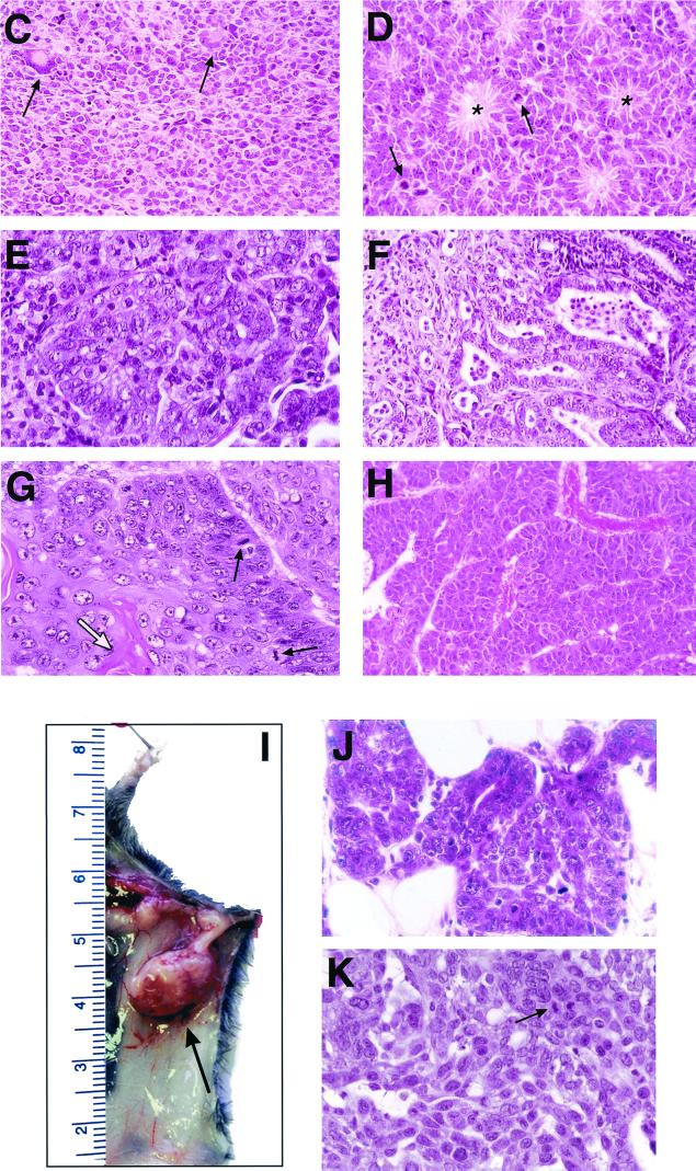
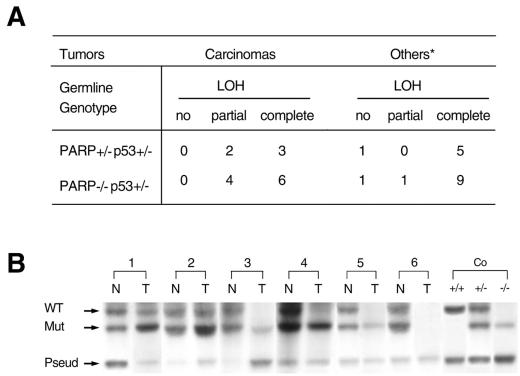
Genomic instability is often caused by mutations in genes that are involved in DNA repair and/or cell cycle checkpoints, and it plays an important role in tumorigenesis. Poly(ADP-ribose) polymerase (PARP) is a DNA strand break-sensing molecule that is involved in the response to DNA damage and the maintenance of telomere function and genomic stability. We report here that, compared to single-mutant cells, PARP and p53 double-mutant cells exhibit many severe chromosome aberrations, including a high degree of aneuploidy, fragmentations, and end-to-end fusions, which may be attributable to telomere dysfunction. While PARP(-/-) cells showed telomere shortening and p53(-/-) cells showed normal telomere length, inactivation of PARP in p53(-/-) cells surprisingly resulted in very long and heterogeneous telomeres, suggesting a functional interplay between PARP and p53 at the telomeres. Strikingly, PARP deficiency widens the tumor spectrum in mice deficient in p53, resulting in a high frequency of carcinomas in the mammary gland, lung, prostate, and skin, as well as brain tumors, reminiscent of Li-Fraumeni syndrome in humans. The enhanced tumorigenesis is likely to be caused by PARP deficiency, which facilitates the loss of function of tumor suppressor genes as demonstrated by a high rate of loss of heterozygosity at the p53 locus in these tumors. These results indicate that PARP and p53 interact to maintain genome integrity and identify PARP as a cofactor for suppressing tumorigenesis.


Figures

FIG. 1
FIG. 1
FISH analysis of metaphase chromosomes (DNA stained with 4′,6′-diamidino-2-phenylindole [DAPI] is shown in blue; Cy3-labeled TTAGGG repeats are shown in yellow) of primary MEFs prepared from wild-type (A), PARP−/− p53−/− (B), and PARP+/+ p53−/− (I) embryos. The corresponding DAPI staining of chromosomes in panels C, E, G, and I are shown in panels D, F, H, and J, respectively. Examples of chromosomal abnormalities in PARP−/− p53−/− and PARP+/+ p53−/− MEFs are telomere associations and end-to-end fusions (fu/t), Robertsonian-like configurations (rl), fragmentations (f), dicentrics (d), and ring-like chromosomes (r). Note chromosomes lacking detectable telomere fluorescence (arrows).
FIG. 2
FIG. 2
Q-FISH analyses of telomere length in PARP and p53 double-mutant cells. Frequency distributions of telomere fluorescence values (pooled p- and q-arm values) in metaphase chromosomes from representative primary MEFs with the indicated genotype are shown. The x axis depicts the intensity of each signal, expressed in (TFUs), with each unit representing ∼1 kb of telomere repeats (42); the y axis shows the frequency of telomeres of a given length.
FIG. 3
FIG. 3
Tumor development in PARP p53 double-mutant mice. (A and B) Tumor incidence in PARP p53 double-mutant mice. PARP-deficient p53−/− animals were monitored over a period of 22 weeks (A), and PARP-deficient p53+/− mice were monitored over a period of 20 months (B). Moribund or tumor-bearing mice were autopsied. (C to K) Histopathological characterization of brain tumors and representative carcinomas from PARP p53 double-mutant mice. (C) Undifferentiated primitive neuroectodermal tumor from a 2.5-month-old PARP−/− p53−/− mouse containing megakaryocytes (arrows) and cells with pleomorphic nuclei and mitotic figures. (D) Ependymoma from a 14.5-month-old PARP−/− p53+/− mouse showing a typical rosette arrangement in the tumor (asterisks), with frequent appearance of mitotic cells (arrows). (E) Lung adenocarcinoma from a 15-month-old PARP−/− p53+/− mouse showing gland-like structure. (F) Prostate carcinoma from a 20-month-old PARP+/− p53+/− mouse showing irregular glandular formation and invasion of the stroma. (G) Keratinizing squamous carcinoma originating from an ear exhibiting numerous keratin pearls (white arrow) and frequent mitotic figures (black arrows). (H) Massive hepatocellular carcinoma showing a trabecular pattern of tumor cells. (I to K), Mammary carcinomas in PARP- and p53-deficient mice. (I) Tumors from the left third pair of mammary glands (arrow) of a 13-month-old PARP−/− p53+/− female mouse. A ductal carcinoma in situ (J) and a solid mammary carcinoma (K) show frequent mitotic figures (arrow).
FIG. 3
FIG. 3
Tumor development in PARP p53 double-mutant mice. (A and B) Tumor incidence in PARP p53 double-mutant mice. PARP-deficient p53−/− animals were monitored over a period of 22 weeks (A), and PARP-deficient p53+/− mice were monitored over a period of 20 months (B). Moribund or tumor-bearing mice were autopsied. (C to K) Histopathological characterization of brain tumors and representative carcinomas from PARP p53 double-mutant mice. (C) Undifferentiated primitive neuroectodermal tumor from a 2.5-month-old PARP−/− p53−/− mouse containing megakaryocytes (arrows) and cells with pleomorphic nuclei and mitotic figures. (D) Ependymoma from a 14.5-month-old PARP−/− p53+/− mouse showing a typical rosette arrangement in the tumor (asterisks), with frequent appearance of mitotic cells (arrows). (E) Lung adenocarcinoma from a 15-month-old PARP−/− p53+/− mouse showing gland-like structure. (F) Prostate carcinoma from a 20-month-old PARP+/− p53+/− mouse showing irregular glandular formation and invasion of the stroma. (G) Keratinizing squamous carcinoma originating from an ear exhibiting numerous keratin pearls (white arrow) and frequent mitotic figures (black arrows). (H) Massive hepatocellular carcinoma showing a trabecular pattern of tumor cells. (I to K), Mammary carcinomas in PARP- and p53-deficient mice. (I) Tumors from the left third pair of mammary glands (arrow) of a 13-month-old PARP−/− p53+/− female mouse. A ductal carcinoma in situ (J) and a solid mammary carcinoma (K) show frequent mitotic figures (arrow).
FIG. 4
FIG. 4
LOH in tumors from PARP-deficient p53+/− mice. (A) Summary of LOH frequency in tumors from PARP-deficient p53+/− mice. ∗, includes lymphoma, sarcomas, and brain tumors. (B) Representative Southern blot analysis showing partial (1T and 2T) and complete (3T, 4T, and 5T) LOH of wild-type p53 in tumors from PARP−/−, p53+/− mice. The loss of both wild-type and mutant p53 bands is also evident in some of the tumors (6T). The wild-type p53 (WT), the mutant allele (Mut), and the p53 pseudogene (Pseud) are indicated. N, normal tissues adjacent to the tumor or tail; T, tumor tissue.

Similar articles

Cited by

References

    1. Agarwal M L, Agarwal A, Taylor W R, Wang Z-Q, Wagner E F, Stark G R. Defective induction but normal activation and function of p53 in mouse cells lacking poly-ADP-ribose polymerase. Oncogene. 1997;15:1035–1041. - PubMed
    1. Artandi S E, Chang S, Lee S L, Alson S, Gottlieb G J, Chin L, DePinho R A. Telomere dysfunction promotes non-reciprocal translocations and epithelial cancers in mice. Nature. 2000;406:641–645. - PubMed
    1. Banin S, Moyal L, Shieh S, Taya Y, Anderson C W, Chessa L, Smorodinsky N I, Prives C, Reiss Y, Shiloh Y, Ziv Y. Enhanced phosphorylation of p53 by ATM in response to DNA damage. Science. 1998;281:1674–1677. - PubMed
    1. Bell D W, Varley J M, Szydlo T E, Kang D H, Wahrer D C, Shanon K E, Lubratovich M, Verselis S J, Issebacher K J, Fraumeni J F, Birch J M, Li F P, Garber J E, Haber D A. Heterozygous germ line hCHK2 mutations in Li-Fraumeni syndrome. Science. 1999;286:2528–2531. - PubMed
    1. Blackburn E H. Telomere states and cell fates. Nature. 2000;408:53–56. - PubMed

Publication types

MeSH terms

LinkOut - more resources