. 2001 Jun;21(12):4055-66.
doi: 10.1128/MCB.21.12.4055-4066.2001.

Phospholipase D activity is required for actin stress fiber formation in fibroblasts

Affiliations

Phospholipase D activity is required for actin stress fiber formation in fibroblasts

Y Kam et al. Mol Cell Biol. 2001 Jun.

Abstract

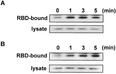
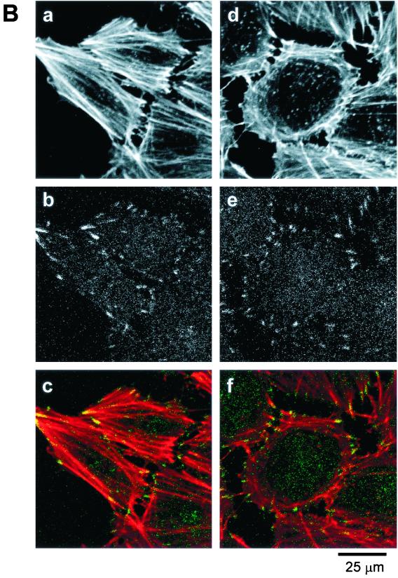
Phospholipase D (PLD) is a ubiquitously expressed enzyme of ill-defined function. In order to explore its cellular actions, we inactivated the rat PLD1 (rPLD1) isozyme by tagging its C terminus with a V5 epitope (rPLD1-V5). This was stably expressed in Rat-2 fibroblasts to see if it acted as a dominant-negative mutant for PLD activity. Three clones that expressed rPLD1-V5 were selected (Rat2V16, Rat2V25, and Rat2V29). Another clone (Rat2V20) that lost expression of rPLD1-V5 was also obtained. In the three clones expressing rPLD1-V5, PLD activity stimulated by phorbol myristate acetate (PMA) or lysophosphatidic acid (LPA) was reduced by ~50%, while the PLD activity of Rat2V20 cells was normal. Changes in the actin cytoskeleton in response to LPA or PMA were examined in these clones. All three clones expressing rPLD1-V5 failed to form actin stress fibers after treatment with LPA. However, Rat2V20 cells formed stress fibers in response to LPA to the same extent as wild-type Rat-2 cells. In contrast, there was no significant change in membrane ruffling induced by PMA in the cells expressing rPLD1-V5. Since Rho is an activator both of rPLD1 and stress fiber formation, the activation of Rho was monitored in wild-type Rat-2 cells and Rat2V25 cells, but no significant difference was detected. The phosphorylation of vimentin mediated by Rho-kinase was also intact in Rat2V25 cells. Rat2V25 cells also showed normal vinculin-containing focal adhesions. However, the translocation of alpha-actinin to the cytoplasm and to the detergent-insoluble fraction in Rat2V25 cells was reduced. These results indicate that PLD activity is required for LPA-induced rearrangement of the actin cytoskeleton to form stress fibers and that PLD might be involved in the cross-linking of actin filaments mediated by alpha-actinin.


Figures

FIG. 1
FIG. 1
V5 epitope tagging of the C terminus inactivates rPLD1. Rat PLD1 or rPLD1-V5 was expressed in COS-7 cells, and the expression and PLD activity were monitored. (A) COS-7 cells were transfected with either pcDNA3.1 (+) vector as a control (lane 1), rPLD1/pcDNA3 (lane 2), or rPLD1-V5/pcDNA3.1/V5-HisA (lane 3). Cells were incubated in DMEM containing 10% fetal bovine serum for 6 h after the transfection and starved in DMEM containing 0.5% BSA for 18 h. Each cell lysate was analyzed by Western blotting by using anti-PLD1 and anti-V5 antibody. (B) PLD activity of COS-7 cells expressing rPLD1 or rPLD1-V5 was measured with or without stimulation with 10 μg of LPA per ml for 5 min or 100 nM PMA for 15 min. Data are representative of two experiments performed in duplicate. (C) The localizations of rPLD1 and rPLD1-V5 were compared by coexpressing Xpress-tagged rPLD1 and EGFP-tagged rPLD1-V5 in COS-7 cells. The expression of Xpress-rPLD1 was visualized by anti-Xpress monoclonal antibody (panel a, red) and compared with the localization of EGFP-rPLD1-V5 (panel b, green). Both images were merged in panel c.
FIG. 1
FIG. 1
V5 epitope tagging of the C terminus inactivates rPLD1. Rat PLD1 or rPLD1-V5 was expressed in COS-7 cells, and the expression and PLD activity were monitored. (A) COS-7 cells were transfected with either pcDNA3.1 (+) vector as a control (lane 1), rPLD1/pcDNA3 (lane 2), or rPLD1-V5/pcDNA3.1/V5-HisA (lane 3). Cells were incubated in DMEM containing 10% fetal bovine serum for 6 h after the transfection and starved in DMEM containing 0.5% BSA for 18 h. Each cell lysate was analyzed by Western blotting by using anti-PLD1 and anti-V5 antibody. (B) PLD activity of COS-7 cells expressing rPLD1 or rPLD1-V5 was measured with or without stimulation with 10 μg of LPA per ml for 5 min or 100 nM PMA for 15 min. Data are representative of two experiments performed in duplicate. (C) The localizations of rPLD1 and rPLD1-V5 were compared by coexpressing Xpress-tagged rPLD1 and EGFP-tagged rPLD1-V5 in COS-7 cells. The expression of Xpress-rPLD1 was visualized by anti-Xpress monoclonal antibody (panel a, red) and compared with the localization of EGFP-rPLD1-V5 (panel b, green). Both images were merged in panel c.
FIG. 2
FIG. 2
The stable expression of V5-tagged rPLD1 in Rat-2 fibroblasts reduces PLD activity. Four G418-resistant clones were selected. (A) The expression of C-terminal V5-tagged rPLD1 was visualized by Western blotting using anti-V5 antibody or an antibody to the C terminus of rPLD1. Lane 1, not Rat-2; lane 2, Rat2V16; lane 3, Rat2V20; lane 4, Rat2V25; lane 5, Rat2V29. The Western blot of endogenous rPLD1 is shown in the lower panel. (B) PLD activity was measured in three clones of Rat2V16, Rat2V25, and Rat2V29 expressing rPLD1-V5 with or without treatment with 10 μg of LPA per ml or 100 nM PMA for 5 or 15 min, respectively. Data are representative of three independent experiments. (C) PLD activity was measured in the Rat2V20 clone, which does not express rPLD1-V5, incubated with or without 10 μg of LPA per ml or 100 nM PMA. Data are representative of two experiments performed in duplicate. Ctrl, control; wt, wild type.
FIG. 3
FIG. 3
Stress fiber formation induced by LPA and membrane ruffling induced by PMA in Rat-2 clones. Wild-type Rat-2 cells (A, B, and C), Rat2V16 cells (D, E, and F), Rat2V20 cells (G, H, and I), Rat2V25 cells (J, K, and L), and Rat2V29 cells (M, N, and O) were grown on coverslips and were serum starved. Filamentous actin was stained with Texas red X-phalloidin and analyzed by confocal microscopy after treatment with 10 μg of LPA per ml for 3 min (B, E, H, K, and N) or with 100 nM PMA for 15 min (C, F, I, L, and O). Untreated control cells are shown in A, D, G, J, and M. The scale bar represents 25 μm.
FIG. 4
FIG. 4
Activation of RhoA in wild-type Rat-2 and Rat2V25 cells by LPA. GTP-bound RhoA was pulled down from whole lysates of wild-type Rat-2 (A) or Rat2V25 (B) cells, using GST-RBD, before and after treatment with 10 μg of LPA per ml for the times indicated. RBD-bound RhoA was visualized by Western blotting (upper panel) and compared with the amount of RhoA in whole-cell lysates (lower panel).
FIG. 5
FIG. 5
The phosphorylation of vimentin by Rho-kinase is intact in Rat2V25 cells. Vimentin was immunoprecipitated with antivimentin monoclonal antibody from Rat-2 and Rat2V25 cells after labeling with 32PO4. The results of an autoradiogram of the immunoprecipitated vimentin (A) and a Coomassie blue stain of vimentin (B) are presented.
FIG. 6
FIG. 6
Vinculin-containing focal adhesions are well formed in both wild-type Rat-2 and Rat2V25 cells. (A) Wild-type Rat-2 (panels a and b) and Rat2V25 cells (panels c and d) were stained with monoclonal antivinculin antibody with (b and d) or without (a and c) treatment with 10 μg of LPA per ml for 3 min. (B) Rat-2 (panels a, b, and c) and Rat2V25 (panels d, e, and f) cells were double-labeled by using Texas red X-phalloidin (a and d) and antivinculin antibody (b and e) after the treatment with LPA. The fluorescence images of actin filaments (red) and vinculin (green) were merged in panels c and f.
FIG. 6
FIG. 6
Vinculin-containing focal adhesions are well formed in both wild-type Rat-2 and Rat2V25 cells. (A) Wild-type Rat-2 (panels a and b) and Rat2V25 cells (panels c and d) were stained with monoclonal antivinculin antibody with (b and d) or without (a and c) treatment with 10 μg of LPA per ml for 3 min. (B) Rat-2 (panels a, b, and c) and Rat2V25 (panels d, e, and f) cells were double-labeled by using Texas red X-phalloidin (a and d) and antivinculin antibody (b and e) after the treatment with LPA. The fluorescence images of actin filaments (red) and vinculin (green) were merged in panels c and f.
FIG. 7
FIG. 7
α-Actinin translocation is reduced in Rat2V25 cells. (A) The Triton X-100-insoluble fraction (see Materials and Methods) of Rat-2 and Rat2V25 cells was isolated and analyzed by Western blotting using anti-α-actinin antibody. LPA (10 μg/ml) and Y-27632 (30 μM) were treated 3 and 30 min before the isolation, respectively. (B) To visualize the change in localization of α-actinin, wild-type Rat-2 (panels a and b) and Rat2V25 cells (panels c and d) were stained with anti-α-actinin antibody, with (b and d) or without (a and c) treatment with 10 μg of LPA per ml for 3 min. (C) Rat-2 cells were stained with anti-α-actinin antibody with (panels b, c, and d) or without (panel a) the stimulation of LPA. Cells were treated with 10 μg of LPA per ml for 3 min without (b) or with pretreatment with 0.5% butan-1-ol (c) or butan-2-ol (d) for 10 min.
FIG. 7
FIG. 7
α-Actinin translocation is reduced in Rat2V25 cells. (A) The Triton X-100-insoluble fraction (see Materials and Methods) of Rat-2 and Rat2V25 cells was isolated and analyzed by Western blotting using anti-α-actinin antibody. LPA (10 μg/ml) and Y-27632 (30 μM) were treated 3 and 30 min before the isolation, respectively. (B) To visualize the change in localization of α-actinin, wild-type Rat-2 (panels a and b) and Rat2V25 cells (panels c and d) were stained with anti-α-actinin antibody, with (b and d) or without (a and c) treatment with 10 μg of LPA per ml for 3 min. (C) Rat-2 cells were stained with anti-α-actinin antibody with (panels b, c, and d) or without (panel a) the stimulation of LPA. Cells were treated with 10 μg of LPA per ml for 3 min without (b) or with pretreatment with 0.5% butan-1-ol (c) or butan-2-ol (d) for 10 min.

Similar articles

Cited by

References

    1. Amano M, Chihara K, Kimura K, Fukata Y, Nakamura N, Maatsuura Y, Kaibuchi K. Formation of actin stress fibers and focal adhesions enhanced by Rho-kinase. Science. 1997;275:1308–1311. - PubMed
    1. Bi K, Roth M G, Ktistakis N T. Phosphatidic acid formation by phospholipase D is required for transport from the endoplasmic reticulum to the Golgi complex. Curr Biol. 1997;7:301–307. - PubMed
    1. Blanchard A, Ohanian V, Critchley D. The structure and function of α-actinin. J Muscle Res Cell Motil. 1989;10:280–289. - PubMed
    1. Bokoch G M, Bohl B P, Chuang T-H. Guanine nucleotide exchange regulates membrane translocation of Rac/Rho GTP-binding proteins. J Biol Chem. 1994;269:31674–31679. - PubMed
    1. Bremser M, Nickel W, Schweikert M, Ravazzola M, Amhevdt M, Hughes C A, Sollner T H, Rothman J E, Wieland F T. Coupling of coat assembly and vesicle budding to packaging of putative cargo receptors. Cell. 1999;96:495–506. - PubMed

Publication types

MeSH terms

LinkOut - more resources