. 2001 May;12(5):1199-213.
doi: 10.1091/mbc.12.5.1199.

UV-induced hyperphosphorylation of replication protein a depends on DNA replication and expression of ATM protein

Affiliations
Free PMC article

UV-induced hyperphosphorylation of replication protein a depends on DNA replication and expression of ATM protein

G G Oakley et al. Mol Biol Cell. 2001 May.
Free PMC article

Abstract

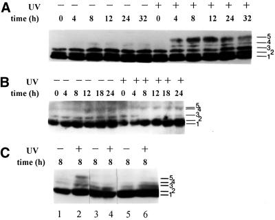
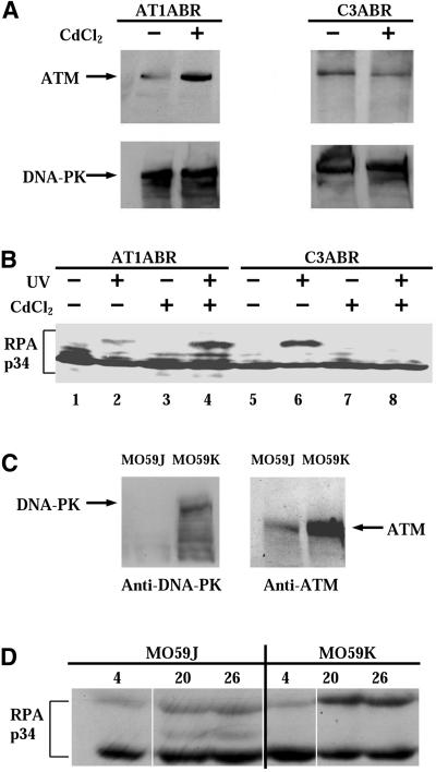
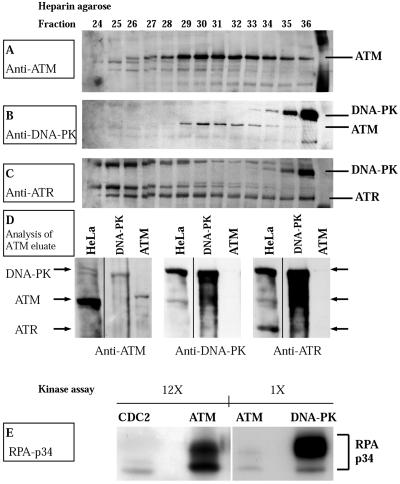
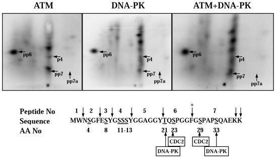
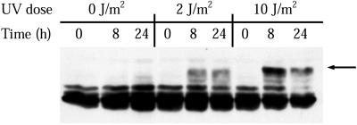
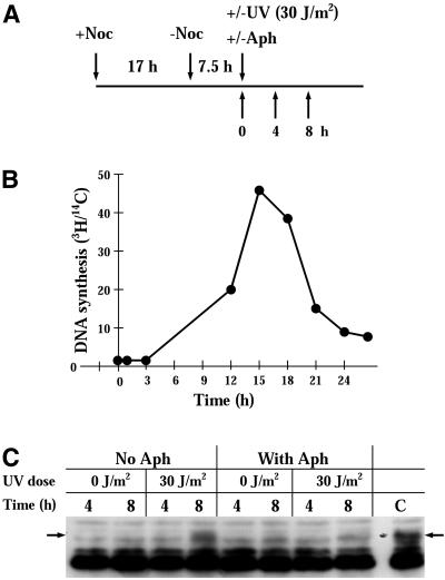
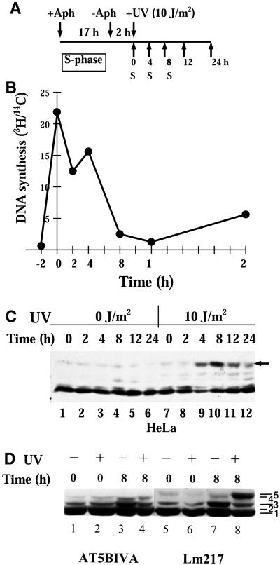
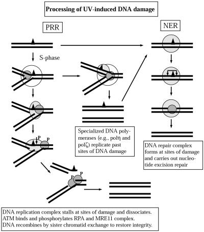
Exposure to DNA-damaging agents triggers signal transduction pathways that are thought to play a role in maintenance of genomic stability. A key protein in the cellular processes of nucleotide excision repair, DNA recombination, and DNA double-strand break repair is the single-stranded DNA binding protein, RPA. We showed previously that the p34 subunit of RPA becomes hyperphosphorylated as a delayed response (4-8 h) to UV radiation (10-30 J/m(2)). Here we show that UV-induced RPA-p34 hyperphosphorylation depends on expression of ATM, the product of the gene mutated in the human genetic disorder ataxia telangiectasia (A-T). UV-induced RPA-p34 hyperphosphorylation was not observed in A-T cells, but this response was restored by ATM expression. Furthermore, purified ATM kinase phosphorylates the p34 subunit of RPA complex in vitro at many of the same sites that are phosphorylated in vivo after UV radiation. Induction of this DNA damage response was also dependent on DNA replication; inhibition of DNA replication by aphidicolin prevented induction of RPA-p34 hyperphosphorylation by UV radiation. We postulate that this pathway is triggered by the accumulation of aberrant DNA replication intermediates, resulting from DNA replication fork blockage by UV photoproducts. Further, we suggest that RPA-p34 is hyperphosphorylated as a participant in the recombinational postreplication repair of these replication products. Successful resolution of these replication intermediates reduces the accumulation of chromosomal aberrations that would otherwise occur as a consequence of UV radiation.


Figures

Figure 1
Figure 1
RPA-p34 phosphorylation in asynchronous normal and A-T cells exposed to 10 J/m2 UVC. Cell lysates (A and B) or nuclear extracts (C) were prepared from asynchronously growing cells at indicated times after 10 J/m2 UV radiation or mock exposure. The p34 subunit of RPA was detected by Western immunoblotting with monoclonal antibody 34A. Phosphorylation causes a slower migration on the gel; at least five different forms of the p34 subunit of RPA can be visualized. (A) RPA-p34 in LM217 cells; (B) RPA-p34 in AT5BIVA cells; (C) RPA-p34 in nuclear lysates of GM00637 (lanes 1 and 2), AT5BIVA (lanes 3 and 4), and AT3BISV (lanes 5 and 6) cells. Band 1 is unphosphorylated RPA-p34; bands 2 and 3 are cell cycle–dependent phosphorylated forms; and band 5 is the DNA damage–induced hyperphosphorylated form.
Figure 2
Figure 2
(A) Expression of ATM and DNA-PK proteins in pMAT1 stably transfected A-T cells (AT1ABR) and pMAT2 stably transfected normal C3ABR cells. Cells were treated with 5 μM CdCl2 for 8 h. Whole-cell lysates (100 μg) were analyzed by SDS-PAGE and immunoblotted with anti-ATM (Ab-3; Oncogene Science) antibodies (top panels). AT1ABR is the parental A-T cell line containing a homozygous 9-bp in-frame deletion and is capable of producing near full-length but nonfunctional protein. Upon induction of the transfected line, a functional full-length recombinant ATM protein is produced. C3ABR is a control lymphoblastoid cell line expressing normal ATM. Induction of the transfected line reduces ATM expression. Membranes were reblotted with anti-DNA-PKcs antibody (Ab-1; Oncogene Science; bottom panels). (B) Phosphorylation of RPA-p34 after exposure of cells to UVC radiation. AT1ABR cells, transfected with a CdCl2-inducible ATM cDNA expression vector pMAT1, and C3ABR cells, transfected with an inducible antisense ATM cDNA expression vector pMAT2, were treated with 5 μM CdCl2 for 8 h (+) or mock-treated (−). After 8 h of CdCl2 treatment, cells were UVC irradiated (30 J/m2) or mock-irradiated. Eight hours after UV treatment, whole-cell lysates were prepared and RPA-p34 was examined by gel electrophoresis, followed by immunoblotting with anti-RPA-p34 (Ab-3; Oncogene Science) antibody. (C) DNA-PKcs (left) and ATM (right) expression in MO59J and MO59K cells detected in whole-cell lysates by immunoblotting. (D) MO59J and MO59K cells were irradiated with 30 J/m2 UV, and whole-cell lysates were prepared at the indicated times after UV irradiation. RPA-p34 hyperphosphorylation was detected by immunoblotting using anti-RPA-p34 (Ab-3; Oncogene Science) antibody. To ensure equal loading and transfer of the protein samples, the gels and the PVDF membranes were stained with Coomassie blue and colloidal gold solution, respectively.
Figure 3
Figure 3
Purification and kinase activity of ATM. (A) Western immunoblot of fractions from heparin agarose affinity column. A 100-μl aliquot of the indicated fractions was analyzed on a 6% SDS-polyacrylamide gel, transferred to PVDF membrane, and probed with antibody to ATM (Novus Biologicals). (B) The PVDF membrane was reprobed without stripping with antibody to DNA-PKcs (Ab-1; Oncogene Science). (C) ATR was detected with anti-ATR (Ab-1; Oncogene Science) without stripping the membrane. (D) Western immunoblot of HeLa cell nuclear extract (70 μg), DNA-PKcs/Ku (100 U; Promega), and purified ATM (35 μl). The samples were analyzed on a 6% SDS-polyacrylamide gel, transferred to PVDF membrane, and probed with antibody to ATM (left; Novus Biologicals). The PVDF membrane was reprobed without stripping with antibody to DNA-PKcs (center, Ab-1; Oncogene Science), and ATR was detected with anti-ATR (right, Ab-1; Oncogene Science). (E) Phosphorylation of RPA by Cdc2p34/cyclin B (New England Biolabs) purified ATM and DNA-PKcs/Ku (Promega). Purified recombinant RPA heterotrimer (0.5 μg) was incubated with either Cdc2p34/cyclin B (20 U), purified ATM or DNA-PKcs/Ku (10 U) in a 30-μl reaction containing 20 mM HEPES (pH 7.4), 10 mM MgCl2, 100 μM ATP, 2 mM DTT, 0.2 μg of single-stranded φX174 virion circular DNA, and 10 μCi [γ-32P]ATP. Samples were incubated for 30 min at 37°C. The kinase reaction was stopped by the addition of 1× Laemmli sample loading buffer, and samples were separated on 12% SDS-polyacrylamide gels followed by autoradiography. The designations 1× and 12× refer to the relative exposure time of the films shown.
Figure 4
Figure 4
Chymotryptic/tryptic phosphopeptide maps of in vitro–phosphorylated recombinant RPA-p34. (Top) Five hundred nanograms of purified recombinant RPA complex was phosphorylated by either purified ATM or 10 U of DNA-PKcs/Ku for 30 min. Hyperphosphorylated RPA-p34 was separated by SDS-PAGE and transferred to a PVDF membrane. The hyperphosphorylated form of RPA-p34 was detected by phosphorimager analysis and excised from the membrane. The hyperphosphorylated RPA-p34 was digested twice with 10 μg of chymotrypsin/trypsin and oxidized with performic acid. The digested peptides were loaded onto TLC plates and separated by electrophoresis at pH 1.9 in the first dimension, followed by ascending chromatography in the second dimension. The labeled peptides were detected by phosphorimager analysis. To verify identical and unique peptides, equal ra-dioactivity from digests of DNA-PKcs/Ku and ATM-phosphorylated RPA-p34 were loaded onto the same chromatography plate and subjected to two-dimensional separation. Numbered tryptic/chymotryptic peptides indicated on the maps with arrows refer to the peptide sequence number (lower panel) with a letter “p” designating a phosphorylated serine or threonine on the peptide. (bottom) The amino acid sequence of the N-terminus of RPA-p34 is shown along with the sites for cleavage by trypsin/chymotrypsin (↓), sites phosphorylated by Cdc2p34/cyclin B and consensus sites for DNA-PKcs/Ku. The asterisk denotes the cleavage site that is blocked by adjacent phosphorylated amino acids.
Figure 5
Figure 5
Time course of RPA phosphorylation after irradiation of XP12BE cells (XP complementation group A). XPA cells were either mock-irradiated or treated with the indicated doses of UVC. At the indicated times after treatment, cell lysates were prepared and the RPA-p34 phosphorylation pattern was analyzed by gel electrophoresis, followed by immunoblotting. The arrow to the right indicates the major hyperphosphorylated form of RPA-p34.
Figure 6
Figure 6
UVC-induced RPA-p34 hyperphosphorylation in the presence of aphidicolin. (A) Protocol for treatment. HeLa cells were treated with 0.3 μM nocodazole for 16 h. Mitotic cells were collected by shaking the cells off the dish and pelleting them. To obtain cells synchronized in G1/S phase, mitotic cells were released from nocodazole treatment for 7.5 h in fresh medium. Cells were either mock-irradiated or irradiated with 30 J/m2 UVC in the presence or absence of aphidicolin (6 μM). (B) The rate of DNA synthesis after release from nocodazole treatment was measured as 3H-thymidine incorporation during a 30-min pulse with 10 μCi/ml 3H-thymidine (NEN Life Science Products, Inc.). Cells were prelabeled with 0.01 μCi/ml 14C-thymidine (NEN Life Science Products, Inc.) for 24 h at 37°C, followed by 3H-thymidine pulse labeling. After labeling, cells were washed with PBS twice and lysed with 500 μl of 0.2 M NaOH per dish. The radioactivity of each sample was counted by dual-label liquid scintillation, and the ratio of 3H/14C reflected the DNA synthesis activity. (C) RPA-p34 phosphorylation in nocodazole-synchronized HeLa cells exposed to UVC. Cells in G1/S phase were either mock-irradiated or irradiated with 30 J/m2 UVC in the presence or absence of aphidicolin (6 μM). After preparation of whole-cell lysates, RPA-p34 was visualized by immunoblotting with anti-RPA-p34 antibody and ECL chemiluminescent detection.
Figure 7
Figure 7
UV-induced RPA-p34 phosphorylation in aphidicolin-synchronized cells. (A) Protocol for treatment. Cells were treated with 6 μM of aphidicolin for 16–20 h. After the medium containing aphidicolin was removed; the cells were washed twice in serum-free medium and further incubated in serum-containing medium without aphidicolin for 2–4 h. After 2–4 h in fresh medium, the cells were mock-irradiated or irradiated with 10 J/m2 and then incubated in the same medium for the indicated times. (B) The rate of DNA synthesis after release from aphidicolin treatment was measured as 3H-thymidine incorporation during a 30-min pulse with 10 μCi/ml 3H-thymidine (NEN Life Science Products, Inc.). Cells were prelabeled with 0.01 μCi/ml 14C-thymidine (NEN Life Science Products, Inc.) for 24 h at 37°C followed by 3H-thymidine pulse labeling. After labeling, cells were washed with PBS twice and lysed with 500 μl of 0.2 M NaOH per dish. The radioactivity of each sample was counted by dual-label liquid scintillation and the ratio of 3H/14C reflected the DNA synthesis activity. (C) Time course of RPA-p34 phosphorylation in HeLa cells. Whole cell lysates were prepared at various times after mock irradiation or irradiation with 10 J/m2 UVC. Proteins were separated on a 12% denaturing polyacrylamide gel. After transfer of the proteins to a PVDF membrane RPA-p34 phosphorylation pattern was analyzed by immunoblotting using an antibody specific for RPA-p34 (monoclonal antibody 34A). (D) RPA-p34 phosphorylation in aphidicolin synchronized normal Lm217 and A-T cells exposed to UVC. Cells were irradiated with 10 J/m2 UVC or mock-irradiated 2 h after release from the aphidicolin block. Cell lysates were prepared 0 or 8 h after irradiation. The p34 subunit of RPA was analyzed by immunoblotting.
Figure 8
Figure 8
Model for cellular processing of UV-induced DNA damage. UV radiation induces mainly pyrimidine cyclobutane dimers and 6-4 photoproducts. The majority of these are repaired by nucleotide excision repair (NER), which involves recognition of the site of damage by XPA with the assistance of RPA and the recruitment of additional repair proteins that incise the DNA on either side of the damage, release a single-stranded oligonucleotide containing the damage and fill in the resulting gap. If cells are in S phase and DNA replication occurs before all the damage is repaired, the DNA replication fork may encounter sites of damage. One of several specialized DNA polymerases may facilitate replication past the damage. If replication becomes stalled at sites of DNA damage, mechanisms of postreplication repair (PRR) are activated. This activation appears to involve the ATM kinase, which phosphorylates RPA and NBS1. The MRE11/RAD50/NBS1 complex may participate in processing the incompletely replicated DNA, facilitating recombinational repair.

Similar articles

Cited by

References

    1. Abramova NA, Russell J, Botchan M, Li R. Interaction between replication protein A and p53 is disrupted after UV damage in a DNA repair-dependent manner. Proc Natl Acad Sci USA. 1997;94:7186–7191. - PMC - PubMed
    1. Andrews NC, Faller DV. A rapid micropreparation technique for extraction of DNA-binding proteins from limiting numbers of mammalian cells. Nucleic Acids Res. 1991;19:2499–2499. - PMC - PubMed
    1. Bender MA, Griggs HG, Bedford JS. Mechanisms of chromosomal aberration production. 3. Chemicals and ionizing radiation. Mutat Res. 1974;23:197–212. - PubMed
    1. Bender MA, Griggs HG, Walker PL. Mechanisms of chromosomal aberration production. I. Aberration induction by ultraviolet light. Mutat Res. 1973;20:387–402. - PubMed
    1. Bierne H, Michel B. When replication forks stop. Mol Microbiol. 1994;13:17–23. - PubMed

Publication types

MeSH terms