. 2001 May;12(5):1215-26.
doi: 10.1091/mbc.12.5.1215.

Yeast rab GTPase-activating protein Gyp1p localizes to the Golgi apparatus and is a negative regulator of Ypt1p

Affiliations
Free PMC article

Yeast rab GTPase-activating protein Gyp1p localizes to the Golgi apparatus and is a negative regulator of Ypt1p

L L Du et al. Mol Biol Cell. 2001 May.
Free PMC article

Abstract

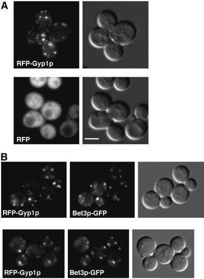
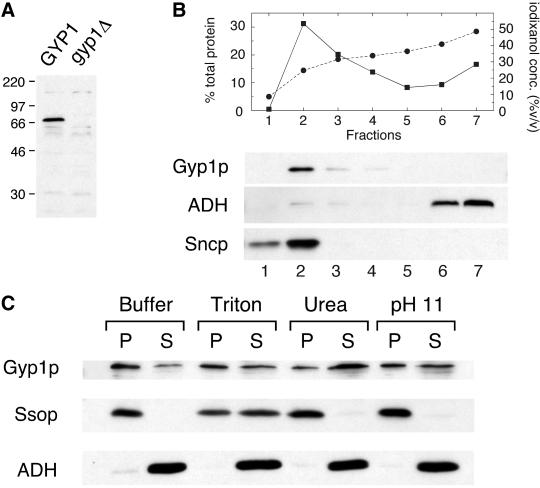
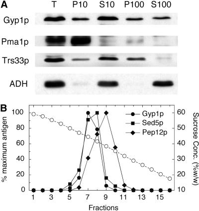
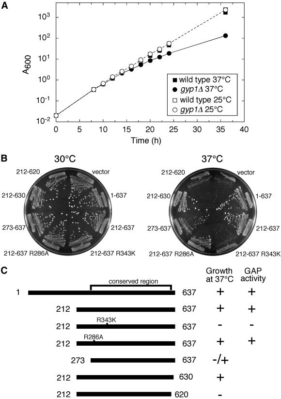
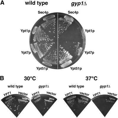
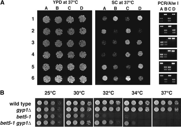
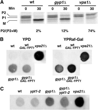
A family of related proteins in yeast Saccharomyces cerevisiae is known to have in vitro GTPase-activating protein activity on the Rab GTPases. However, their in vivo function remains obscure. One of them, Gyp1p, acts on Sec4p, Ypt1p, Ypt7p, and Ypt51p in vitro. Here, we present data to reveal its in vivo substrate and the role that it plays in the function of the Rab GTPase. Red fluorescent protein-tagged Gyp1p is concentrated on cytoplasmic punctate structures that largely colocalize with a cis-Golgi marker. Subcellular fractionation of a yeast lysate confirmed that Gyp1p is peripherally associated with membranes and that it cofractionates with Golgi markers. This localization suggests that Gyp1p may only act on Rab GTPases on the Golgi. A gyp1Delta strain displays a growth defect on synthetic medium at 37 degrees C. Overexpression of Ypt1p, but not other Rab GTPases, strongly inhibits the growth of gyp1Delta cells. Conversely, a partial loss-of-function allele of YPT1, ypt1-2, can suppress the growth defect of gyp1Delta cells. Furthermore, deletion of GYP1 can partially suppress growth defects associated with mutants in subunits of transport protein particle complex, a complex that catalyzes nucleotide exchange on Ypt1p. These results establish that Gyp1p functions on the Golgi as a negative regulator of Ypt1p.


Figures

Figure 1
Figure 1
Localization of RFP-Gyp1p. (A) RFP-Gyp1p localizes to punctate structures in live cells. RFP-Gyp1p (top) or RFP (bottom) was expressed from a CEN plasmid in gyp1Δ cells (NY2291). The same cells were imaged in fluorescence and differential interference contrast modes. Bar, 4 μm. (B) RFP-Gyp1p partially colocalizes with a cis-Golgi marker, Bet3p-GFP, in live cells. RFP-Gyp1p and Bet3p-GFP were expressed from CEN plasmids in bet3Δ cells (NY2294). The same cells were imaged in fluorescence mode with trimethylrhodamine B isothiocyanate filter and fluorescein isothiocyanate filter and in differential interference contrast mode.
Figure 2
Figure 2
Gyp1p is peripherally associated with membranes. (A) Antibody detection of Gyp1p. Equal amounts of total cell extracts prepared from GYP1 pep4 (NY2295) and gyp1Δ pep4 (NY2296) strains were resolved by SDS-PAGE, blotted, and probed with affinity-purified polyclonal antibody raised against recombinant Gyp1p. The positions of size markers (kDa) are indicated. (B) Lysate of NY2295 was loaded at the bottom of a tube containing 35% iodixanol, and centrifuged in a TLA 120.2 rotor at 120,000 rpm for 3 h. Seven fractions were collected from the top. The percentage of total protein in each fraction (▪) and iodixanol concentration of each fraction (●) was determined. The amount of Gyp1p, ADH, and Sncp in each fraction was determined by Western blot. (C) Lysate of NY2295 was mixed with lysis buffer and lysis buffer containing Triton X-100, urea, and sodium carbonate so that the final concentrations were 2% Triton X-100, 4 M urea, and 0.1 M sodium carbonate (pH 11), respectively. The mixtures were incubated on ice for 40 min and then centrifuged at 100,000 × g for 30 min. Equal amounts of pellet and supernatant fractions were analyzed by SDS-PAGE and Western blot. Urea treatment shifted the distribution of Gyp1p toward the supernatant but did not affect the distribution of the integral membrane protein Ssop.
Figure 3
Figure 3
Subcellular fractionation of Gyp1p. (A) Differential centrifugation of yeast lysate. Total lysate of NY2295 (T) was centrifuged at 10,000 × g to generate supernatant S10 and pellet P10. The S10 supernatant was centrifuged at 100,000 × g to obtain supernatant S100 and pellet P100. Equal amounts of samples were prepared for electrophoresis, separated by SDS-PAGE, and transferred to nitrocellulose. Western blots were probed with polyclonal antibodies against Gyp1p, Pma1p, Trs33p, and ADH. (B) Sucrose gradient fractionation of yeast lysate. Lysate of NY2295 was loaded on the top of a linear 20–60% (wt/wt) sucrose gradient and centrifuged at 120,000 × g for 20 h. Fractions were collected from the bottom. Sucrose concentration (circles) in each fraction was determined using a refractometer. The amounts of Gyp1p, Sed5p, and Pep12p in each fraction were determined by Western blot and densitometry.
Figure 4
Figure 4
GAP activity is essential for the in vivo function of Gyp1p. (A) gyp1Δ has a growth defect in SC medium at 37°C. Wild-type (NY1210) and gyp1Δ (NY2292) cells were grown in SC medium at 25°C to log phase. They were inoculated into 25°C SC medium and 37°C SC medium, so that the final absorbance at 600 nm (A600) was 0.02. The growth was monitored by A600. The cultures were diluted 10 times in fresh medium when the A600 exceeded 1.0. The A600 in the figure reflects the cell density in the original volume, having taken the dilution into consideration. (B) Plate assay to test the ability of different constructs to complement the SC medium growth defect of gyp1Δ. CEN LEU2 plasmids expressing different Gyp1p proteins from the GYP1 promoter were introduced into gyp1Δ cells (NY2292). The transformants were streaked onto SC-LEU plates and incubated at 30 and 37°C. Photos were taken after 4 d. (C) The complementation activity of different Gyp1p proteins correlates with their in vitro GAP activity. The colony size of the full-length Gyp1p construct transformant was scored as +. The colony size of the vector transformant was scored as −. The in vitro GAP activity of Gyp1p(1–637) and Gyp1p(212–637) was determined by measuring the activity of purified recombinant glutathione S-transferase fusion proteins (our unpublished observation). The activity of the R286A and R343K mutants was based on published results (Albert et al., 1999).
Figure 5
Figure 5
Overexpression of Ypt1p specifically inhibits the growth of gyp1Δ. (A) Wild-type (NY1210) and gyp1Δ (NY2291) cells were transformed with a 2 μ URA3 plasmid containing the GYP1 gene, and a CEN HIS3 plasmid overproducing either Sec4p, Ypt1p, Ypt7p, or Ypt51p, from a GPD promoter. Transformants were first streaked on a minimal medium plate containing uracil and then restreaked on minimal medium plate containing 5-fluoroorotic acid. Photos were taken after 4-d incubation at 30°C. (B) Growth of gyp1Δ is inhibited by an extra copy of the YPT1 gene. Wild-type (NY1210) and gyp1Δ (NY2291) cells were transformed with a CEN plasmid containing the YPT1 gene. Transformants were streaked on a minimal medium plate and incubated at 30 and 37°C. Photos were taken after 4 d.
Figure 6
Figure 6
(A) ypt1-2 suppresses the growth defect of gyp1Δ. Tetrads from a gyp1Δ/gyp1Δ YPT1/ypt1-2 diploid (NY2297) were dissected. The same number of cells of each progeny was spotted on YPD and SC plates and incubated at 37°C for 2 d. The 630-bp YPT1 open reading frame from each progeny was amplified by PCR. The PCR products were digested by Alw I. PCR product from YPT1 cells can be digested to produce two bands of 380 and 250 bp. PCR product from ypt1-2 cells cannot be digested. (B) gyp1Δ partially suppresses the growth defect of bet5-1. Tenfold serial dilutions of wild-type (NY1211), gyp1Δ (NY2293), bet5-1 (NY2298), and gyp1Δ bet5-1 (NY2299) were spotted on YPD plates and grown for 36 h at 25, 30, 32, 34, and 37°C.
Figure 7
Figure 7
gyp1Δ missorts CPY to the cell surface. (A) Cells were labeled with 35S for 10 min at 30°C in minimal medium and chased with unlabeled methionine and cysteine for 30 min. CPY was immunoprecipitated from total lysates, separated on SDS-PAGE, and quantified by phosphorimaging. P1, ER form; P2, Golgi form; M, mature form. (B) Overexpression of Ypt1p enhances the CPY missorting phenotype of gyp1Δ. The same number of cells was spotted on YPD and YP-raffinose-galactose plates. Wet nitrocellulose membranes were overlaid on top of the plates. The plates were incubated at 30°C overnight. The amount of CPY absorbed on the membrane was determined by Western blot. (C) ypt1-2 suppresses the CPY missorting phenotype of gyp1Δ. The same number of cells was spotted on YPD plate at 30°C. CPY secretion was determined by the overlay assay.

Similar articles

Cited by

References

    1. Albert S, Gallwitz D. Two new members of a family of Ypt/Rab GTPase activating proteins. Promiscuity of substrate recognition. J Biol Chem. 1999;274:33186–33189. - PubMed
    1. Albert S, Gallwitz D. Msb4p, a protein involved in Cdc42p-dependent organization of the actin cytoskeleton, is a Ypt/Rab-specific GAP. Biol Chem. 2000;381:453–456. - PubMed
    1. Albert S, Will E, Gallwitz D. Identification of the catalytic domains and their functionally critical arginine residues of two yeast GTPase-activating proteins specific for Ypt/Rab transport GTPases. EMBO J. 1999;18:5216–5225. - PMC - PubMed
    1. Azuma Y, Dasso M. The role of Ran in nuclear function. Curr Opin Cell Biol. 2000;12:302–307. - PubMed
    1. Bacon RA, Salminen A, Ruohola H, Novick P, Ferro-Novick S. The GTP-binding protein Ypt1 is required for transport in vitro: the Golgi apparatus is defective in ypt1 mutants. J Cell Biol. 1989;109:1015–1022. - PMC - PubMed

Publication types

MeSH terms