. 2001 May;12(5):1257-74.
doi: 10.1091/mbc.12.5.1257.

Regulation of initiation of S phase, replication checkpoint signaling, and maintenance of mitotic chromosome structures during S phase by Hsk1 kinase in the fission yeast

Affiliations
Free PMC article

Regulation of initiation of S phase, replication checkpoint signaling, and maintenance of mitotic chromosome structures during S phase by Hsk1 kinase in the fission yeast

T Takeda et al. Mol Biol Cell. 2001 May.
Free PMC article

Abstract

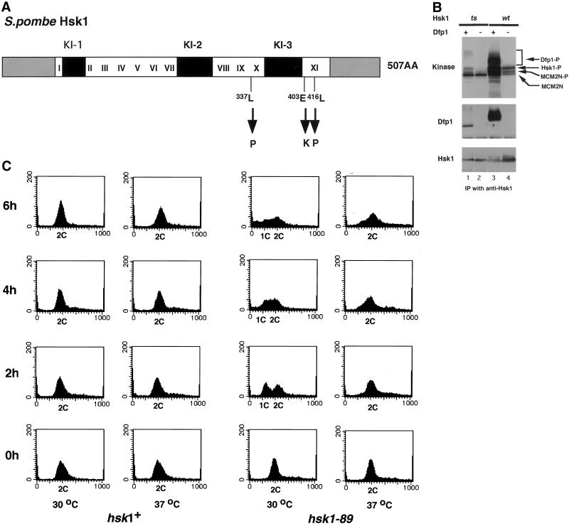
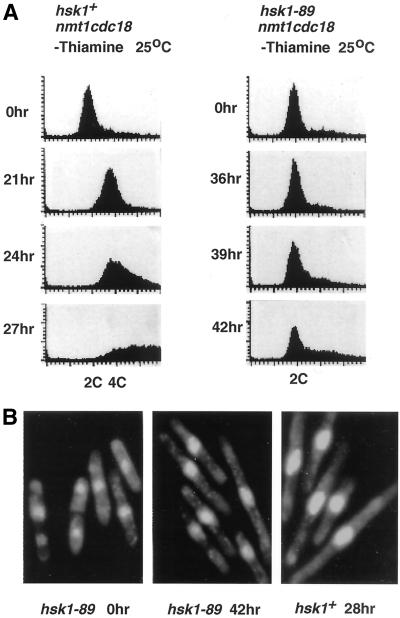
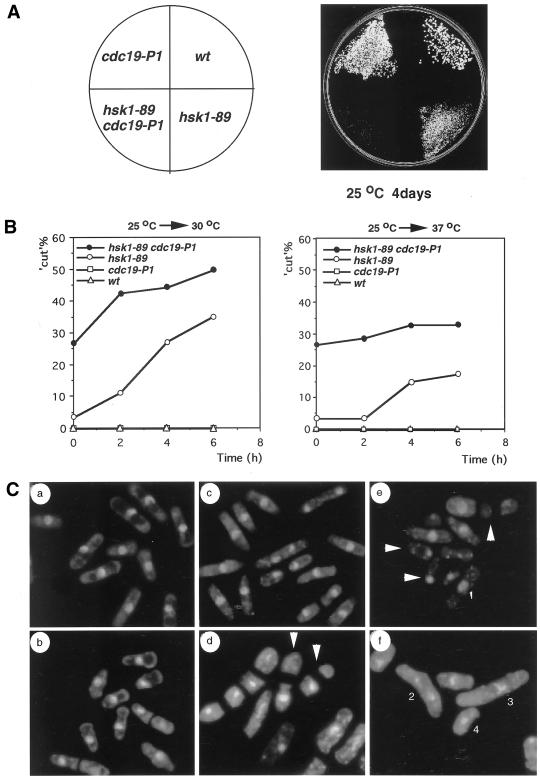
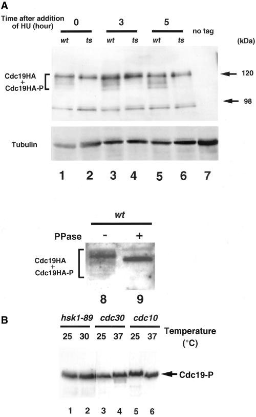
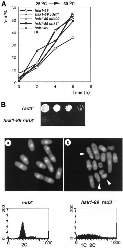
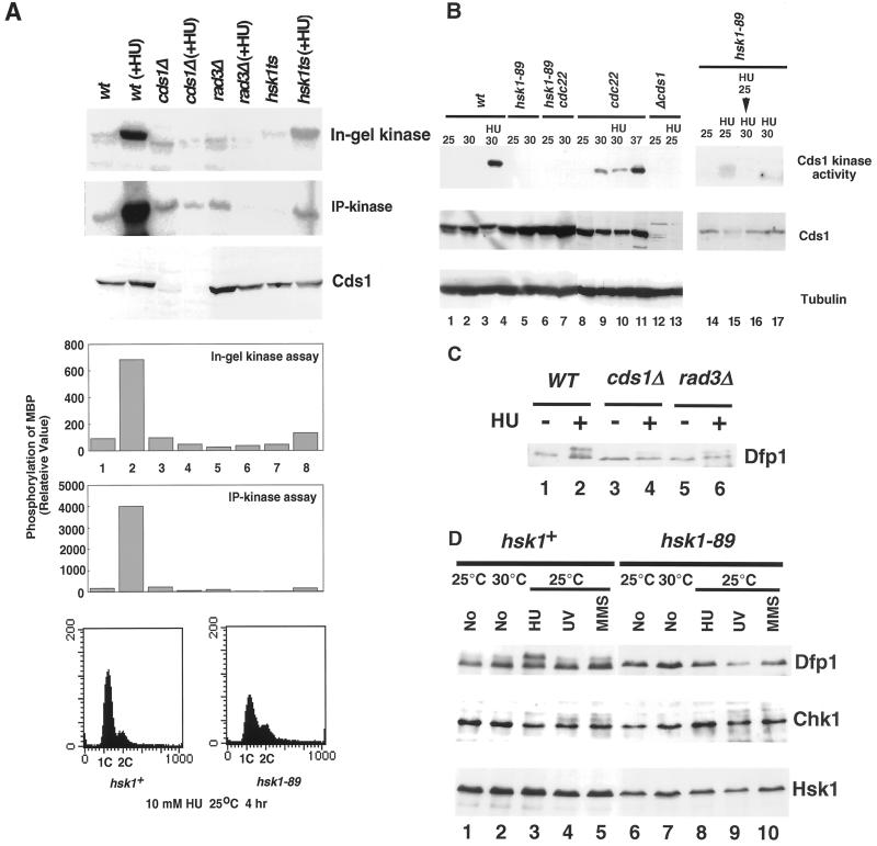
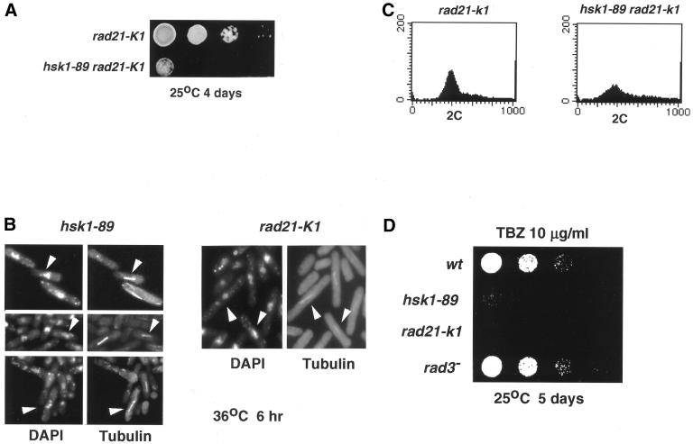
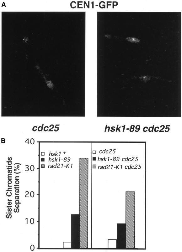
Hsk1, Saccharomyces cerevisiae Cdc7-related kinase in Shizosaccharomyces pombe, is required for G1/S transition and its kinase activity is controlled by the regulatory subunit Dfp1/Him1. Analyses of a newly isolated temperature-sensitive mutant, hsk1-89, reveal that Hsk1 plays crucial roles in DNA replication checkpoint signaling and maintenance of proper chromatin structures during mitotic S phase through regulating the functions of Rad3 (ATM)-Cds1 and Rad21 (cohesin), respectively, in addition to expected essential roles for initiation of mitotic DNA replication through phosphorylating Cdc19 (Mcm2). Checkpoint defect in hsk1-89 is indicated by accumulation of cut cells at 30 degrees C. hsk1-89 displays synthetic lethality in combination with rad3 deletion, indicating that survival of hsk1-89 depends on Rad3-dependent checkpoint pathway. Cds1 kinase activation, which normally occurs in response to early S phase arrest by nucleotide deprivation, is largely impaired in hsk1-89. Furthermore, Cds1-dependent hyperphosphorylation of Dfp1 in response to hydroxyurea arrest is eliminated in hsk1-89, suggesting that sufficient activation of Hsk1-Dfp1 kinase is required for S phase entry and replication checkpoint signaling. hsk1-89 displays apparent defect in mitosis at 37 degrees C leading to accumulation of cells with near 2C DNA content and with aberrant nuclear structures. These phenotypes are similar to those of rad21-K1 and are significantly enhanced in a hsk1-89 rad21-K1 double mutant. Consistent with essential roles of Rad21 as a component for the cohesin complex, sister chromatid cohesion is partially impaired in hsk1-89, suggesting a possibility that infrequent origin firing of the mutant may affect the cohesin functions during S phase.


Figures

Figure 1
Figure 1
Characterization of hsk1-89 strain and Hsk1-89 mutant protein. (A) Schematic drawing of the structure of Hsk1 protein and location of mutations in Hsk1-89. Amino acids, L337 (CTC), E403 (GAG), and L416 (CTC) were changed to P (CCC), K (AAG), and P (CCC), respectively, in Hsk1-89 mutant protein as indicated. White, black, and striped boxes indicate the conserved kinase domains (I-XI), nonconserved kinase inserts (KI-1, KI-2, and KI-3), and N-terminal and C-terminal tail regions, respectively. (B) Extracts were prepared from insect cells expressing Hsk1 (wild-type or mutant form) with (+) or without (−) Dfp1 protein as indicated in the figure, and immunoprecipitates prepared by anti-Hsk1 antibody were assayed in in vitro kinase reactions by using GST-SpMCM2N protein as a substrate (Takeda et al., 1999; top). The immunoprecipitates were also analyzed by Western blotting to detect Dfp1 and Hsk1 protein (middle and bottom). Hsk1-P, Dfp1-P, and MCM2N-P represent phosphorylated forms of Hsk1, Dfp1, and GST-SpMCM2N proteins, respectively, and MCM2N indicates the nonshifted (but phosphorylated) form of GST-MCM2N protein. (C) Wild-type (hsk1+) or hsk1-89 cells grown at 25°C were shifted to 30°C or 37°C and aliquots of cells were taken every 2 h and fixed. DNA contents of cells were analyzed by FACS.
Figure 2
Figure 2
Inhibition of Cdc18-induced overreplication in hsk1-89 mutant. (A) Cdc18 expression was induced from nmt1 promoter-driven cdc18+ integrated on the chromosome in hsk1+ or hsk1-89 at 25°C (Nishitani and Nurse, 1995). Aliquots were taken at the times indicated after shift to medium lacking thiamine and then analyzed by FACS. (B) hsk1-89 cells (center) at 42 h and hsk1+ cells (right) at 28 h after induction of Cdc18 protein were stained with DAPI and subjected to fluorescence microscopy to visualize nuclei. The extent of elongation of cell sizes is similar between the wild type and mutant. hsk1-89 cells (left) before induction of expression.
Figure 3
Figure 3
Genetic interaction of hsk1-89 with cdc19-P1. (A) Growth of wild-type, cdc19-P1, hsk1–89, and hsk1-89 cdc19-P1 double mutant. Vegetatively growing cells of each strain were streaked on a rich plate and incubated at 25°C for 4 d. (B) Frequencies of cut cells in hsk1-89 and hsk1-89 cdc19-P1 after shift from 25 to 30°C (left) or to 37°C (right) were determined every 2 h. (C) Morphology of mutant cells stained with DAPI. (a) wild-type, 25°C, (b) cdc19-P1, 25°C, (c) hsk1-89, 25°C, (d) hsk1-89, cdc19-P1, 25°C, (e) hsk1-89, 6 h at 30°C, (f) hsk1-89, 6 h at 37°C. White arrows in d and e indicate cut cells. Abnormal chromosome structures (assigned by 1–4) in hsk1-89 at nonpermissive temperatures are indicated in e and f. Similar abnormal chromosomal morphologies of hsk1-89 are also shown in Figure 7B.
Figure 4
Figure 4
Inability of Hsk1-89 to phosphorylate Cdc19/MCM2 protein in vivo. (A) Phosphorylation of Cdc19/MCM2 protein in vivo. Vegetatively growing (5 × 10⁶ cells/ml) hsk1+ (NI319; lanes 1, 3, 5, 8, and 9) or hsk1-89 (NI486; lanes2, 4, 6, 10, and 11) carrying HA-tagged cdc19+ on the chromosome under its own promoter was arrested with 10 mM HU for 3 h (lanes 3 and 4) or for 5 h (lanes 5, 6, 8, and 9). Cell extracts were prepared by the regular glass bead method and run on an 8% SDS-PAGE. Cdc19 protein was immunoprecipitated by anti-HA antibody and was treated with λ-phosphatase before electrophoresis (lane 9) or untreated (lane 8). The extract from the wild-type (nontagged) cells (NT145) was run as a negative control (lane 7). Blotting was conducted by using anti-HA antibody. In lanes 8 and 9, the samples were run on a long gel (30 cm in length) at 500 V for 12 h at 4°C to maximize the separation of the phosphorylated forms. The lower panel for lanes 1–7 is the blotting with anti-tubulin antibody for loading control on a separate gel on which the same amount of each extract was run. (B) Phosphorylation of Mcm2 (Cdc19) in cdc mutants and hsk1-89 arrested at the nonpermissive temperature. hsk1-89 cells grown at 25°C (lane 1) were arrested at 30°C for 2 h (lane 2) and cdc30ts or cdc10ts cells vegetatively growing at 25°C (lanes 3 and 5) were arrested at 37°C for 4 h (lanes 4 and 6). Cell extracts were prepared and phosphorylation of Mcm2 (Cdc19) was analyzed as described in A, except that anti-Cdc19 antibody (a gift from Dr. S. Forsburg) was used.
Figure 5
Figure 5
Defect of hsk1-89 in DNA replication checkpoint control. (A) Enhancement of cut cell formation in hsk1-89 by the presence of an additional mutation or by early S phase arrest with HU. Cells from strains indicated were grown at 25°C and were shifted to 30°C for 6 h (○, □, ▵, ●). hsk1-89 cells were shifted to 30°C in the presence of 10 mM HU (x; hsk1–89 HU). The number of cut cells was counted as described in Figure 3B. (B) Genetic interaction of hsk1-89 with rad3 deletion. rad3 cells were grown to 5 × 10⁶ cells/ml at 25°C in YES liquid medium. hsk1-89 rad3 cells, which had doubling time of >6 h in YES at 25°C, were grown to 5 × 10⁵ cells/ml, and then were concentrated to 5 × 10⁶ cells/ml by centrifugation. Fivefold serial dilution of each cell suspension (5 × 10⁶ cells/ml) was spotted onto a rich plate, which was incubated at the permissive temperature for 4 d (top). Cells were fixed, stained with DAPI, and examined under fluorescent microscopy (middle), or harvested and analyzed for DNA content by FACS (bottom). Arrowheads indicate examples of cut cells.
Figure 6
Figure 6
Defect of hsk1-89 in Cds1 kinase activation in response to nucleotide deprivation. (A) Cds1 kinase activity in various strains in the presence or absence of HU. (Top) Extracts were prepared by the boiling method (Takeda et al., 1999) from the strains indicated, which were grown at 25°C with or without 10 mM HU-treatment for 4 h, and in-gel kinase assays were conducted as described in MATERIALS AND METHODS. (Middle) Extracts were prepared from the same set of the strains at a low salt by glass beads disruption. Cds1 protein was immunoprecipitated by anti-Cds1 protein antibody from the extracts in the presence of 0.5 M NaCl, and the immunoprecipitates were used for in vitro kinase assays with 10 μg of MBP as a substrate. The reaction mixtures were run on 10% SDS-PAGE and phosphorylated MBP are shown. (Bottom) Extracts used for in-gel kinase assays were analyzed by Western blotting using anti-Cds1 antibody to detect Cds1 protein. In the upper graph, the extent of phosphorylation in in-gel kinase assay was quantified. In the middle graph, the extent of MBP phosphorylation in IP-kinase assay was quantified. The presence of approximately the same amount of Cds1 protein in the immunoprecipitates was confirmed (our unpublished results). Weak phosphorylation of MBP observed in cds1Δ and rad3Δ extracts is due to the presence of nonspecific kinases in the immunoprecipitates. In the bottom panel, DNA contents of HU-arrested hsk1+ or hsk1-89 cells were analyzed by FACS. (B) Cds1 kinase activity was measured by in-gel kinase assays in strains indicated, which were grown at the temperature shown (upper panel). Cells were arrested with 10 mM HU for 4 h (lanes 3, 10, 13. 15, 16, and 17). In lane 16, cells were grown at 25°C for 2 h and then shifted to 30°C for 2 h. Cds1 or α-tubulin protein was analyzed by immunoblotting (middle and lower panels). (C) HU-induced hyperphosphorylation of Dfp1 protein in various strains. Strains indicated were grown at 30°C. Ten millimolar HU was added to half of the cells and incubation was continued for another 4 h. Dfp1 protein was analyzed by Western blotting. (D) hsk1+ or hsk1-89 cells were grown at the temperature indicated without any treatment (No; lanes 1, 2, 6, and 7), treated with 10 mM HU for 4 h (HU; lanes 3 and 8), exposed to UV at 150 J/m2 followed by incubation in rich medium for 1 h (UV; lanes 4 and 9), or treated with 0.05% MMS for 4 h followed by incubation in rich medium for 1 h (MMS, lanes 5 and 10). Dfp1 protein (upper panel), Chk1 protein (middle panel; HA-tagged), or Hsk1 protein (lower panel) was detected by Western blotting. In C and D, extracts were prepared by the boiling method.
Figure 7
Figure 7
Requirement of Hsk1 in maintenance of proper chromatin structures. (A) Genetic interaction between hsk1-89 and rad21-K1. Fivefold serial dilution of exponentially growing cultures indicated were spotted onto a rich plate, which was incubated at the permissive temperature. (B) Abnormal nuclear structures and tublin distributions in hsk1-89 (left) and rad21-K1 (right). Vegetatively growing cells were arrested at 36°C for 6 h and indirect immunofluorescence was conducted using anti-tublin. DAPI staining was conducted to visualize nuclei. Upper, middle, and lower panels of hsk1-89 represent aberrant nuclear structures such as condensed chromosomes with short spindles, abnormally stretched chromosomes, and those unequally separated by elongated spindles, respectively (indicated by white arrows). In this experiment, cells were shifted to 36°C to visualize the immunostained cells more clearly. Patterns of immunostaining and nuclear morphology at 36°C were very similar to those at 37°C (Figure 3C, f). (C) DNA content in rad21-K1 and hsk1-89 rad21-K1 double mutant. Cells grown at 25°C were harvested and analyzed for DNA content by FACS. (D) TBZ sensitivity of hsk1-89 mutant. Fivefold serial dilution of growing cells indicated were spotted onto a rich plate containing TBZ (10 μg/ml) and were incubated at the permissive temperature.
Figure 8
Figure 8
Premature sister chromatid separation in hsk1-89. Vegetatively growing hsk1+ or hsk1-89 cells carrying the GFP-LacI-NLS, which can bind to the lacO repeats integrated near the cen1 (Nabeshima et al., 1998), were arrested at 36°C for 5 h, fixed with methanol, and then subjected to fluorescence microscopy. (A) Nonsplit cen1-GFP signal in cdc25 cell (left) and a prematurely split cen1-GFP signals in hsk1-89 cdc25 cells (right). (B) Fractions of cells with split cen1-GFP signals are plotted for each strain indicated. The GFP signals in >100 nuclei for each cdc25+ strain (left), and those in >300 nuclei for each strain carrying cdc25 mutation (right) were examined. Populations of binucleate cells after cdc25 block (right) in hsk1+, hsk1–89, and rad21-K1 were 9.0, 2.4, and 3.9%, respectively, consistent with synchronistic arrest in G2/M boundary in these cdc25 strains under this condition. The above-mentioned experiments were conducted at 36°C for clearer visualization of cen1-GFP signals.

Similar articles

Cited by

References

    1. Aparicio OM, Weinstein DM, Bell SP. Components and dynamics of DNA replication complexes in S. cerevisiae: redistribution of MCM proteins and Cdc45 during S phase. Cell. 1997;91:59–69. - PubMed
    1. Bell S, Stillman B. Nucleotide-dependent recognition of chromosomal origins of DNA replication by a multiprotein complex. Nature. 1992;357:128–134. - PubMed
    1. Bhaumik D, Wang TS-F. Mutational effect of fission yeast Polα on cell cycle events. Mol Biol Cell. 1998;9:2107–2123. - PMC - PubMed
    1. Boddy MN, Furnari B, Mondesert O, Russell P. Replication checkpoint enforced by kinase Cds1 and Chk1. Science. 1998;280:909–912. - PubMed
    1. Bousset K, Diffley JF. The Cdc7 protein kinase is required for origin firing during S phase. Genes Dev. 1998;12:480–490. - PMC - PubMed

MeSH terms