. 2001 May;12(5):1315-28.
doi: 10.1091/mbc.12.5.1315.

Tetraploid state induces p53-dependent arrest of nontransformed mammalian cells in G1

Affiliations
Free PMC article

Tetraploid state induces p53-dependent arrest of nontransformed mammalian cells in G1

P R Andreassen et al. Mol Biol Cell. 2001 May.
Free PMC article

Abstract

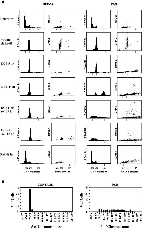
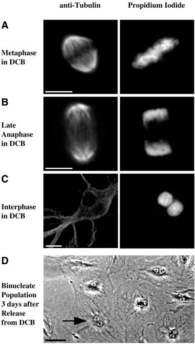
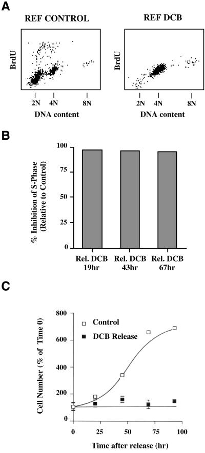
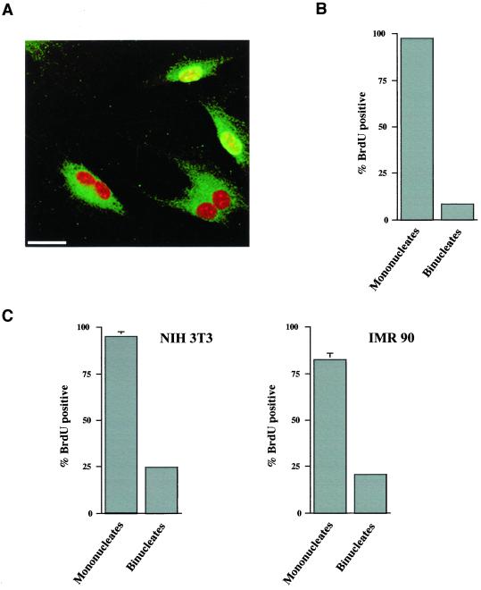
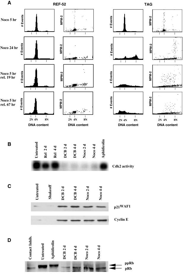
A "spindle assembly" checkpoint has been described that arrests cells in G1 following inappropriate exit from mitosis in the presence of microtubule inhibitors. We have here addressed the question of whether the resulting tetraploid state itself, rather than failure of spindle function or induction of spindle damage, acts as a checkpoint to arrest cells in G1. Dihydrocytochalasin B induces cleavage failure in cells where spindle function and chromatid segregation are both normal. Notably, we show here that nontransformed REF-52 cells arrest indefinitely in tetraploid G1 following cleavage failure. The spindle assembly checkpoint and the tetraploidization checkpoint that we describe here are likely to be equivalent. Both involve arrest in G1 with inactive cdk2 kinase, hypophosphorylated retinoblastoma protein, and elevated levels of p21(WAF1) and cyclin E. Furthermore, both require p53. We show that failure to arrest in G1 following tetraploidization rapidly results in aneuploidy. Similar tetraploid G1 arrest results have been obtained with mouse NIH3T3 and human IMR-90 cells. Thus, we propose that a general checkpoint control acts in G1 to recognize tetraploid cells and induce their arrest and thereby prevents the propagation of errors of late mitosis and the generation of aneuploidy. As such, the tetraploidy checkpoint may be a critical activity of p53 in its role of ensuring genomic integrity.


Figures

Figure 1
Figure 1
REF-52 cells, but not transformed TAG cells, arrest in G1 following tetraploidization by treatment with DCB. (A) Both untreated REF-52 (left) and TAG (right) cells had similar cell cycle distributions, as shown by histograms of DNA content determined by flow cytometry. Mitotically synchronized (mitotic shakeoff) REF-52 and TAG cells were in mitosis as determined by 4N DNA content (1st and 3rd columns) and elevated MPM-2 signals (2nd and 4th columns). Both REF-52 and TAG cells resumed cycling and had DNA content profiles similar to untreated cells 48 h following release from mitotic synchronization (Rel. 48 h). REF-52 and TAG cells exited mitosis, as determined by minimal MPM-2 signals (2nd and 4th columns, respectively), following release of mitotically synchronized cells into medium containing 10 μM DCB, an inhibitor of the actin assembly required for cytokinesis, for 5 h (DCB 5 h). Tetraploid REF-52 cells remained arrested in G1with 4N DNA content (1st column) and minimal MPM-2 signal (2nd column) either when maintained in DCB for 24 h following mitotic synchronization, or when released from 5 h treatment with DCB for either 19 or 67 h. In contrast, transformed TAG cells continued cycling and developed 8N DNA content with elevated MPM-2 signal when maintained in DCB for 24 h following mitotic synchronization. When released from 5 h treatment with DCB following mitotic synchronization for either 19 or 67 h, TAG cells became aneuploid with elevated MPM-2 signal and a corresponding DNA content ranging from 2N to 8N. (B) As determined by chromosome counts, transformed TAG cells rapidly become aneuploid following tetraploidization by inhibition of cytokinesis with DCB. Control cells had 78 ± 0.9 chromosomes, whereas chromosome counts prepared from chromosome spreads of DCB-treated cells ranged from 76 to 156. Control cells were released from mitotic synchronization into drug-free medium for 24 h and were then arrested in mitosis by treatment with 0.5 μg/ml nocodazole for 12 h. DCB-treated cells were made tetraploid by 5 h exposure to 10 μM DCB following mitotic synchronization. These cells were then also released into drug-free medium for 19 h and then accumulated in mitosis with 0.5 μg/ml nocodazole for 12 h. A total of 40 cells was counted both for controls and DCB-treated cells.
Figure 2
Figure 2
REF-52 cells form a normal mitotic spindle and undergo normal chromosome segregation, but become binucleate, in the presence of DCB. (A) Metaphase REF-52 cells in 10 μM DCB formed a normal mitotic spindle, as determined by immunofluorescence microscopy with anti-tubulin antibodies (left column). Chromosomes aligned normally, indicating that the spindle functioned normally, as determined by counterstaining of chromosomes with propidium iodide (right column). The chromosomes are positioned equidistant between the poles of the mitotic spindle. (B) At late anaphase, the mitotic spindle detected by anti-tubulin antibodies (left column) elongated normally and chromosomes detected by staining with propidium iodide (right column) were completely segregated. (C) At 5 h treatment with 10 μM DCB following mitotic synchronization cells became binucleate, as determined by counterstaining with propidium iodide (right column). The anti-tubulin image (left) shows that the two nuclei were present in a single cell. (D) A phase contrast image of a field of REF-52 cells 3 d following mitotic synchronization and tetraploidization with 10 μM DCB shows that cells were binucleate. An arrow indicates a typical binucleate cell. Bars, 10 μm (A–C) and 50 μm (D).
Figure 3
Figure 3
REF-52 cells are inhibited from entering S phase and do not proliferate following tetraploidization induced by exposure to DCB. (A) REF-52 cells, released from mitotic synchronization into DCB for 5 h, and then released into drug-free medium for 19 h, did not enter S phase by 24 h after mitosis, as determined by BrdU incorporation measured by two-dimensional flow cytometry. By contrast, controls released into drug-free medium for 24 h following mitotic synchronization reentered S phase and incorporated BrdU. (B) REF-52 cells made tetraploid by 5 h treatment with DCB following mitotic synchronization showed >95% inhibition of S phase entry through 67 h following release from DCB, relative to controls, measured 24 h following release from mitotic synchronization. Inhibition of S phase was quantitated for 10,000 cells each from two-dimensional FACS histograms to obtain the number of cells incorporating BrdU. (C) Counts of cell number show that REF-52 cells did not proliferate following tetraploidization induced by treatment with DCB. Cells were sychronized in mitosis, selectively detached, and then released from mitotic synchronization into me dium containing 10 μM DCB for 5 h. Cells were then fixed (0 time) or were released in drug-free medium for up to 96 h. Cells were fixed at the time points indicated and cell numbers counted using a hemacytometer. By contrast, mitotically synchronized cells replated in drug-free medium resumed proliferation. Each point is an average of eight counts, and error bars represent SDs.
Figure 4
Figure 4
DCB induces cell cycle arrest only in cells that become binucleate following short-term exposure. (A) REF-52 cells were exposed to 10 μM DCB for 5 h without pre-synchronization, and they were then cultured for 25 h in medium containing 10 μM BrdU but not DCB. As shown by an immunofluorescence image, mononucleate, but not binucleate, cells incorporate BrdU (green) following 5 h treatment with DCB. Mononucleate cells are distinguishable from binucleate cells by a counterstain of nuclei with propidium iodide (red). FITC background was enhanced to better distinguish between mononucleate and binucleate cells. Bar, 25 μm. (B) Quantitation of the percentage of mononucleate and binucleate REF-52 cells that incorporate BrdU in 25 h following a 5 h exposure to DCB. (C) Similar analysis of random cycling NIH3T3 and IMR-90 cells was conducted following exposure to DCB for 10 h and then BrdU for 24 h. Incorporation is specifically suppressed in binucleate cells, indicating cell cycle arrest in cells that become tetraploid due to inhibition of cytokinesis by DCB. For each condition, at least 300 cells were counted. Values shown are mean values of three counts. SDs were as stated in the text.
Figure 5
Figure 5
Aberrant exit from mitosis in the presence of nocodazole yields an arrest similar to that obtained by cleavage failure in DCB. (A) Both REF-52 and TAG cells exited mitosis in the presence of 0.04 μg/ml nocodazole for 5 h following mitotic synchronization (noco 5 h) and displayed minimal MPM-2 signal with 4N DNA content. REF-52 cells remained arrested in interphase as shown by a continued 4N DNA content (1st column) and minimal MPM-2 signal (2nd column) both in 0.04 μg/ml nocodazole for 24 h or following release from 5 h treatment with nocodazole for 19 or 67 h after aberrant exit from mitosis. By contrast, TAG cells did not arrest, and in the presence of 0.04 μg/ml nocodazole for 24 h had 8N DNA content with elevated MPM-2 signal. But when released from 5-h treatment with nocodazole for either 19 or 67 h cells developed an aneuploid DNA content yielding mitotic cells that continued to cycle. Tetraploidization by either aberrant exit frommitosis in the presence of nocodazole or inhibition of cytokinesis by DCB results in identical profiles of effectors of G1 progression. Cells were treated with either 10 μM DCB or 0.04 μg/ml nocodazole for 5 h following mitotic synchronization, and then following tetraploidization were released in drug-free medium for the indicated period of time. (B) Cdk2 activity, which is required for G1-S progression (Pagano et al., 1993; Tsai et al., 1993), was suppressed by tetraploidization of REF-52 cells with either DCB or nocodazole relative to untreated cells, and also suppressed relative to cells arrested in S phase by treatment with 5 μM aphidicolin for 24 h, or to cells released from mitotic synchronization for 2 or 4 d. (C) Both the cdk2 inhibitor p21WAF1 and the cdk2 activator cyclin E were similarly present at elevated levels in cells made tetraploid by exposure to either nocodazole or DCB relative to untreated cells or mitotic cells synchronized by selective detachment (mitotic shakeoff). (D) pRb was in a hypophosphorylated state during G1 arrest following tetraploidization of cells by either nocodazole or DCB, as it was in cells arrested in G1 by contact inhibition. By contrast, pRb was in a hyperphosphorylated state in untreated cells and those arrested in S phase by treatment with aphidicolin.
Figure 6
Figure 6
p53 is required for tetraploid arrest whether induced by inhibition of cytokinesis or by exit from mitosis without the formation of a mitotic spindle. p53 was inactivated in REF-52 (p53DD) by expression of a dominant-negative mutant (Shaulian et al., 1992). (A) Inactivation of p53 in cells expressing p53DD resulted in their inability to induce p21WAF1 following exposure to gamma irradiation, as demonstrated by Western blots. By contrast, control REF-52 cells (p53+/+) induced p21WAF1 following irradiation. (B) FACSscan analysis showed that both p53+/+ and p53DD cells had similar cell cycle profiles when untreated and had the 4N DNA content expected of cells following mitotic synchronization (mitotic shakeoff) and subsequent tetraploidization by exposure to 10 μM DCB for 5 h (DCB 5 h). Control p53+/+ cells remained arrested with 4N DNA content following exposure to DCB, whether released into drug-free medium for 19 or 67 h, or released into 0.04 μg/ml nocodazole for 19 h. By contrast, p53DD cells continued cycling when made tetraploid by exposure to DCB. Cells accumulated with 8N DNA content when released from DCB into nocodazole and became aneuploid when released from DCB into drug-free medium for 19 or 67 h. (C) Similarly, p53DD cells continued cycling when made tetraploid by aberrant exit from mitosis in the presence of 0.04 μg/ml nocodazole, whereas control p53+/+ cells arrested with 4N DNA content. p53DD cells accumulated a subpopulation of 8N cells when exposed to 0.04 μg/ml nocodazole for 24 h following mitotic synchronization, and they became aneuploid when mitoticallysynchronized, then treated with 0.04 μg/ml nocodazole for 5 h, and finally released into drug-free medium for either 19 or 67 h. (D) p21WAF1 was induced in a p53-dependent manner in cells made tetraploid with DCB, relative to controls or actively cycling cells 2 d after release from mitotic synchronization. Western blots show p21WAF1 was induced in p53+/+, but not p53DD cells following mitotic synchronization and then treatment with DCB for 5 h to obtain a tetraploid G1 population.

References

    1. Andreassen PR, Margolis RL. Microtubule dependency of p34cdc2 inactibation and mitotic exit in mammalian cells. J Cell Biol. 1994;127:789–802. - PMC - PubMed
    1. Andreassen PR, Martineau SN, Margolis RL. Chemical induction of mitotic checkpoint override in mammalian cells results in aneuploidy following a transient tetraploid state. Mutat Res. 1996;372:181–194. - PubMed
    1. Aubin JE, Osborn M, Weber K. Inhibition of cytokinesis and altered contractile ring morphology induced by cytochalasins in synchronized PtK2 cells. Exp Cell Res. 1981;136:63–79. - PubMed
    1. Baker SJ, Markowitz S, Fearon ER, Willson JK, Vogelstein B. Suppression of human colorectal carcinoma cell growth by wild-type p53. Science. 1990;249:912–915. - PubMed
    1. Banin S, Moyal L, Shieh S, Taya Y, Anderson CW, Chessa L, Smorodinsky NI, Prives C, Reiss Y, Shiloh Y, Ziv Y. Enhanced phosphorylation of p53 by ATM in response to DNA damage. Science. 1998;281:1674–1677. - PubMed

Publication types

MeSH terms