. 2001 May 21;193(10):1135-47.
doi: 10.1084/jem.193.10.1135.

Role of the parasite-derived prostaglandin D2 in the inhibition of epidermal Langerhans cell migration during schistosomiasis infection

Affiliations

Role of the parasite-derived prostaglandin D2 in the inhibition of epidermal Langerhans cell migration during schistosomiasis infection

V Angeli et al. J Exp Med. .

Abstract

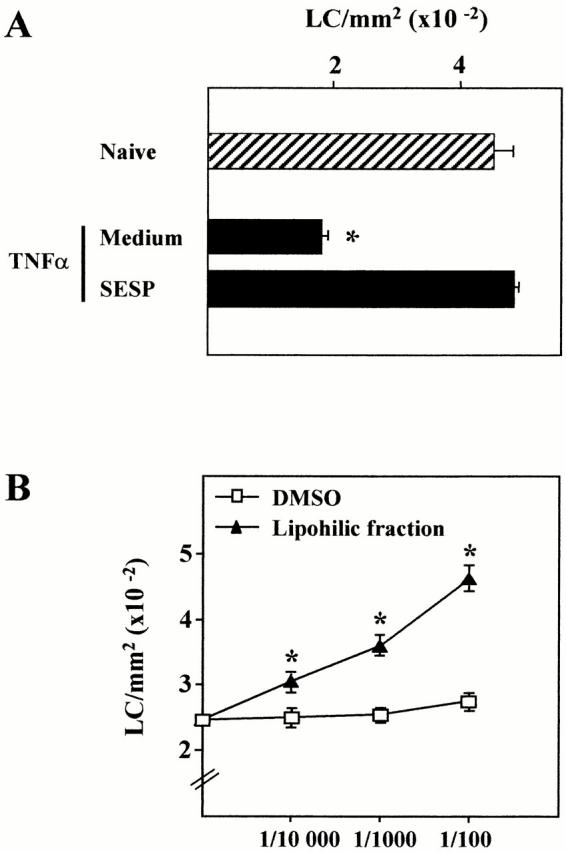
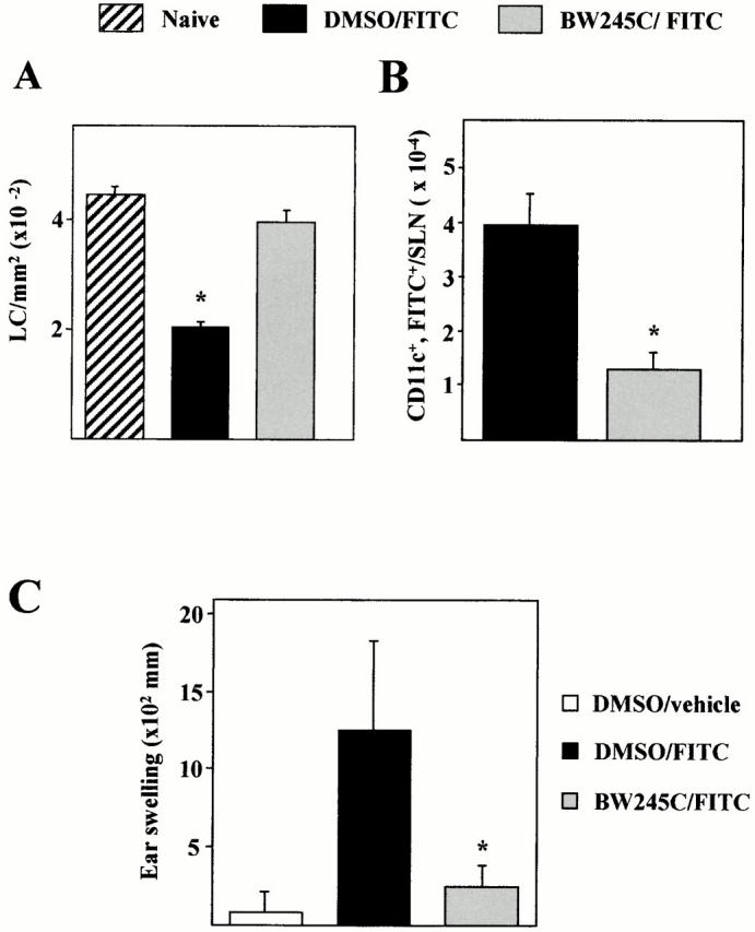
Epidermal Langerhans cells (LCs) play a key role in immune defense mechanisms and in numerous immunological disorders. In this report, we show that percutaneous infection of C57BL/6 mice with the helminth parasite Schistosoma mansoni leads to the activation of LCs but, surprisingly, to their retention in the epidermis. Moreover, using an experimental model of LC migration induced by tumor necrosis factor (TNF)-alpha, we show that parasites transiently impair the departure of LCs from the epidermis and their subsequent accumulation as dendritic cells in the draining lymph nodes. The inhibitory effect is mediated by soluble lipophilic factors released by the parasites and not by host-derived antiinflammatory cytokines, such as interleukin-10. We find that prostaglandin (PG)D2, but not the other major eicosanoids produced by the parasites, specifically impedes the TNF-alpha-triggered migration of LCs through the adenylate cyclase-coupled PGD2 receptor (DP receptor). Moreover, the potent DP receptor antagonist BW A868C restores LC migration in infected mice. Finally, in a model of contact allergen-induced LC migration, we show that activation of the DP receptor not only inhibits LC emigration but also dramatically reduces the contact hypersensitivity responses after challenge. Taken together, we propose that the inhibition of LC migration could represent an additional stratagem for the schistosomes to escape the host immune system and that PGD2 may play a key role in the control of cutaneous immune responses.


Figures

Figure 1
Figure 1
Immunohistochemical staining of murine epidermal sheets after transcutaneous infection by S. mansoni. Epidermal sheets were prepared either from noninfected or from S. mansoni–infected mice and LCs were stained for MHC class II (A and B) or for CD86 (C and D). The arrow indicates the “ghost” of parasite. The isotype control mAb did not reveal any reactivity (not shown). Original magnification: ×400.
Figure 2
Figure 2
Effect of S. mansoni infection on the migration of epidermal LCs in vivo (A) and ex vivo (B). (A) Epidermal sheets were prepared at different times after the infection (1 to 120 h) and the number of LC/mm2 was determined after anti-MHC class II staining. Controls included epidermal sheets from naive mice and from mice exposed to water without parasites (Non-infected). Results are expressed as means ± SD and are representative of four independent experiments (n = 7). (B) Skin explants were obtained from ears of noninfected or S. mansoni–infected mice (6, 24, 48, and 120 h). The number of LC/mm2 was determined in the epidermis from the explants after 24 h of culture and compared with the epidermis from fresh skin (naive). Results are expressed as means ± SD and are representative of three independent experiments (n = 4). Significant differences are designated by * (P < 0.001).
Figure 4
Figure 4
(A) RT-PCR analysis of mRNAs specific for pro- and antiinflammatory cytokines in the epidermis of S. mansoni–infected mice. Epidermal sheets were prepared from noninfected (0 h) or infected (1, 6, 24, and 120 h) mice, total RNA extracted, and RT-PCR was carried out using the primers shown in Table . Representative results of three independent experiments are shown. (B) Role of IL-10 in the inhibition of LC migration. IL-10 KO or WT mice were infected (or not) and 24 h after infection, epidermal sheets were prepared and the number of LCs/mm2 determined by immunohistochemistry. Before infection, WT mice were treated with neutralizing anti–IL-10 or isotype-matched mAbs (IgG1). As a positive control, TNF-α was intradermally injected 1 h before the analysis. Significant differences are designated by * (P < 0.001).
Figure 3
Figure 3
Effect of S. mansoni infection on the TNF-α–induced LC migration in vivo. (A) 6, 24, and 120 h after infection, mice (four mice/time point) were intradermally injected with 30 μl of PBS/BSA (carrier) containing or not containing 50 ng TNF-α into both ear pinnae. Ears were removed 1 h later, epidermal sheets were prepared, and the number of LC/mm2 was determined by immunohistochemistry. In TNF-α–treated S. mansoni-infected mice, we noted that LCs remained interdigitated among surrounding KCs and still expressed E-cadherin (not shown). The experiment shown is representative of five experiments (n = 8) and values are means ± SD. (B) Detection of CD11c-expressing cells in SLNs from TNF-α–treated mice previously (24 h), or not, infected with S. mansoni (original magnification: ×200).
Figure 3
Figure 3
Effect of S. mansoni infection on the TNF-α–induced LC migration in vivo. (A) 6, 24, and 120 h after infection, mice (four mice/time point) were intradermally injected with 30 μl of PBS/BSA (carrier) containing or not containing 50 ng TNF-α into both ear pinnae. Ears were removed 1 h later, epidermal sheets were prepared, and the number of LC/mm2 was determined by immunohistochemistry. In TNF-α–treated S. mansoni-infected mice, we noted that LCs remained interdigitated among surrounding KCs and still expressed E-cadherin (not shown). The experiment shown is representative of five experiments (n = 8) and values are means ± SD. (B) Detection of CD11c-expressing cells in SLNs from TNF-α–treated mice previously (24 h), or not, infected with S. mansoni (original magnification: ×200).
Figure 6
Figure 6
HPLC analysis of S. mansoni schistosomula eicosanoid production. 1 μl of the lipophilic fraction was injected, fractions were collected every 30 s, and monitored using a densitometer (wavelength: 195, 230, and 270 nm). The elution position of external standards are indicated. Note that the scales of the arbitrary values are different in each panel.
Figure 5
Figure 5
Effect of SESP on the TNF-α–induced LC migration in vivo. (A) The supernatant of a 4-h culture of schistosomula (SESP) or (B) increasing amounts of the lipophilic fraction from the SESP (diluted in DMSO) were intradermally injected to mice. After 20 min, mice were treated with 50 ng of TNF-α and the epidermal sheets were analyzed 1 h after for the determination of LC density. These data are representative of three experiments (n = 4). Significant differences are designated by * (P < 0.001).
Figure 9
Figure 9
Effect of the PGD2 analogue BW245C on the FITC-induced migration of LCs and on CHS responses. Mice were injected intradermally into ear pinnae with BW245C (100 nM) 15 min before and 5 h after FITC topical application. (A) Epidermal LC density was analyzed 18 h after FITC painting and (B) the number of CD11c+FITC+ cells present in the SLNs determined 24 h after FITC application. (C) 5 d after sensitization, mice were challenged and 24 h later, ear thickness was measured. Results are expressed as means ± SD and are representative of three independent experiments (n = 7). Significant differences are designated by * (P < 0.001) for A and C and (P < 0.05) for B.
Figure 7
Figure 7
Effect of (A) the major schistosomula ES eicosanoids and (B) of the PGD2 analogue BW245C and the cAMP-elevating agent forskolin on the TNF-α–induced LC migration in vivo. (B) Mice received increasing concentrations of PGF, PGE2, PGD2, 15-HETE, 5-HETE, or vehicle alone (DMSO), or (B) they received BW245C (10 nM), forskolin (10 μM), or DMSO. After 20 min, mice were treated with 50 ng of TNF-α and epidermal sheets were prepared 1 h later. The number of LC/mm2 was determined by immunohistochemistry. This data is representative of three experiments (n = 4). Significant differences are designated by * (P < 0.001).
Figure 8
Figure 8
(A) Expression of mRNA for the DP receptor in total epidermal cells and in the LC (XS52) and KC (Pam212) lines as assessed by RT-PCR. (B) Effect of the DP receptor antagonist BW A868C on LC emigration after infection with S. mansoni. 15 min before the infection, mice were injected intradermally with increasing amounts of BW A868C. 6 h later, epidermis were stained using anti-MHC class II Abs. This data is representative of three experiments (n = 4). Significant differences are designated by * (P < 0.001).

References

    1. Banchereau J., Steinman R.M. Dendritic cells and the control of immunity. Nature. 1998;392:245–252. - PubMed
    1. Tang A., Amagai M., Granger L.G., Stanley J.R., Udey M.C. Adhesion of epidermal Langerhans cells to keratinocytes is mediated by E-cadherin. Nature. 1993;361:82–85. - PubMed
    1. Kimber I., Dearman R.J., Cumberbatch M., Huby R.J. Langerhans cells and chemical allergy. Curr. Opin. Immunol. 1998;10:614–619. - PubMed
    1. Moodycliffe A.M., Kimber I., Norval M. Role of TNFα in ultraviolet B light-induced dendritic cell migration and suppression of contact hypersensitivity. Immunology. 1992;81:79–84. - PMC - PubMed
    1. Ban E., Dupre I., Hermann E., Rohn W., Vendeville C., Quatannens B., Ricciardi-Castagnoli P., Capron A., Riveau G. CpG motifs induce Langerhans cell migration in vivo . Int. Immunol. 2000;12:737–744. - PubMed

Publication types

MeSH terms