. 2001 May 21;193(10):1213-20.
doi: 10.1084/jem.193.10.1213.

Induction of M3-restricted cytotoxic T lymphocyte responses by N-formylated peptides derived from Mycobacterium tuberculosis

Affiliations

Induction of M3-restricted cytotoxic T lymphocyte responses by N-formylated peptides derived from Mycobacterium tuberculosis

T Chun et al. J Exp Med. .

Abstract

Major histocompatibility complex (MHC) class I-restricted CD8(+) T cells play a critical role in the protective immunity against Mycobacterium tuberculosis (Mtb). However, only a few Mtb peptides recognized by MHC class Ia-restricted CD8(+) T cells have been identified. Information on epitopes recognized by class Ib-restricted T cells is even more limited. M3 is an MHC class Ib molecule that preferentially presents N-formylated peptides to CD8(+) T cells. Because bacteria initiate protein synthesis with N-formyl methionine, the unique binding specificity of M3 makes it especially suitable for presenting these particular bacterial epitopes. We have scanned the full sequence of the Mtb genome for NH2-terminal peptides that share features with other M3-binding peptides. Synthetic peptides corresponding to these sequences were tested for their ability to bind to M3 in an immunofluorescence-based peptide-binding assay. Four of the N-formylated Mtb peptides were able to elicit cytotoxic T lymphocytes (CTLs) from mice immunized with peptide-coated splenocytes. The Mtb peptide-specific, M3-restricted CTLs lysed the Mtb-infected macrophages effectively, suggesting that these N-formylated Mtb peptides are presented as the naturally processed epitopes by Mtb-infected cells. Furthermore, T cells from Mtb-infected lungs, spleen, and lymph nodes responded to N-formylated Mtb peptides in an M3-restricted manner. Taken together, our data suggest that M3-restricted T cells may participate in the immune response to Mtb.


Figures

Figure 1
Figure 1
N-formylated Mtb peptides increase surface expression of M3. P388-M3 transfectants were incubated overnight with varying concentrations of N-formylated peptides and stained for M3 expression. The range of concentrations and the corresponding hatchmarks are shown. The bars represent mean fluorescence intensity after staining with mAb130 as described. The results are representative of two experiments.
Figure 2
Figure 2
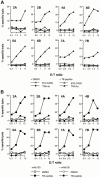
Mtb peptide-specific CTLs lyse targets in an M3-restricted, peptide-dependent manner. (A) The M3wt-transfected TR8.4a (H2-M3wt) and untransfected B10.CAS2 (H2-M3cas) fibroblast cell lines were incubated overnight with or without 10 μM of Mtb peptides and used as targets in a 51Cr-release assay. (B) The macrophage cell line P388 (H-2d, H2-M3wt) was incubated overnight with or without 10 μM of Mtb peptides and used as target. The Mtb-specific CTL lines were incubated with target cells in the presence of either 100 μl of mAb130 hybridoma supernatant or 100 μl of RPMI 10 in a 4-h 51Cr-release assay. The E/T ratios are shown. (C) P388 was incubated overnight with 10 μM of various N-formylated peptides and used as target. The E/T ratio for all CTLs is 3:1. Results were comparable in two experiments.
Figure 2
Figure 2
Mtb peptide-specific CTLs lyse targets in an M3-restricted, peptide-dependent manner. (A) The M3wt-transfected TR8.4a (H2-M3wt) and untransfected B10.CAS2 (H2-M3cas) fibroblast cell lines were incubated overnight with or without 10 μM of Mtb peptides and used as targets in a 51Cr-release assay. (B) The macrophage cell line P388 (H-2d, H2-M3wt) was incubated overnight with or without 10 μM of Mtb peptides and used as target. The Mtb-specific CTL lines were incubated with target cells in the presence of either 100 μl of mAb130 hybridoma supernatant or 100 μl of RPMI 10 in a 4-h 51Cr-release assay. The E/T ratios are shown. (C) P388 was incubated overnight with 10 μM of various N-formylated peptides and used as target. The E/T ratio for all CTLs is 3:1. Results were comparable in two experiments.
Figure 3
Figure 3
Mtb peptide–specific, M3-restricted CTLs recognize Mtb-infected macrophages and are primed in infected mice. (A) Bone marrow–derived macrophages were either maintained in medium alone or acutely infected with Mtb. After 48 h, adherent cells were harvested, labeled with 51Cr, and used as targets in a CTL assay. Black bars represent the specific lysis of infected macrophages by various Mtb peptide–specific CTLs, and the lysis of uninfected macrophages (white bars) was included for comparison. The E/T ratio was 30:1 for CTL4B and 10:1 for the rest of the CTL lines. Results shown are the means from triplicate wells and the standard errors were <5%. Results were comparable in three experiments. (B) IFN-γ production by lymphocytes from Mtb-infected mice in response to Mtb peptide stimulation. Lymphocytes from lung and lymph nodes (LN) and spleen of KbDb KO or B6 mice were harvested at 21 d after infection and stimulated with a mixture of TB peptides (TB1–TB7, 2 μM each). After 4 d, the amounts of IFN-γ in the culture supernatants were measured by ELISA. Results shown are the means from triplicate wells, and the standard errors are shown. (C) Reactivity of splenocytes from Mtb-infected mice to various TB peptides. Splenocytes from mice at 21 d after infection were stimulated with each TB peptide (10 μM) in the presence or absence of an anti-M3 Ab (mAb130). After 4 d, the levels of IFN-γ in the culture supernatants were quantitated by ELISA. Splenocytes from uninfected mice did not secrete detectable amounts of IFN-γ in response to TB peptide stimulation. For B and C, three KbDb KO and two B6 mice were used for each experiment, and the results were comparable in two experiments. Cells from mice infected for 10 d gave similar results but with lower IFN-γ production.
Figure 3
Figure 3
Mtb peptide–specific, M3-restricted CTLs recognize Mtb-infected macrophages and are primed in infected mice. (A) Bone marrow–derived macrophages were either maintained in medium alone or acutely infected with Mtb. After 48 h, adherent cells were harvested, labeled with 51Cr, and used as targets in a CTL assay. Black bars represent the specific lysis of infected macrophages by various Mtb peptide–specific CTLs, and the lysis of uninfected macrophages (white bars) was included for comparison. The E/T ratio was 30:1 for CTL4B and 10:1 for the rest of the CTL lines. Results shown are the means from triplicate wells and the standard errors were <5%. Results were comparable in three experiments. (B) IFN-γ production by lymphocytes from Mtb-infected mice in response to Mtb peptide stimulation. Lymphocytes from lung and lymph nodes (LN) and spleen of KbDb KO or B6 mice were harvested at 21 d after infection and stimulated with a mixture of TB peptides (TB1–TB7, 2 μM each). After 4 d, the amounts of IFN-γ in the culture supernatants were measured by ELISA. Results shown are the means from triplicate wells, and the standard errors are shown. (C) Reactivity of splenocytes from Mtb-infected mice to various TB peptides. Splenocytes from mice at 21 d after infection were stimulated with each TB peptide (10 μM) in the presence or absence of an anti-M3 Ab (mAb130). After 4 d, the levels of IFN-γ in the culture supernatants were quantitated by ELISA. Splenocytes from uninfected mice did not secrete detectable amounts of IFN-γ in response to TB peptide stimulation. For B and C, three KbDb KO and two B6 mice were used for each experiment, and the results were comparable in two experiments. Cells from mice infected for 10 d gave similar results but with lower IFN-γ production.

Comment in

References

    1. Enarson D.A., Murray J.F. Global epidemiology of tuberculosis. In: Rom W.M., Gary S.M., editors. Tuberculosis. Little, Brown and Company; New York: 1996. pp. 57–75.
    1. Mazzaccaro R.J., Gedde M., Jensen E.R., van Santen H.M., Ploegh H.L., Rock K.L., Bloom B.R. Major histocompatibility class I presentation of soluble antigen facilitated by Mycobacterium tuberculosis infection. Proc. Natl. Acad. Sci. USA. 1996;93:11786–11791. - PMC - PubMed
    1. Teitelbaum R., Cammer M., Maitland M.L., Freitag N.E., Condeelis J., Bloom B.R. Mycobacterial infection of macrophages results in membrane-permeable phagosomes. Proc. Natl. Acad. Sci. USA. 1999;96:15190–15195. - PMC - PubMed
    1. Canaday D.H., Ziebold C., Noss E.H., Chervenak K.A., Harding C.V., Boom W.H. Activation of human CD8+ αβ TCR+ cells by Mycobacterium tuberculosis via an alternate class I MHC antigen-processing pathway. J. Immunol. 1999;162:372–379. - PubMed
    1. Orme I.M., Collins F.M. Adoptive protection of the Mycobacterium tuberculosis-infected lung. Dissociation between cells that passively transfer protective immunity and those that transfer delayed-type hypersensitivity to tuberculin. Cell. Immunol. 1984;84:113–120. - PubMed

Publication types