. 2001 Jun;10(6):1113-23.
doi: 10.1110/ps.41701.

Environmentally induced reversible conformational switching in the yeast cell adhesion protein alpha-agglutinin

Affiliations

Environmentally induced reversible conformational switching in the yeast cell adhesion protein alpha-agglutinin

H Zhao et al. Protein Sci. 2001 Jun.

Abstract

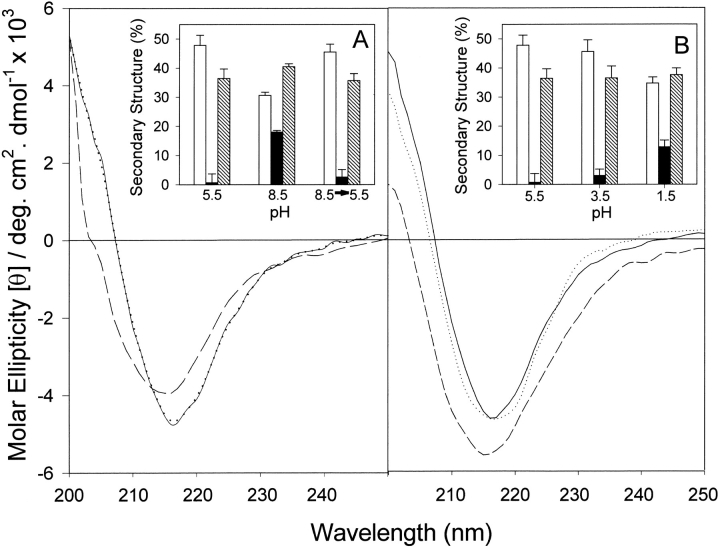
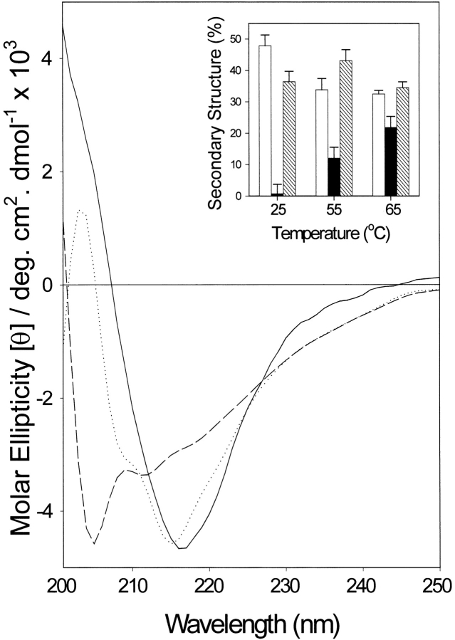
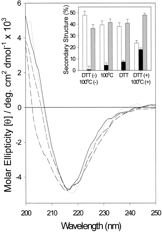
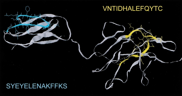
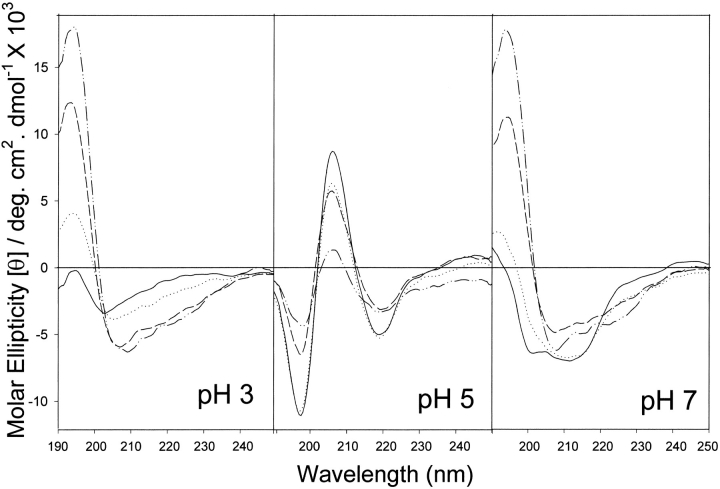
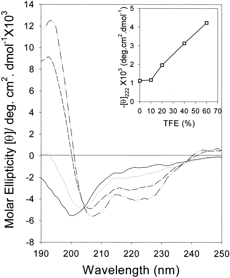
The yeast cell adhesion protein alpha-agglutinin is expressed on the surface of a free-living organism and is subjected to a variety of environmental conditions. Circular dichroism (CD) spectroscopy shows that the binding region of alpha-agglutinin has a beta-sheet-rich structure, with only approximately 2% alpha-helix under native conditions (15-40 degrees C at pH 5.5). This region is predicted to fold into three immunoglobulin-like domains, and models are consistent with the CD spectra as well as with peptide mapping and site-specific mutagenesis. However, secondary structure prediction algorithms show that segments comprising approximately 17% of the residues have high alpha-helical and low beta-sheet potential. Two model peptides of such segments had helical tendencies, and one of these peptides showed pH-dependent conformational switching. Similarly, CD spectroscopy of the binding region of alpha-agglutinin showed reversible conversion from beta-rich to mixed alpha/beta structure at elevated temperatures or when the pH was changed. The reversibility of these changes implied that there is a small energy difference between the all-beta and the alpha/beta states. Similar changes followed cleavage of peptide or disulfide bonds. Together, these observations imply that short sequences of high helical propensity are constrained to a beta-rich state by covalent and local charge interactions under native conditions, but form helices under non-native conditions.


Figures

Fig. 1.
Fig. 1.
Effect of pH on far-UV CD of α-agglutinin20–351. (A) Basic pH: Spectra were taken at 25°C in 30 mM sodium acetate at pH 5.5 (solid line), or in 100 mM sodium phosphate at pH 8.5 (dashed line). The spectrum of the reconstituted sample pH (8.5→5.5), which had been preincubated in 100 mM sodium phosphate at pH 8.5 for 2 h and 30 mM sodium acetate at pH 5.5 for 30 min, was measured at 25°C in 30 mM sodium acetate at pH 5.5 (dotted line). (Inset) Secondary structure content calculated by curve fitting: (open bar) β-sheet; (solid bar) α-helix; (hatched bar) aperiodic structures. Error bars: see text. All spectra contained ∼16% turn (not shown). (B) Acidic pH: Spectra were measured at 25°C in 30 mM sodium acetate at pH 5.5 (solid line); 30 mM sodium acetate at pH 3.5 (dotted line); or 100 mM sodium phosphate at pH 1.5 (dashed line). (Inset) Secondary structure content calculated by curve fitting.
Fig. 2.
Fig. 2.
Effect of temperature on far-UV CD of α-agglutinin20–351. Samples were equilibrated at various temperatures for 20 min and spectra were measured in 30 mM sodium acetate at pH 5.5 at 15°, 25°, 35°, and 45°C (solid line); 55°C (dotted line); or 65°C (dashed line). (Inset) Secondary structure content at different temperatures calculated by curve fitting: (open bar) β-sheet; (solid bar) α-helix; (hatched bar) aperiodic structures. Error bars: see text. All spectra contained ∼16% turn (not shown).
Fig. 3.
Fig. 3.
Effect of DTT and brief heat treatment at 100°C on far-UV CD of α-agglutinin20–351. Spectra were taken at 25°C in 30 mM sodium acetate at pH 5.5; without any treatment (solid line); with 10 mM DTT treatment (dotted line); with heat treatment at 100°C for 5 min (dashed line); or DTT treatment plus heat treatment at 100°C for 5 min (dash/dot line). (Inset) Secondary structure content calculated by curve fitting: (open bar) β-sheet;; (solid bar) α-helix; (hatched bar) aperiodic structures. Error bars: see text. All spectra contained ∼16% turn (not shown).
Fig. 4.
Fig. 4.
Three-dimensional orientation of two peptides of high helical propensity derived from the proposed β-strands regions of α-agglutinin20–351. Model of domains II and III of α-agglutinin, with peptide I (C–C` strand region of domain II) shown in cyan and peptide II (E–F strand region of domain III) shown in yellow. The models were based on similarity to the CD2/CD4 subfamily of the Ig superfamily, and were tested by peptide mapping and site-directed mutagenesis (Lipke et al. 1995; Grigorescu et al. 2000). The peptide sequences are shown in single letter code, and their helical propensities are summarized in Table 1.
Fig. 5.
Fig. 5.
Effect of TFE on far-UV CD of peptide I at different pH values. Spectra were measured in 10 mM sodium phosphate at pH 2–10 in 10% TFE (solid line); 20% TFE (dotted line); 40% TFE (dashed line), or 60% TFE (dash/dot line). The spectra below pH 3 were similar to one another, as were those above 8. The spectra at pH 3, 5, and 7 are shown.
Fig. 6.
Fig. 6.
Effect of TFE on far-UV CD of peptide II. Spectra were measured in 10 mM sodium phosphate at pH 2–10 in 0% TFE (dash/dot line); 10% TFE (solid line); 20% TFE (dotted line); 40% TFE (dashed line), or 60% TFE (dash/dot/dot line). Because the spectra at all pH values were similar, only the curves at pH 5 are shown. (Inset) Molar ellipticity at 222 mn versus concentration of TFE.

References

    1. Baldwin, E.P., Hajiseyedjavadi, O., Baase, W.A., and Matthews, B.W. 1993. The role of backbone flexibility in the accommodation of variants that repack the core of T4 lysozyme. Science 262 1715–1718. - PubMed
    1. Baskakov, I.V., Aagaard, C., Mehlhorn, I., Wille, H., Groth, D., Baldwin, M.A., Prusiner, S.B., and Cohen, F.E. 2000. Self-assembly of recombinant prion protein of 106 residues. Biochemistry 39 2792–2804. - PubMed
    1. Bullough, P.A., Hughson, F.M., Skehel, J.J., and Wiley, D.C. 1994. Structure of influenza haemagglutinin at the pH of membrane fusion. Nature 371 37–43. - PubMed
    1. Cappellaro, C., Hauser, K., Mrsa, V., Watzele, M., Watzele, G., Gruber, C., and Tanner, W. 1991. Saccharomyces cerevisiae a- and alpha-agglutinin: Characterization of their molecular interaction. EMBO J. 10 4081–4088. - PMC - PubMed
    1. Carr, C.M. and Kim, P.S. 1993. A spring-loaded mechanism for the conformational change of influenza hemagglutinin. Cell 73 823–832. - PubMed

Publication types

MeSH terms