

Ocular pulse amplitude is reduced in patients with advanced retinitis pigmentosa
- PMID: 11371487
- PMCID: PMC1724009
- DOI: 10.1136/bjo.85.6.678
Ocular pulse amplitude is reduced in patients with advanced retinitis pigmentosa
Abstract
Background/aims: The choroid, a low resistance vascular structure carrying 85% of the ocular blood flow, provides nourishment to and removal of potential toxic waste products from the adjacent non-vascularised outer layers of the retina, macula, and optic disc regions. Choroidal perfusion may be reduced in retinitis pigmentosa (RP) and might contribute to retinal pigment epithelium (RPE) degeneration. The aim of this study was to determine whether choroidal perfusion is reduced in RP and whether this is correlated with the stage of disease.
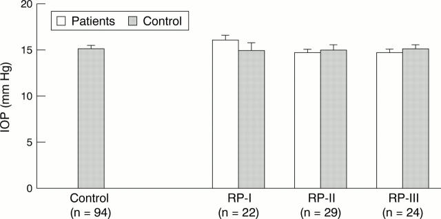
Methods: Ocular pulse amplitude (OPA) evaluated with the ocular blood flow (OBF) system, applanation intraocular pressure (IOP), visual fields, blood pressure (BP), and heart rate (HR) were measured in 75 RP patients having stage RP-I (stage I: visual field size: 7.85-14.67 cm(2); n = 22), stage RP-II (stage II: visual field size: 2.83-7.84 cm(2); n = 29), or stage RP-III (stage III: visual field size: 0.52-2.82 cm(2); n = 24) were compared with matched healthy controls and each other.
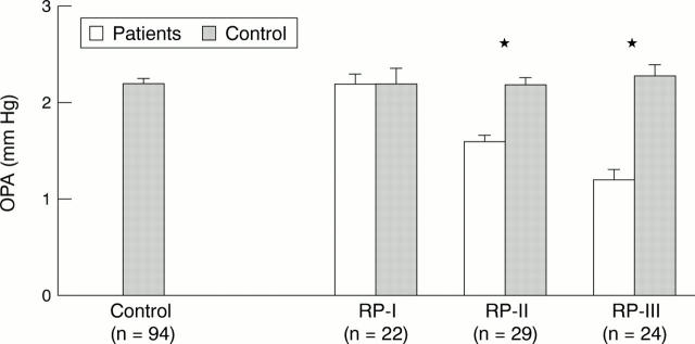
Results: Neither IOP nor systemic perfusion parameters were significantly (p >0.1) altered, but OPA (mm Hg) in RP patients beginning with stage RP-II (1.6 (0.1), 27.3%, p<0.0001), and RP-III (1.2 (0.1), 45.5%, p<0.0001) was significantly reduced when compared with matched subgroups from a pool of healthy controls (2.2 (0.1), n = 94).
Conclusions: OPA can be used neither for early clinical detection of RP nor to follow the natural course of the disease. However, our data show that in advanced stages of RP not only the retina but also the choroidal circulation is affected.
Figures
References
MeSH terms
LinkOut - more resources
Full Text Sources