. 2001 Jun;183(12):3574-81.
doi: 10.1128/JB.183.12.3574-3581.2001.

Bacillus subtilis locus encoding a killer protein and its antidote

Affiliations

Bacillus subtilis locus encoding a killer protein and its antidote

E Adler et al. J Bacteriol. 2001 Jun.

Abstract

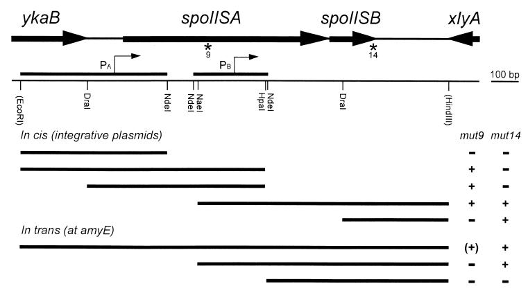
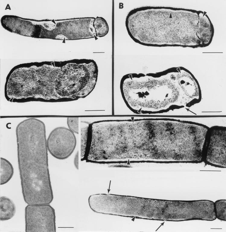
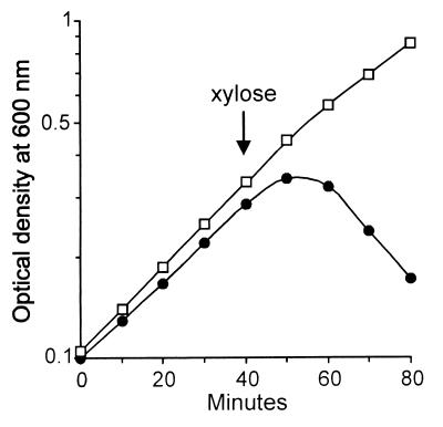
We have isolated mutations that block sporulation after formation of the polar septum in Bacillus subtilis. These mutations were mapped to the two genes of a new locus, spoIIS. Inactivation of the second gene, spoIISB, decreases sporulation efficiency by 4 orders of magnitude. Inactivation of the first gene, spoIISA, has no effect on sporulation but it fully restores sporulation of a spoIISB null mutant, indicating that SpoIISB is required only to counteract the negative effect of SpoIISA on sporulation. An internal promoter ensures the synthesis of an excess of SpoIISB over SpoIISA during exponential growth and sporulation. In the absence of SpoIISB, the sporulating cells show lethal damage of their envelope shortly after asymmetric septation, a defect that can be corrected by synthesizing SpoIISB only in the mother cell. However, forced synthesis of SpoIISA in exponentially growing cells or in the forespore leads to the same type of morphological damage and to cell death. In both cases protection against the killing effect of SpoIISA can be provided by simultaneous synthesis of SpoIISB. The spoIIS locus is unique to B. subtilis, and since it is completely dispensable for sporulation its physiological role remains elusive.


Figures

FIG. 1
FIG. 1
Characterization of the spoIIS locus. The genetic organization of the spoIIS region is shown at the top, with partial or complete open reading frames displayed as thick arrows. Asterisks indicate the locations of the mut9 and mut14 mutations. In the simplified physical map, the bordering restriction sites originate from the plasmids used for cloning the region, either from the vector backbone (EcoRI) or from an additional genomic fragment present in the insert (HindIII). The two fragments fused to lacZ for monitoring promoter activity are shown with the presumed positions of the transcription starts (thin arrows). Thick bars in the bottom part of the figure indicate the extents of the DNA fragments that were used for complementation analysis of the two spoIIS mutants, with the results shown in the right-side columns. When these fragments were cloned in integrative plasmids, correction of the sporulation defect (indicated as +) was observed in a variable proportion of the recombinants, depending on the location of the mutation relative to the fragments. Conversely, introducing these DNA fragments at the ectopic amyE locus led to a homogeneous population of transformants. Partial restoration of sporulation of the mut9 strain is indicated by (+).
FIG. 2
FIG. 2
The two SpoIIS proteins. A schematic representation of the SpoIISA and SpoIISB proteins (248 and 56 residues, respectively) is shown with the coordinates of the three putative transmembrane domains of SpoIISA. The topological model for SpoIISA is based on the predictions of the TopPred II program (6) and includes the presence of six positively charged residues between the first two transmembrane segments.
FIG. 3
FIG. 3
Expression of spoIIS-lacZ. The specific activity of β-galactosidase was monitored in a wild-type strain containing a transcriptional spoIIS-lacZ fusion, either from the Pa promoter (●) or from the Pb promoter (○), as defined in Fig. 1. Both fusions were inserted at the amyE locus. Bacteria were induced to sporulate by exhaustion in DS medium at 37°C, with the onset of sporulation defined as the time when cultures deviate from exponential growth.
FIG. 4
FIG. 4
Death of spoIIS mutants in stationary phase. The optical density of cells grown in DS medium at 37°C was monitored during exponential growth and sporulation. Strains contained either a wild-type spoIIS locus (▴), the spoIISA(mut9) mutation (■), or the spoIISB null mutation (○). The strain containing the spoIISB(mut14) mutation behaved exactly as the spoIISA(mut9) mutant and, for clarity, its results are not shown.
FIG. 5
FIG. 5
Morphological consequences of SpoIISA activity. (A) spoIISB cells were grown in DS medium and harvested 2 h (top) or 4 h (bottom) after the onset of sporulation. (B) Cells of a wild-type strain containing an extra copy of spoIISA under the control of the forespore-specific spoIIQ promoter were grown in DS medium and harvested 2 h (top) or 4 h (bottom) after the onset of sporulation. (C) Cells of a wild-type strain containing an extra copy of spoIISA under the control of the xylA promoter were grown in LB medium without xylose (left) or in the presence of 5 mM xylose (added at an optical density at 600 nm of 0.5) and harvested 1.5 h after xylose addition (right). Representative examples of cellular morphologies are shown. Arrowheads point to plasmolysis zones where the cytoplasmic membrane appears to be detached from the cell wall. Thin arrows indicate holes in the peptidoglycan layer. Bars represent 0.3 μm.
FIG. 6
FIG. 6
SpoIISA-induced death during exponential growth. Cells containing an extra copy of spoIISA at the amyE locus under the control of the xylA promoter were grown in LB medium at 37°C, and their optical density was monitored before and after addition of 5 mM xylose (arrow). Cells also contained a wild-type spoIIS locus with (□) or without (●) an additional copy of spoIISB at thrC.

References

    1. Arigoni F, Guérout-Fleury A-M, Barák I, Stragier P. The SpoIIE phosphatase, the sporulation septum, and the establishment of forespore-specific transcription in Bacillus subtilis: a reassessment. Mol Microbiol. 1999;31:1407–1415. - PubMed
    1. Bai U, Mandic-Mulec I, Smith I. SinI modulates the activity of SinR, a developmental switch protein of Bacillus subtilis, by protein-protein interaction. Genes Dev. 1993;7:139–148. - PubMed
    1. Barák I, Youngman P. SpoIIE mutants of Bacillus subtilis comprise two distinct phenotypic classes consistent with a dual functional role for the SpoIIE protein. J Bacteriol. 1996;178:4984–4989. - PMC - PubMed
    1. Bishop R E, Leskiw B K, Hodges R S, Kay C M, Weiner J H. The entericidin locus of Escherichia coli and its implications for programmed cell death. J Mol Biol. 1998;280:583–596. - PubMed
    1. Cervin M A, Spiegelman G B, Raether B, Ohlsen K, Perego M, Hoch J A. A negative regulator linking chromosome segregation to developmental transcription in Bacillus subtilis. Mol Microbiol. 1998;29:85–95. - PubMed

Publication types

Substances

LinkOut - more resources