. 2001 Jun;183(12):3680-8.
doi: 10.1128/JB.183.12.3680-3688.2001.

Stability of Mycoplasma pneumoniae cytadherence-accessory protein HMW1 correlates with its association with the triton shell

Affiliations

Stability of Mycoplasma pneumoniae cytadherence-accessory protein HMW1 correlates with its association with the triton shell

M F Balish et al. J Bacteriol. 2001 Jun.

Abstract

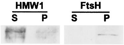
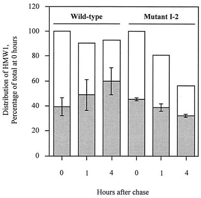
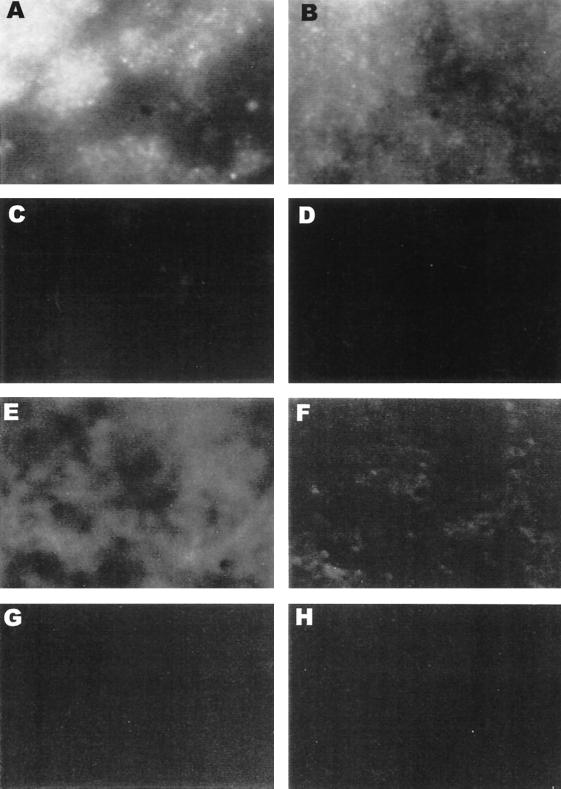
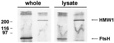
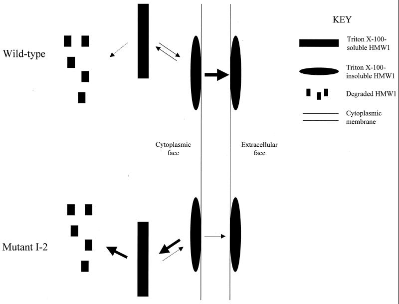
Mycoplasma pneumoniae adsorbs to host respiratory epithelium primarily by its attachment organelle, the proper function of which depends upon mycoplasma adhesin and cytoskeletal proteins. Among the latter are the cytadherence-associated proteins HMW1 and HMW2, whose specific roles in this process are unknown. In the M. pneumoniae cytadherence mutant I-2, loss of HMW2 results in accelerated turnover of HMW1 and other cytadherence-accessory proteins, probably by proteolysis. However, both the mechanism of degradation and the means by which these proteins are rendered susceptible to it are not understood. In this study, we addressed whether HMW1 degradation is a function of its presence among specific subcellular fractions and established that HMW1 is a peripheral membrane protein that is antibody accessible on the outer surfaces of both wild-type and mutant I-2 M. pneumoniae but to a considerably lesser extent in the mutant. Quantitation of HMW1 in Triton X-100-fractionated extracts from cells pulse-labeled with [(35)S]methionine indicated that HMW1 is synthesized in a Triton X-100-soluble form that exists in equilibrium with an insoluble (cytoskeletal) form. Pulse-chase analysis demonstrated that over time, HMW1 becomes stabilized in the cytoskeletal fraction and associated with the cell surface in wild-type M. pneumoniae. The less efficient transition to the cytoskeleton and mycoplasma cell surface in mutant I-2 leads to accelerated degradation of HMW1. These data suggest a role for HMW2 in promoting export of HMW1 to the cell surface, where it is stable and fully functional.


Figures

FIG. 1
FIG. 1
The deduced amino acid sequence of HMW1 suggests that there are three domains (4). Domain I (the N-terminal 170 amino acids of HMW1; white box) is predicted to consist of mostly β-strand structure. Domain II (residues 171 to 522; black box [this boundary has been revised from that in reference upon closer examination]) is the acidic, proline-rich (APR) domain common to several M. pneumoniae cytoskeletal proteins (see text). Domain III (the remainder of HMW1; medium gray box) includes a C-terminal domain postulated to be involved in targeting for proteolytic degradation (25). The dark gray box within domain I represents the location of the EAGR box (residues 106 to 136; see Discussion and Fig. 8). The light gray boxes within domain III represent the locations of predicted coiled-coil domains (residues 778 to 818 and 842 to 881; see Discussion).
FIG. 2
FIG. 2
Immunoblot analysis of wild-type M. pneumoniae subcellular fractions. Anti-P1 serum (1:1,000) (A), anti-EF-G serum (1:1,000) (B), anti-HMW1 serum (1:10,000) (C), and anti-HMW2 serum (1:2,000) (D) were used. Lane 1, lysate; lane 2, postlysis supernatant (cytosol); lane 3, major membrane band collected from the sucrose gradient; lane 4, pooled material above the major gradient band; lane 5, pooled material below the major gradient band. Each lane contains 11 μg of protein. The migration positions (in kilodaltons) of molecular mass markers are shown to the left of the gels.
FIG. 3
FIG. 3
Immunoblot analysis of susceptibility of M. pneumoniae HMW1 and FtsH to alkali solubilization. Lanes S, supernatant (alkali-soluble peripheral membrane fraction) lanes P, pellet (alkali-insoluble integral membrane fraction). Anti-HMW1 serum (1:10,000) and anti-FtsH serum (1:500) were used.
FIG. 4
FIG. 4
Accumulation of HMW1 in the triton shell in wild-type M. pneumoniae but not in mutant I-2. Each time point is the average of three experiments. Zero-hour totals were independently normalized to 100% for the wild type and mutant 1-2; all data in the graph are in relation to these zero-hour values. For the wild type, the standard deviation (indicated by error bars) for the total amount of HMW1 compared with 0 h is 12.0% for 1 h and 6.2% for 4 h. For mutant 1-2, these values are 17.1% for 1 h and 10.2% for 4 h. Gray bars show the percentages of total HMW1 at 0 h that is present in the triton shell (TX insoluble); white bars show the percentages of total HMW1 at 0 h that is TX soluble. Gray bars plus white bars are the total HMW1 (insoluble plus soluble) compared to the 0-h time point.
FIG. 5
FIG. 5
Indirect immunofluorescence of whole wild-type (A to D) and mutant I-2 (E to H) M. pneumoniae colonies. Anti-P1 serum (A and E), anti-HMW1 serum (B and F), anti-EF-G serum (C and G), and rabbit preimmune serum (D and H) were used.
FIG. 6
FIG. 6
Immunoprecipitation of M. pneumoniae HMW1 and FtsH from lysates and whole cells. Whole-cell RIP and lysate RIP were performed. For each RIP, immunoprecipitation with anti-FtsH serum (left lane) and with anti-HMW1 serum (right lane) was done. The migration positions (in kilodaltons) of molecular mass markers are shown to the left of the gel.
FIG. 7
FIG. 7
Model for HMW1 trafficking in wild-type and mutant I-2 M. pneumoniae. In wild-type M. pneumoniae, newly synthesized HMW1 equilibrates between a TX-soluble pool susceptible to degradation and a TX-insoluble pool which is likely membrane associated and awaiting export to the cell surface, where it remains insoluble. Little HMW1 is degraded (dotted arrow) in wild-type cells. In the absence of HMW2 (mutant 1-2), HMW1 is inefficiently exported, as evidenced by the decreased amount detected by immunofluorescence (Fig. 5) or whole-cell RIP (data not shown), so preexport HMW1 accumulates in the soluble pool, where it is degraded, accounting for the low steady-state levels of HMW1 in the mutant.
FIG. 8
FIG. 8
CLUSTAL W 1.8 alignment of EAGR boxes (named EAGR for enriched in aromatic and glycine residues) from M. pneumoniae HMW1, P200, and MP119 and their M. genitalium orthologs. Except for MP119 (M. pneumoniae) and MG200 (M. genitalium), M. pneumoniae and M. genitalium sequences are indicated with the letter P or G after the name of the protein, respectively. M. pneumoniae P200 EAGR boxes are numbered from the N terminus to the C terminus; M. genitalium P200 ortholog EAGR boxes are numbered according to their positional counterparts in M. pneumoniae, but this protein lacks an EAGR box corresponding to the third box in M. pneumoniae P200. Consensus sequence symbols: @, aromatic residues; 0, nonpolar residues; !, polar residues; dots, any residue. Numbers surrounding EAGR box sequences indicate the amino acid position within the protein. The M. genitalium homologs of HMW1 and P200 are MG312 and MG386, respectively.

References

    1. Baseman J B, Cole R M, Krause D C, Leith D K. Molecular basis for cytadsorption of Mycoplasma pneumoniae. J Bacteriol. 1982;151:1514–1522. - PMC - PubMed
    1. Dandekar T, Huynen M, Regula J T, Ueberle B, Zimmermann C U, Andrade M A, Doerks T, Sánchez-Pulido L, Snel B, Suyama M, Yuan Y P, Herrmann R, Bork P. Re-annotating the Mycoplasma pneumoniae genome sequence: adding value, function, and reading frames. Nucleic Acids Res. 2000;28:3278–3288. - PMC - PubMed
    1. Dirksen L B, Krebes K A, Krause D C. Phosphorylation of cytadherence-accessory proteins in Mycoplasma pneumoniae. J Bacteriol. 1994;176:7499–7505. - PMC - PubMed
    1. Dirksen L B, Proft T, Hilbert H, Plagens H, Herrmann R, Krause D C. Sequence analysis and characterization of the hmw gene cluster of Mycoplasma pneumoniae. Gene. 1996;171:19–25. - PubMed
    1. Feldner J, Göbel U, Bredt W. Mycoplasma pneumoniae adhesin is located to the tip structure by monoclonal antibody. Nature (London) 1982;298:765–767. - PubMed

Publication types

LinkOut - more resources