. 2001 Jun 5;98(12):6935-40.
doi: 10.1073/pnas.111001898. Epub 2001 May 22.

Intramembrane charge movements and excitation- contraction coupling expressed by two-domain fragments of the Ca2+ channel

Affiliations

Intramembrane charge movements and excitation- contraction coupling expressed by two-domain fragments of the Ca2+ channel

C A Ahern et al. Proc Natl Acad Sci U S A. .

Abstract

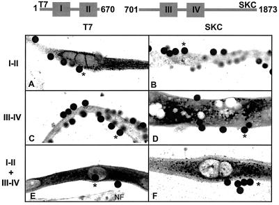
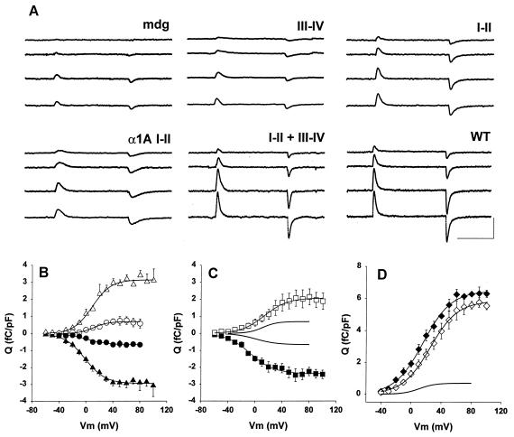
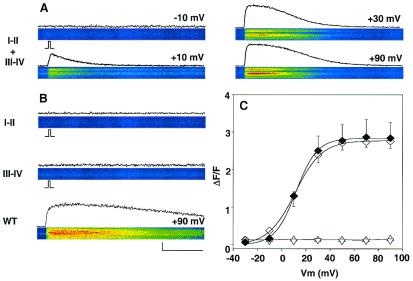
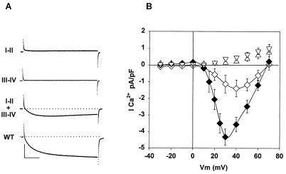
To investigate the molecular basis of the voltage sensor that triggers excitation-contraction (EC) coupling, the four-domain pore subunit of the dihydropyridine receptor (DHPR) was cut in the cytoplasmic linker between domains II and III. cDNAs for the I-II domain (alpha1S 1-670) and the III-IV domain (alpha1S 701-1873) were expressed in dysgenic alpha1S-null myotubes. Coexpression of the two fragments resulted in complete recovery of DHPR intramembrane charge movement and voltage-evoked Ca(2+) transients. When fragments were expressed separately, EC coupling was not recovered. However, charge movement was detected in the I-II domain expressed alone. Compared with I-II and III-IV together, the charge movement in the I-II domain accounted for about half of the total charge (Q(max) = 3 +/- 0.23 vs. 5.4 +/- 0.76 fC/pF, respectively), and the half-activation potential for charge movement was significantly more negative (V(1/2) = 0.2 +/- 3.5 vs. 22 +/- 3.4 mV, respectively). Thus, interactions between the four internal domains of the pore subunit in the assembled DHPR profoundly affect the voltage dependence of intramembrane charge movement. We also tested a two-domain I-II construct of the neuronal alpha1A Ca(2+) channel. The neuronal I-II domain recovered charge movements like those of the skeletal I-II domain but could not assist the skeletal III-IV domain in the recovery of EC coupling. The results demonstrate that a functional voltage sensor capable of triggering EC coupling in skeletal myotubes can be recovered by the expression of complementary fragments of the DHPR pore subunit. Furthermore, the intrinsic voltage-sensing properties of the alpha1A I-II domain suggest that this hemi-Ca(2+) channel could be relevant to neuronal function.


Figures

Figure 1
Figure 1
Confocal immunofluorescence of dysgenic myotubes expressing DHPR fragments. Confocal images (42 × 18 μm) show details of the intracellular distribution of the expressed fragments. Cells were transfected with the CD8 cDNA plus the following: I-II cDNA (A and B), III-IV cDNA (C and D), and I-II + III-V cDNAs (E and F). Cells were incubated with CD8 antibody beads, fixed, and stained with T7 (A, C, and E) or SKC (B, D, and F) primary/fluorescein-conjugated secondary antibodies. Pixel intensity was converted to a 16-level inverted gray scale, with high-intensity pixels in black. Asterisks show on-focus CD8 antibody beads (diameter 4.5 μm) bound to cells expressing the indicated fragment(s). NF indicates a nontransfected myotube in the same focal plane of the transfected cell.
Figure 2
Figure 2
Expression of intramembrane charge movements by skeletal and neuronal two-domain fragments. (A) Gating-type currents in myotubes expressing the indicated fragment, nontransfected (mdg), and full-length α1S (WT). The 50-ms step potentials are −10, 10, 50, and 70 mV. The cell capacitance was (in pF) 281 (mdg), 290 (I-II), 317 (III-IV), 242 (I-II + III-IV), 260 (full-length α1S, WT), and 260 (α1A I-II). (Calibration bars are 25 ms and 0.5 nA.) (B–D) Q-V curves for mdg (B, ○, ●), I-II (B, ▵, ▴), α1A I-II (C, □, ■), I-II + III-IV (D, ⋄), and WT (D, ♦). In B and C, the ON charge is positive and the OFF charge is negative. In D, only the OFF charge is shown. Curves correspond to a Boltzmann fit of the population mean Q-V curve. Parameters of the fit are (B) Qmax ON = 0.7, 3.1 fC/pF; Qmax OFF = −0.66, −3 fC/pF; V1/2 ON = 1.1, 9 mV; V1/2 OFF = 1.3, 1.6 mV; k ON = 10.5, 15.2 mV, k OFF = 15.5, 13.8 mV for mdg and I-II, respectively. (C) Qmax ON = 2.1 fC/pF; Qmax OFF = −2.4 fC/pF; V1/2 ON = 6.8 mV; V1/2 OFF = −3.6 mV; k ON = 17.2 mV, k OFF = 17.7 mV for α1A I-II. (D) Qmax OFF = 5, 5.3 fC/pF; V1/2 OFF = 25, 22 mV; k OFF = 18.2, 18.5 mV for I-II + III-IV and WT, respectively. (C, D) The curve without symbols is a fit of the Q-V curve of mdg cells.
Figure 3
Figure 3
Recovery of skeletal-type EC coupling by coexpression of skeletal two-domain fragments. The confocal line-scan images show fluo-4 fluorescence across myotubes in response to a 50-ms depolarization from a holding potential of −40 mV. Traces immediately above each line scan show the time course of the fluorescence change in resting units (ΔF/F). (A) Ca2+ transients for I-II + III-IV at the indicated potentials. (B) Absence of Ca2+ transients for single fragments and Ca2+ transient for full-length α1S (WT) at +90 mV. (C) The average ΔF/F at the peak of the transient was plotted as a function of voltage for cells expressing I-II + III-IV (⋄, 15 cells), WT (♦, six cells), I-II (▵, eight cells), and III-IV (▿, 10 cells). Curves are a Boltzmann fit of the population mean ΔF/F-V curve. Parameters of the fit are ΔF/Fmax = 2.84, 2.78; V1/2 = 11.7, 11.02 mV; and k = 8.7, 11.03 mV for WT and for I-II + III-V, respectively. Line-scan images have a constant temporal dimension of 2.05 s (horizontal) and a variable spatial dimension (vertical) depending on the cell length. The cell dimension in the line scans was (in μm): 88 (A), 50 (B, I-II), 90 (B, III-IV), and 59 (B, full-length α1S, WT). (Bars = 500 ms and 1 ΔF/F.)
Figure 4
Figure 4
Absence of Ca2+ currents in myotubes expressing individual DHPR fragments. (A) Absence or presence of whole-cell Ca2+ currents in myotubes expressing the indicated fragments. The depolarizing potential was +20 mV for 500 ms from a holding potential of −40 mV. (B) Current–voltage curves of the Ca2+ current of myotubes expressing I-II + III-IV (◊, 7 cells), full-length α1S (♦, 9 cells), I-II (▵, 11 cells), and III-IV (▿, 10 cells). The cell capacitance was (in pF) 154 (I-II), 186 (III-IV), 226 (I-II + III-IV), and 207 (full-length α1S, WT). (Calibration bars are 100 ms and 1 nA.)

Similar articles

Cited by

References

    1. Catterall W A. Annu Rev Biochem. 1995;64:493–531. - PubMed
    1. Bezanilla F. Physiol Rev. 2000;80:555–592. - PubMed
    1. Rios E, Pizarro G. Physiol Rev. 1991;71:849–908. - PubMed
    1. Huang C L-H. Physiol Rev. 1988;68:1197–1247. - PubMed
    1. Melzer W, Schneider M F, Simon B J, Szucs G. J Physiol (London) 1986;373:481–511. - PMC - PubMed

Publication types

LinkOut - more resources