. 2001 Jun 5;98(12):6698-703.
doi: 10.1073/pnas.111092198. Epub 2001 May 22.

Haploinsufficiency of Sox9 results in defective cartilage primordia and premature skeletal mineralization

Affiliations

Haploinsufficiency of Sox9 results in defective cartilage primordia and premature skeletal mineralization

W Bi et al. Proc Natl Acad Sci U S A. .

Abstract

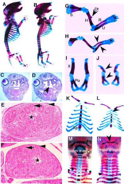
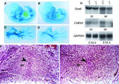
In humans, SOX9 heterozygous mutations cause the severe skeletal dysmorphology syndrome campomelic dysplasia. Except for clinical descriptions, little is known about the pathogenesis of this disease. We have generated heterozygous Sox9 mutant mice that phenocopy most of the skeletal abnormalities of this syndrome. The Sox9(+/-) mice died perinatally with cleft palate, as well as hypoplasia and bending of many skeletal structures derived from cartilage precursors. In embryonic day (E)14.5 heterozygous embryos, bending of radius, ulna, and tibia cartilages was already prominent. In E12.5 heterozygotes, all skeletal elements visualized by using Alcian blue were smaller. In addition, the overall levels of Col2a1 RNA at E10.5 and E12.5 were lower than in wild-type embryos. We propose that the skeletal abnormalities observed at later embryonic stages were caused by delayed or defective precartilaginous condensations. Furthermore, in E18.5 embryos and in newborn heterozygotes, premature mineralization occurred in many bones, including vertebrae and some craniofacial bones. Because Sox9 is not expressed in the mineralized portion of the growth plate, this premature mineralization is very likely the consequence of allele insufficiency existing in cells of the growth plate that express Sox9. Because the hypertrophic zone of the heterozygous Sox9 mutants was larger than that of wild-type mice, we propose that Sox9 also has a role in regulating the transition to hypertrophic chondrocytes in the growth plate. Despite the severe hypoplasia of cartilages, the overall organization and cellular composition of the growth plate were otherwise normal. Our results suggest the hypothesis that two critical steps of the chondrocyte differentiation pathway are sensitive to Sox9 dosage. First, an early step presumably at the stage of mesenchymal condensation of cartilage primordia, and second, a later step preceding the transition of chondrocytes into hypertrophic chondrocytes.


Figures

Figure 1
Figure 1
Expression of Sox9 in primary chondrocytes isolated from wild-type (+/+) and heterozygous (+/−) neonates. (A) Northern analysis. Ten micrograms of total RNA was hybridized with a Sox9 probe from the deleted region. The same membrane then was hybridized with GAPDH as an RNA-loading control for the quantity of RNA. (B) Western blotting with SOX9 antibody. β-actin antibody (Sigma) was used on the same membrane as a protein-loading control.
Figure 2
Figure 2
Skeletal malformations in Sox9+/− embryos. (A and B) Lateral view of the skeletal preparation of wild-type (A) and heterozygous (B) newborns. (C and D) Hematoxylin/eosin-stained coronal sections of the heads at the level of molars of E18.5 wild-type (C) and Sox9+/− (D) embryos. The arrow indicates the cleft palate in the mutants. (E and F) Transverse sections through testes of E14.5 Sox9+/− embryos. (E) Wild-type testis (Swiss) showed seminiferous cords (arrow) enclosing the germ cells. Fetal Leydig cells comprise the interstitium (asterisk) between the seminiferous cords, and a tunica albuginea (arrowhead) surrounds the testis. (F) Sox9+/− testis (129SvEv/C57B6) was histologically normal. (GN) Dissected skeletal elements of wild-type controls (G, I, K, and M) and Sox9+/− mutants (H, J, L, and N). (G and H) E18.5 forelimbs. The deltoid tuberosity (Dt) of the humerus was missing in the mutants. Arrowheads indicate the bending of the radius and ulna. (I and J) E18.5 pelvic bones. Arrowheads indicate a bending ilium and pubic bone. (K and L) E18.5 rib cages. Arrowhead indicates a missing manubrium sterna. (M and N) Newborn trachea. Arrowhead indicates the bending of the hyoid bone in the Sox9+/− mutants. S, scapula; H, humerus; R, radius; U, ulna; Il, ilium; Pu, pubic; Xp, xiphoid process; Hy, hyoid bone; Tc, thyroid cartilage; Cc, cricoid cartilage; Tr, tracheal rings.
Figure 3
Figure 3
Abnormalities in the cartilaginous elements in Sox9+/− embryos at E14.5. (AH) Skeletal preparation of wild-type (A, CE, and G) and Sox9+/− embryos (BD, F, and H). Arrowhead in H indicates that the bending site of the ulna was outside the ossification center. (IL) Cartilage staining of wild-type (I and K) and Sox9+/− (J and L) embryos. Meckel's cartilage was interrupted (arrow) and bowing (arrowhead) in the mutants (J). (K and L) Scapulae. Arrowheads indicate a missing spine and arrow indicates the hypoplastic blade in the mutants. (M and N) Sections of E15.5 sternum of wild-type (M) and Sox9+/− embryos (N). The cartilage primordia of the sternum were malformed in the mutants as indicated by arrows. Me, Meckel's cartilage; S, scapula; Sp, spine of scapula; H, humerus; R, radius; U, ulna; F, femur; Fi, fibula; T, tibia.
Figure 4
Figure 4
Alcian blue staining of wild-type (A and C) and Sox9+/− (B and D) embryos at E12.5. (A and B) Whole embryos. (C and D) Forelimbs. (E and F) Hematoxylin/eosin staining of transverse sections through equivalent thoracic regions. Cells in the sclerotome surrounding the notochord (arrowhead) appeared like mesenchymal cells in the Sox9+/− mutants (F), but were typical cobblestone-like prechondrocytic cells in wild-type controls (E). (G) Northern analysis with total RNA from wild-type (+/+) and Sox9+/− mutant (+/−) embryos at E10.5 and E12.5. The expression levels of Sox9 and Col2a1 were measured by densitometry in a PhosphorImager (Molecular Dynamics) and the percent expression levels of the mutant relative to wild type are shown. S, scapula; H, humerus; R, radius; U, ulna; F, femur; Fi, fibula; T, tibia; Pv, prevertebrae.
Figure 5
Figure 5
Premature mineralization in the Sox9+/− mutants. (AH) Skeletal preparation of wild-type (A, C, E, and G) and Sox9+/− (B, D, F, and H) newborns. (A and B) Middle ears. (C and D) Dorsal view of the skull. (E and F) Seventh cervical vertebrae. (G and H) Third thoracic vertebrae. (I and J) Von Kossa staining of sections of fifth thoracic vertebrae. The cartilaginous regions between the vertebral body and the pedicles of dorsal arches in wild-type control (I) were mineralized in E18.5 Sox9+/− mutants (J). (K and L) Hematoxylin/eosin staining of sections of equivalent caudal vertebrae from E15.5 wild-type (K) and Sox9+/− (L) embryos. The area of hypertrophic chondrocytes are indicated by arrowheads. Ip, interparietal bone; So, supraoccipital bone; Vb, vertebral body; Da, dorsal arch; Id, intervertebral discs.
Figure 6
Figure 6
Enlarged hypertrophic zone in Sox9+/− epiphyseal growth plate of the proximal tibia. (A and B) Alcian blue staining of the epiphyseal growth plates of wild-type (A) and Sox9+/− (B) embryos at E18.5. (CJ) RNA in situ hybridization analysis of the expression of Col10a1 (E and F), parathyroid hormone receptor (PTHrP-R; G and H), and Indian hedgehog (Ihh; I and J) in the tibia epiphyseal growth plates of wild-type (C, E, G, and I) and Sox9+/− (D, F, H, and J) neonates. (C and D) Bright field. H, hypertrophic zone.

References

    1. Tacchetti C, Tracella S, Dozin B, Quarto R, Robino G, Cancedda R. Exp Cell Res. 1992;200:26–33. - PubMed
    1. Cancedda R, Descalzi Cancedda F, Castagnola P. Int Rev Cytol. 1995;159:265–358. - PubMed
    1. Bi W, Deng J M, Zhang Z, Behringer R R, de Crombrugghe B. Nat Genet. 1999;22:85–89. - PubMed
    1. Wright E, Hargrave M R, Christiansen J, Cooper L, Kun J, Evans T, Gangadharan U, Greenfield A, Koopman P. Nat Genet. 1995;9:15–20. - PubMed
    1. Zhao Q, Eberspaecher H, Lefebvre V, de Crombrugghe B. Dev Dyn. 1997;209:377–386. - PubMed

Publication types