. 2001 Jun 5;98(12):6611-6.
doi: 10.1073/pnas.111099998. Epub 2001 May 22.

O-linkage of N-acetylglucosamine to Sp1 activation domain inhibits its transcriptional capability

Affiliations

O-linkage of N-acetylglucosamine to Sp1 activation domain inhibits its transcriptional capability

X Yang et al. Proc Natl Acad Sci U S A. .

Abstract

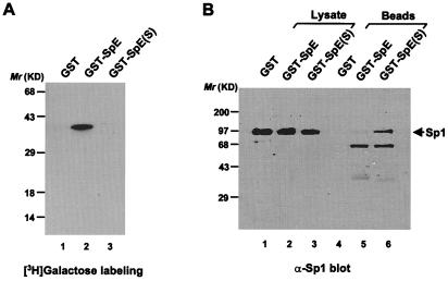
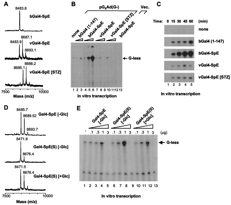
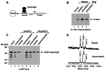
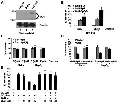
The posttranslational modification of eukaryotic intracellular proteins by O-linked N-acetylglucosamine (O-GlcNAc) monosaccharides is essential for cell viability, yet its precise functional roles are largely unknown. O-GlcNAc transferase utilizes UDP-GlcNAc, the end product of hexosamine biosynthesis, to catalyze this modification. The availability of UDP-GlcNAc correlates with glycosylation levels of intracellular proteins as well as with transcriptional levels of some genes. Meanwhile, transcription factors and RNA polymerase II can be modified by O-GlcNAc. A linkage between transcription factor O-GlcNAcylation and transcriptional regulation therefore has been postulated. Here, we show that O-GlcNAcylation of a chimeric transcriptional activator containing the second activation domain of Sp1 decreases its transcriptional activity both in an in vitro transcription system and in living cells, which is in concert with our observation that O-GlcNAcylation of Sp1 activation domain blocks its in vitro and in vivo interactions with other Sp1 molecules and TATA-binding protein-associated factor II 110. Furthermore, overexpression of O-GlcNAc transferase specifically inhibits transcriptional activation by native Sp1 in cells. Thus, our studies provide direct evidence that O-GlcNAcylation of transcription factors is involved in transcriptional regulation.


Figures

Figure 1
Figure 1
O-GlcNAc blocks interaction between SpE and the full-length Sp1 in vivo. (A) Approximately equivalent amounts (0.5 μg) of GST fusion proteins were subjected to in vitro [3H]galactose labeling by galactosyltransferase. (B) The full-length Sp1 coprecipitated with the wild-type and mutant GST-SpE was analyzed by immunoblot with anti-Sp1 antiserum. One percent of whole-cell lysates were analyzed as well.
Figure 2
Figure 2
Transcriptional activation by Gal4-SpE chimeras with diverse glycosylation status. (A) Mass spectra of tryptic digests of Gal4-SpE fusions. The GST fusions were produced in bacteria (bGal4-SpE) and in BSC40 cells infected with recombinant vaccinia virus (vGal4-SpE). Some infected cells were treated with streptozotocin (vGal4-SpE [STZ]). (B) Dose-response of Gal4-SpE fusions in activating transcription in vitro. Gal4-SpE proteins (0.3, 1.0, and 3.0 μg) were added to reactions with linearized DNA template pG5Ad(G−). Gal4 (1) (0.3 and 3.0 μg) was added to reactions as a control (lanes 2 and 3). bGal4-SpE (1.0 μg) was added to a reaction with an empty vector (Vec.) (lane 13). (C) Time course of Gal4-SpE-activated transcription in vitro. One microgram of each fusion was used. (D) Mass spectra of tryptic digests of the wild-type and mutant Gal4-SpE, which were expressed in BSC40 cells grown either in glucose-free ([−Glc]) or glucose-containing ([+Glc]) medium. (E) Dose-dependent transcriptional activation by the wild-type and mutant Gal4-SpE. Fusion proteins (0.1, 0.3, 1.0, and 3.0 μg) were added to transcription reactions with the linearized DNA template pG5Ad(G−).
Figure 3
Figure 3
GST-Gal4-SpE chimera remains glycosylated in an in vitro transcription system. (A) Schematic diagram of isolation of DNA-bound GST-Gal4-SpE. (B) In vitro transcription on immobilized DNA templates. Equal amounts of empty vector (Vector), Gal4-binding site-deleted template [pAd(G−)], or pG5Ad(G−) were coupled to Dynabeads mixed with vGST-Gal4-SpE in reactions. A reaction with dissolved pG5Ad(G−) template (Free) served as positive control. (C) The binding of vGST-Gal4-SpE to immobilized DNA templates was analyzed by immunoblot with anti-GST antibody. (D) Mass spectra of tryptic digests of vGST-Gal4-SpE either bound to DNA or in supernatant.
Figure 4
Figure 4
Transcriptional regulation in vivo by O-GlcNAc. (A) Northern blot analysis of endogenous OGT expression in distinct cell lines. (B and C) Distinct cell lines were cotransfected with 20 μg of G5-Luc reporter and 10 μg of expression vectors for Gal4(1–94) alone, the wild type, and mutant Gal4-SpE, respectively, and then treated with glucose as indicated. (D) HeLa or HepG2 cells were cotransfected with 20 μg of G5-Luc reporter and 10 μg of the wild type or mutant Gal4-SpE expression vector, together with 10 μg of the empty vector or OGT expression vector. The cells were grown in normal glucose (5.5 mM) medium. (E) Overexpression of OGT represses native Sp1-activated transcription. HepG2 cells were cotransfected with 20 μg of GC6-Luc reporter or 20 μg of G5-Luc reporter plus 10 μg of the expression vector for wild-type Gal4 (Gal4 wt), together with increasing amounts of OGT expression vector. Cells were transfected with equal amounts of DNA by inclusion of the parental expression vector.

References

    1. Hart G W. Annu Rev Biochem. 1997;66:315–335. - PubMed
    1. Shafi R, Iyer S P, Ellies L G, O'Donnell N, Marek K W, Chui D, Hart G W, Marth J D. Proc Natl Acad Sci USA. 2000;97:5735–5739. . (First Published May 9, 2000; 10.1073/pnas.100471497) - PMC - PubMed
    1. Liu K, Paterson A J, Chin E, Kudlow J E. Proc Natl Acad Sci USA. 2000;97:2820–2825. - PMC - PubMed
    1. Yki-Jarvinen H, Virkamaki A, Daniels M C, McClain D, Gottschalk W K. Metabolism. 1998;47:449–455. - PubMed
    1. Sayeski P P, Kudlow J E. J Biol Chem. 1996;271:15237–15243. - PubMed

Publication types

LinkOut - more resources