. 2001 May 22;98(11):6021-6.
doi: 10.1073/pnas.111150298.

The structures of anthranilate synthase of Serratia marcescens crystallized in the presence of (i) its substrates, chorismate and glutamine, and a product, glutamate, and (ii) its end-product inhibitor, L-tryptophan

Affiliations

The structures of anthranilate synthase of Serratia marcescens crystallized in the presence of (i) its substrates, chorismate and glutamine, and a product, glutamate, and (ii) its end-product inhibitor, L-tryptophan

G Spraggon et al. Proc Natl Acad Sci U S A. .

Abstract

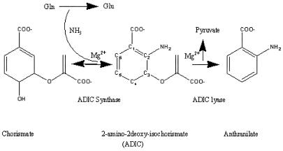
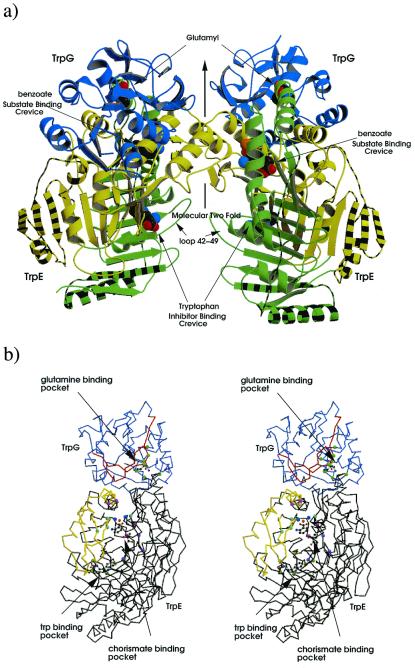
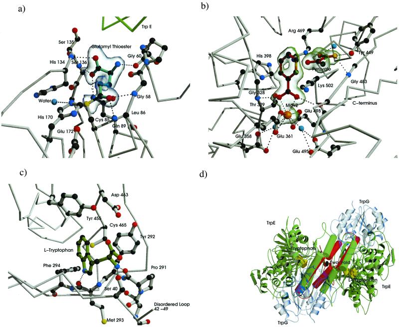
The crystal structure of anthranilate synthase (AS) from Serratia marcescens, a mesophilic bacterium, has been solved in the presence of its substrates, chorismate and glutamine, and one product, glutamate, at 1.95 A, and with its bound feedback inhibitor, tryptophan, at 2.4 A. In comparison with the AS structure from the hyperthermophile Sulfolobus solfataricus, the S. marcescens structure shows similar subunit structures but a markedly different oligomeric organization. One crystal form of the S. marcescens enzyme displays a bound pyruvate as well as a putative anthranilate (the nitrogen group is ambiguous) in the TrpE subunit. It also confirms the presence of a covalently bound glutamyl thioester intermediate in the TrpG subunit. The tryptophan-bound form reveals that the inhibitor binds at a site distinct from that of the substrate, chorismate. Bound tryptophan appears to prevent chorismate binding by a demonstrable conformational effect, and the structure reveals how occupancy of only one of the two feedback inhibition sites can immobilize the catalytic activity of both TrpE subunits. The presence of effectors in the structure provides a view of the locations of some of the amino acid residues in the active sites. Our findings are discussed in terms of the previously described AS structure of S. solfataricus, mutational data obtained from enteric bacteria, and the enzyme's mechanism of action.


Figures

Scheme 1
Scheme 1
Figure 1
Figure 1
Sequence alignments of AS subunits from S. marcescens and S. solfataricus calculated from aligned structures of the two molecules. Structure alignment was performed with o (24) and stamp (36) and the figure produced by alscript (37). Secondary structures were assigned by dssp (38). Numbering is based on the S. marcescens sequence. Conserved hydrophobic residues are shaded yellow, conserved nonhydrophobic residues are shaded green, conserved polar residues are in bold font, conserved small residues in small font, and conserved structural regions are shown boxed. Those residues underlined in a shaded blue box are solvent inaccessible in the heterodimer TrpE:TrpG interface, whereas those in brown are solvent excluded in the heterotetramer interface. β-Sheet regions are shown as light purple arrows, whereas α-helices are in blue. (a) Alignment of TrpGs: residues of the active site are shaded red, whereas those in close contact with the glutamyl residue are shaded gold. (b) Alignment of TrpE: residues in close contact to anthranilate shaded red, whereas those in close contact with pyruvate are in light blue. Residues shaded gold are in close contact with tryptophan.
Figure 2
Figure 2
Structure of the AS of S. marcescens. (a) Ribbon diagram of the AS oligomer, TrpG subunits shown in blue, TrpE subdomain I shown in green subdomain II in yellow. Striped regions correspond to additional structure in S. marcescens compared with that of S. solfataricus. Glutamyl, benzoate, pyruvate, and tryptophan are shown as cpk models. (b) Stereo diagram of the heterodimer; TrpG shown in lilac, TrpE in black; regions of TrpG that move on addition of tryptophan relative the C-crystal are shown in red, whereas those of TrpE are in yellow; residues important to the CA-binding pocket (G328, T329, H398, G485) are shown as light blue balls, residues involved in pyruvate interactions (Y449, R469, G483) are in purple, residues involved in magnesium coordination (E358,361, E495, E498) are colored light purple, magnesium ion in orange, water molecules in dark blue, Trp-binding residues (S40, P291, M293, V453, Y455) are light green, and residues involved in glutamine binding (P57, G58, G60, C85, L86, Q89, S135, S136) are in green. Benzoate, pyruvate, magnesium, and glutamyl are shown as ball-and-stick figures. Produced by bobscript and raster 3d (–42).
Figure 3
Figure 3
Substrate product and Trp-binding sites of the AS molecule. Carbon atoms are dark gray, nitrogen blue, and oxygen red. Electrostatic and hydrogen-bond interactions are shown as black dotted lines. (a) Binding residues for glutamyl thioester intermediate of TrpG. Glutamyl moiety drawn with cyan bonds. A σA-weighted FoFc map contoured at 3.5 SD is shown in transparent blue. (b) CA-binding pocket, anthranilate, and pyruvate are drawn with dark red bonds, magnesium ion in orange, ordered waters in cyan. A σA-weighted FoFc map contoured at 3.0 SD is shown in transparent green. (c) Trp-binding pocket, tryptophan shown in green. (d) Conformational states associated with anthranilate, pyruvate, and Trp-bound forms. The molecule is viewed with the molecular 2-fold perpendicular to the page (i.e., perpendicular to the view in Fig. 2a). TrpG subunits shown in transparent blue, α helixes and loops involved in heterotetramer rearrangement shown as rods, C-crystal representation shown in green rearrangement in the T-crystal, shown in red. Tryptophan is shown as cpk model in yellow. Produced by bobscript and raster 3d (–42).

References

    1. Morollo A A, Bauerle R. Proc Natl Acad Sci USA. 1993;90:9983–9987. - PMC - PubMed
    1. Zalkin H, Hwang L H. J Biol Chem. 1971;246:6899–6907. - PubMed
    1. Zalkin H. Adv Enzymol Relat Areas Mol Biol. 1993;66:203–309. - PubMed
    1. Tesmer J J, Klem T J, Deras M L, Davisson V J, Smith J L. Nat Struct Biol. 1996;3:74–86. - PubMed
    1. Bauerle R, Hess J, French S. Methods Enzymol. 1987;142:366–386. - PubMed

Publication types

MeSH terms