. 2001 May 22;98(11):6051-5.
doi: 10.1073/pnas.111145098.

Solution structure of DFF40 and DFF45 N-terminal domain complex and mutual chaperone activity of DFF40 and DFF45

Affiliations

Solution structure of DFF40 and DFF45 N-terminal domain complex and mutual chaperone activity of DFF40 and DFF45

P Zhou et al. Proc Natl Acad Sci U S A. .

Abstract

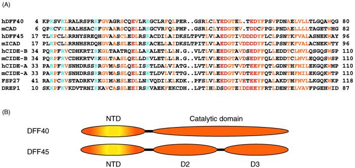
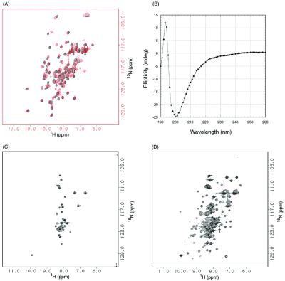
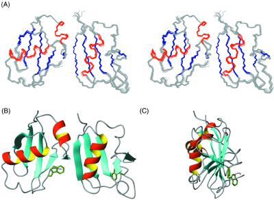
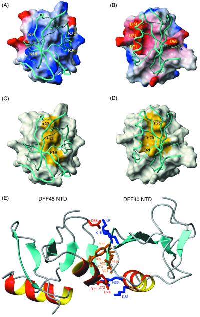
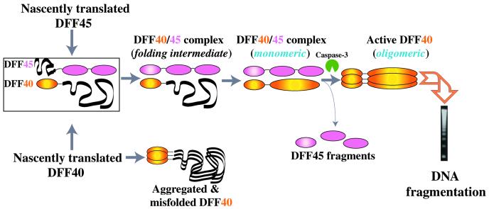
Apoptotic DNA fragmentation is mediated by a caspase-activated DNA fragmentation factor (DFF)40. Expression and folding of DFF40 require the presence of DFF45, which also acts as a nuclease inhibitor before DFF40 activation by execution caspases. The N-terminal domains (NTDs) of both proteins are homologous, and their interaction plays a key role in the proper functioning of this two-component system. Here we report that the NTD of DFF45 alone is unstructured in solution, and its folding is induced upon binding to DFF40 NTD. Therefore, folding of both proteins regulates the formation of the DFF40/DFF45 complex. The solution structure of the heterodimeric complex between NTDs of DFF40 and DFF45 reported here shows that the mutual chaperoning includes the formation of an extensive network of intermolecular interactions that bury a hydrophobic cluster inside the interface, surrounded by intermolecular salt bridges.


Figures

Figure 1
Figure 1
(A) Sequence alignment of CIDE proteins. Residues conserved in the family are colored yellow for hydrophobic residues, blue for basic residues, and red for acidic residues. (B) Domain structures (NTD and the catalytic domain) of DFF40 and (NTD, D2, and D3) of DFF45 are shown schematically.
Figure 2
Figure 2
DFF40 induces folding of DFF45. (A) 1H/15N HSQC spectra of DFF40 NTD (1–80) in the absence (black) and in the presence (red) of DFF45 NTD (1). (B) CD spectra of DFF45 NTD in the absence of DFF40 NTD. (C) 1H/15N HSQC spectra of DFF45 NTD in the absence of DFF40 NTD. (D) 1H/15N HSQC spectra of DFF45 NTD in the presence of DFF40 NTD.
Figure 3
Figure 3
Structure of DFF40/45 CIDE complex. (A) Stereo view of superimposed DFF40/45 homophilic NTD complex with DFF45 NTD (Left) and DFF40 NTD (Right). (B) Ribbon representation of the DFF40/45 NTD complex in the same orientation as A. (C) Ribbon representation of DFF40/45 complex with a 90° rotation along z axis.
Figure 4
Figure 4
Interface of the DFF40/45 CIDE complex. (A) Surface diagram of DFF40 NTD. In this figure, DFF45 NTD is shown in a ribbon diagram. The surface electrostatic potential of DFF40 is colored coded so that regions with electrostatic potential <−8 kBT are red, whereas those >+8 kBT are blue (where kB and T are Boltzmann constant and temperature, respectively). Basic residues important for the interactions are mapped on the surface. (B) Surface diagram of DFF45 NTD. In this figure, DFF40 NTD is shown in a ribbon diagram. The surface electrostatic potential of DFF45 is color coded so that regions with electrostatic potential <−8 kb are red, whereas those >+8 kb are blue (where kb and T are Boltzmann constant and temperature, respectively). Acidic residues important for the interactions are mapped on the surface. (C) Surface diagram of DFF40 NTD (same orientation as in A). In this figure, DFF45 NTD is shown in a ribbon diagram. The hydrophobic surface of DFF40 (Phe-19, Val-21, and Ala-22) is colored yellow. (D) Surface diagram of DFF45 NTD (same orientation as in B). In this figure, DFF40 NTD is shown in a ribbon diagram. The hydrophobic surface of DFF45 (Ile-69, Val-70, and Tyr-75) is colored yellow. (E) The homophilic DFF40/45 interaction involves both hydrophilic and hydrophobic interactions. Residues involved in the binding of DFF40 and DFF45 are color coded so that hydrophobic residues are colored brown, basic residues are colored blue, and acidic residues are colored red.
Figure 5
Figure 5
Model of the mutual chaperoning of DFF40 and DFF45.

References

    1. Thompson C B. Science. 1995;267:1456–1462. - PubMed
    1. Liu X, Li P, Widlak P, Zou H, Luo X, Garrard W T, Wang X. Proc Natl Acad Sci USA. 1998;95:8461–8466. - PMC - PubMed
    1. Enari M, Sakahira H, Yokoyama H, Okawa K, Iwamatsu A, Nagata S. Nature (London) 1998;391:43–50. - PubMed
    1. Sakahira H, Enari M, Nagata S. Nature (London) 1998;391:96–99. - PubMed
    1. Halenbeck R, MacDonald H, Roulston A, Chen T T, Conroy L, Williams L T. Curr Biol. 1998;8:537–540. - PubMed

Publication types

MeSH terms

Associated data