. 2001 May 22;98(11):6186-91.
doi: 10.1073/pnas.111158198.

Remodeling of the postnatal mouse testis is accompanied by dramatic changes in stem cell number and niche accessibility

Affiliations

Remodeling of the postnatal mouse testis is accompanied by dramatic changes in stem cell number and niche accessibility

T Shinohara et al. Proc Natl Acad Sci U S A. .

Abstract

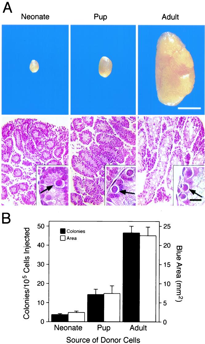
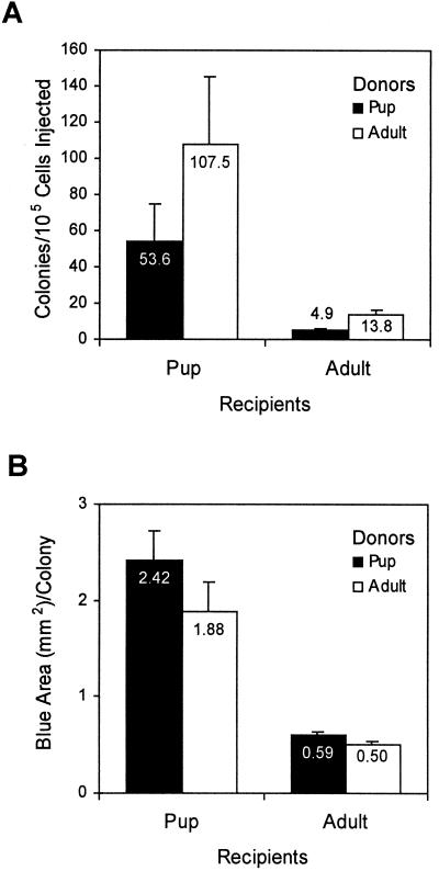
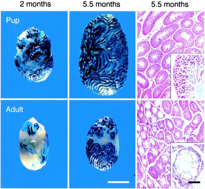
Little is known about stem cell biology or the specialized environments or niches believed to control stem cell renewal and differentiation in self-renewing tissues of the body. Functional assays for stem cells are available only for hematopoiesis and spermatogenesis, and the microenvironment, or niche, for hematopoiesis is relatively inaccessible, making it difficult to analyze donor stem cell colonization events in recipients. In contrast, the recently developed spermatogonial stem cell assay system allows quantitation of individual colonization events, facilitating studies of stem cells and their associated microenvironment. By using this assay system, we found a 39-fold increase in male germ-line stem cells during development from birth to adult in the mouse. However, colony size or area of spermatogenesis generated by neonate and adult stem cells, 2-3 months after transplantation into adult tubules, was similar ( approximately 0.5 mm(2)). In contrast, the microenvironment in the immature pup testis was 9.4 times better than adult testis in allowing colonization events, and the area colonized per donor stem cell, whether from adult or pup, was about 4.0 times larger in recipient pups than adults. These factors facilitated the restoration of fertility by donor stem cells transplanted to infertile pups. Thus, our results demonstrate that stem cells and their niches undergo dramatic changes in the postnatal testis, and the microenvironment of the pup testis provides a more hospitable environment for transplantation of male germ-line stem cells.


Figures

Figure 1
Figure 1
Determination of spermatogonial stem cell number in neonate, pup, and cryptorchid adult ZFlacZ donor testes by transplantation into busulfan-treated adult B6/SJL recipients. (A) Macroscopic (Top) and histological (Bottom) appearance of ZFlacZ donor testes. Neonate (day 0–2 PP), pup (day 6 PP), and cryptorchid adult testes are shown left, middle, and right, respectively. Neonate testes weighed 0.94 ± 0.1 mg, and 0.62 × 10⁶ ± 0.14 cells were recovered per testis. Pup testes weighed 4.1 ± 0.1 mg, and 0.98 × 10⁶ ± 0.17 cells were recovered per testis. Cryptorchid adult testes weighed 24.1 ± 0.9 mg, and 1.82 × 10⁶ ± 0.04 cells were recovered per testis. There are many more interstitial cells in testes as they mature from neonate to adult that are removed during testis cell recovery (see Materials and Methods). Testis weight and cell recovery data were based on n ≥ 6. Testes from all donor ages were similarly devoid of differentiated germ cells and therefore show no X-gal staining. Arrows indicate germ cells. [Bars = 2 mm (Top) and 20 μm (Bottom Inset).] Testes were stained with X-gal, sectioned, and counterstained with nuclear fast red. (B) Colonization of recipient testes by donor testis cells from developmental ages in A. Approximately 1–3 × 10⁵ cells were injected into each testis. Spermatogonial stem cell number and colony expansion are determined by the number of individual blue colonies of spermatogenesis (black bars) and total blue area (open bars) per 105 cells injected, respectively. The number of colonies produced per 105 cells from neonate, pup, and cryptorchid adult were 3.4 ± 0.4, 13.9 ± 2.8, and 46.1 ± 3.5, respectively, and blue area was 2.22 ± 0.30 mm2, 7.21 ± 1.95 mm2, and 22.34 ± 2.23 mm2, respectively (mean ± SEM; n = 22 for neonate and pup; n = 23 for cryptorchid adult).
Figure 2
Figure 2
Evaluation of recipient environment (pup vs. adult) and its effect on donor spermatogonial stem cell colonization. Pup (day 5–12 PP) and cryptorchid adult ROSA26 donor testis cells were transplanted into pup or adult W recipients (Wv/W or W54/Wv). Donor pup testes weighed 2.34 ± 0.37 mg, and 1.18 × 10⁶ ± 0.17 × 10⁶ cells were recovered per testis. Cryptorchid adult donor testes weighed 22.18 ± 0.59 mg, and 1.15 × 10⁶ ± 0.17 × 10⁶ cells were recovered per testis. Testis weight and cell recovery data were based on n ≥ 4. Degree of colonization, 2 months after transplantation, is represented by number of individual blue colonies per 105 cells injected (A), and by blue area (mm2) per colony (B). Black bars represent pup donors and open bars represent adult donors. Note that regardless of donor, pup recipients had more colonies per 105 cells (A) and a greater area (mm2) per colony (B). Values are means ± SEM; n = 5–14.
Figure 3
Figure 3
Macroscopic and histological comparison of pup and adult W recipient testes after transplantation of adult cryptorchid ROSA26 donor cells. (Top) Donor cells were transplanted into the testes of 5–9-day-old pup W recipients. (Bottom) Donor cells were transplanted into the testes of adult W recipients. Recipient testes stained with X-gal 2 months after transplantation of donor cells are shown (Left). Blue color represents donor-derived spermatogenesis. Testes stained with X-gal ≈5.5 months after donor cell transplantation are shown (Center). Note greater X-gal staining and degree of donor-derived spermatogenesis in pup recipient. (Bar = 2 mm.) Histological sections from recipient testes ≈5.5 months after transplantation are displayed (Right). Insets: Note normal appearance and organization of germ cells and the presence of spermatozoa in the center of a pup seminiferous tubule, and the absence of spermatogenesis in a tubule from an adult. Stain, Nuclear Fast red. (Bar = 50 μm.)

Similar articles

Cited by

References

    1. Harrison D E. Blood. 1980;55:77–81. - PubMed
    1. Till J E, McCulloch E A. Radiat Res. 1961;14:213–222. - PubMed
    1. Brinster R L, Avarbock M R. Proc Natl Acad Sci USA. 1994;91:11303–11307. - PMC - PubMed
    1. Brinster R L, Zimmermann J W. Proc Natl Acad Sci USA. 1994;91:11298–11302. - PMC - PubMed
    1. Schofield R. Blood Cells. 1978;4:7–25. - PubMed

Publication types