. 2001 May 22;98(11):6348-53.
doi: 10.1073/pnas.101132598.

Frequent fusion of the JAZF1 and JJAZ1 genes in endometrial stromal tumors

Affiliations

Frequent fusion of the JAZF1 and JJAZ1 genes in endometrial stromal tumors

J I Koontz et al. Proc Natl Acad Sci U S A. .

Abstract

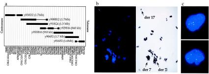
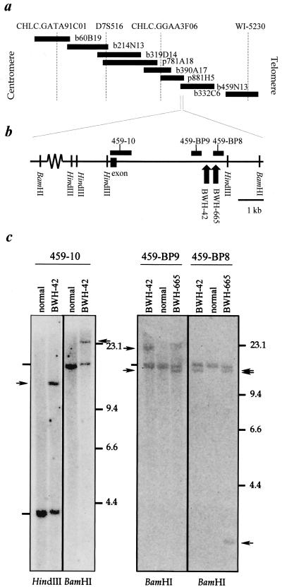
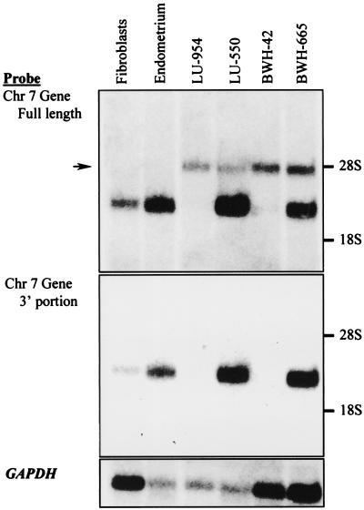
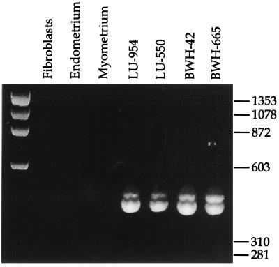
Endometrial stromal tumors are divided into three types: benign stromal nodules, endometrial stromal sarcomas, and undifferentiated endometrial sarcomas. A variety of cytogenetic abnormalities involving chromosome 7 have been reported in endometrial stromal sarcomas, including a recurrent t(7;17)(p15;q21). We have identified two zinc finger genes, which we have termed JAZF1 and JJAZ1, at the sites of the 7p15 and 17q21 breakpoints. Analyses of tumor RNA indicate that a JAZF1/JJAZ1 fusion is present in all types of endometrial stromal tumors; however, the fusion appears to be rarer among endometrial stromal sarcomas that would be considered high-grade according to certain classification schemes. These findings suggest that the less malignant endometrial stromal tumors may evolve toward more malignant types, but that some endometrial stromal sarcomas with relatively abundant mitotic activity may compose a biologically distinct group.


Figures

Figure 1
Figure 1
Analysis of YACs in the vicinity of the 7p15 breakpoint. (a) Map of YAC clones at the 7p15 site. STSs are shown at the top without reference to the genetic distances between them. Solid, boxed areas indicate the presence of the STS within the DNA of the YAC; horizontal lines indicate the absence of the STS, presumably because of a deletion in the YAC insert. (b) FISH analysis of tumor BWH-42 with YAC probes derived from chromosome 7. Chromosomes and cells were hybridized with biotin-labeled DNA, and hybridization was detected with avidin-conjugated FITC. (Left) FISH was performed with YAC y908B12 and the chromosomal DNA counterstained with 4′,6-diamidino-2-phenylindole (DAPI). The hybridization signal is split between two derivative 7 chromosomes–that of the t(7;17) to the left and of the t(7;13) to the right–and one derivative 17 chromosome, as identified in the contrast-enhanced image of the DAPI stain on the right. (c) FISH performed with the y908B12 probe on interphase nuclei isolated from paraffin-embedded tumor in case BWH-665.
Figure 2
Figure 2
Localization of the 7p15 breakpoint to the centromeric end of BAC b459N13. (a) Map of the BAC and PAC clones in the centromeric region of YAC y908B12. STSs are shown at the top of the map at relative positions based on DNA sequence available for the region. (b) Position of Southern blot hybridization probes within DNA near the centromeric end of BAC b459N13. The approximate positions of chromosome 7p15 breakpoints in cases BWH-42 and -665, as determined in the Southern blots in c, are illustrated with upward arrows near the telomeric end of the region shown. (c) Southern blot analyses of DNA derived from the tumors in cases BWH-42 and -665. Normal DNA was extracted from fibroblasts of the patient in case BWH-42. The probes used for each blot are indicated above the autoradiograms; the restriction enzymes used are shown below. Arrowheads indicate the positions of rearranged restriction fragments; dashes the positions of the germline restriction fragments.
Figure 3
Figure 3
Northern blot analyses of poly(A)+ RNA from ESSs and normal control tissues. The source of normal fibroblasts was the same as that in Fig. 2, whereas normal endometrium was obtained from an unrelated patient. (a) Analysis performed with a probe for the 5′ end of EST AA431106 (nucleotides 74 to 1561 of JAZF1 in Fig. 5a). The arrow indicates the position of a band corresponding to a ≈4.5-kb transcript seen only in tumor samples. (b) Analysis performed with a probe for the 3′ end of EST AA431106 (nucleotides 1120 to 2883 of JAZF1 in Fig. 5a). (c) Analysis with a probe for the GAPDH gene used as a control for the quality and amount of RNA on the blot.
Figure 4
Figure 4
RT-PCR analysis of the RNA sequence transcribed across the site of recombination in the t(7;17)(p15;q21). Ethidium bromide-stained agarose gel of RT-PCR products amplified from total RNA of normal tissues and the four low-grade ESSs known to carry the translocation. The source of fibroblasts and normal endometrium was the same as that in Fig. 3; the normal myometrium was obtained from the same specimen as the endometrium.

References

    1. Zaloudek C, Norris H J. In: Blaustein's Pathology of the Female Genital Tract. Kurman R J, editor. New York: Springer; 1994. pp. 487–528.
    1. Norris H J, Taylor H B. Cancer. 1966;19:755–766. - PubMed
    1. Tavassoli F A, Norris H J. Histopathology. 1981;5:1–10. - PubMed
    1. Evans H L. Cancer. 1982;50:2170–2182. - PubMed
    1. Chang K L, Crabtree G S, Lim-Tan S K, Kempson R L, Hendrickson M R. Am J Surg Pathol. 1990;14:415–438. - PubMed

Publication types

MeSH terms

LinkOut - more resources