. 2001 May;107(10):1235-44.
doi: 10.1172/JCI11685.

Osteoprotegerin inhibits prostate cancer-induced osteoclastogenesis and prevents prostate tumor growth in the bone

Affiliations

Osteoprotegerin inhibits prostate cancer-induced osteoclastogenesis and prevents prostate tumor growth in the bone

J Zhang et al. J Clin Invest. 2001 May.

Abstract

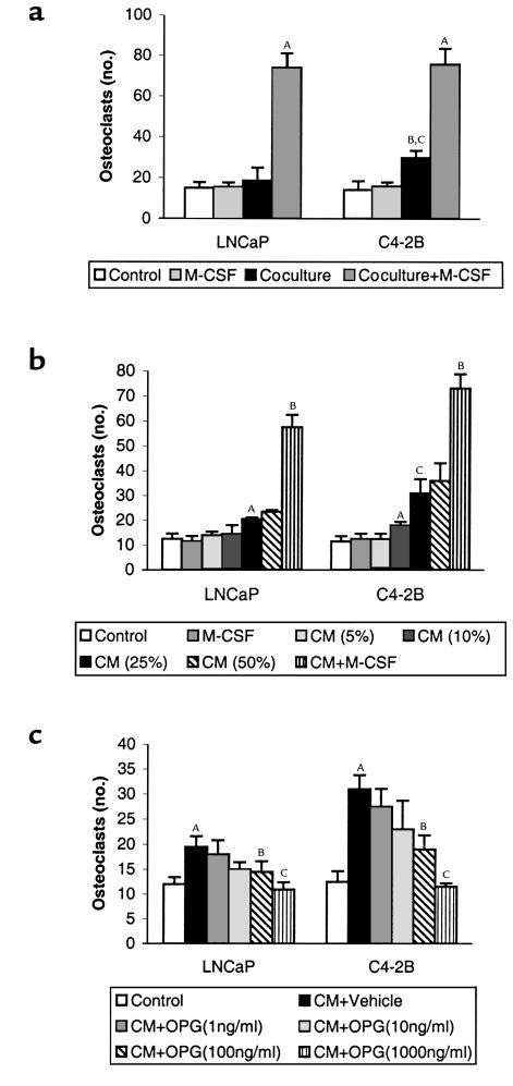
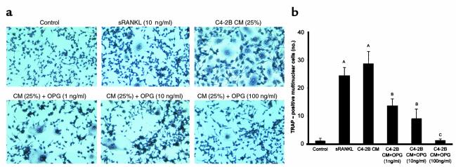
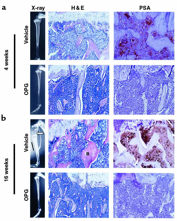
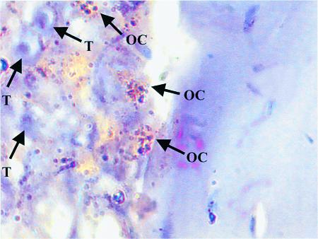
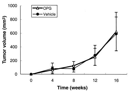
Prostate cancer (CaP) forms osteoblastic skeletal metastases with an underlying osteoclastic component. However, the importance of osteoclastogenesis in the development of CaP skeletal lesions is unknown. In the present study, we demonstrate that CaP cells directly induce osteoclastogenesis from osteoclast precursors in the absence of underlying stroma in vitro. CaP cells produced a soluble form of receptor activator of NF-kappaB ligand (RANKL), which accounted for the CaP-mediated osteoclastogenesis. To evaluate for the importance of osteoclastogenesis on CaP tumor development in vivo, CaP cells were injected both intratibially and subcutaneously in the same mice, followed by administration of the decoy receptor for RANKL, osteoprotegerin (OPG). OPG completely prevented the establishment of mixed osteolytic/osteoblastic tibial tumors, as were observed in vehicle-treated animals, but it had no effect on subcutaneous tumor growth. Consistent with the role of osteoclasts in tumor development, osteoclast numbers were elevated at the bone/tumor interface in the vehicle-treated mice compared with the normal values in the OPG-treated mice. Furthermore, OPG had no effect on CaP cell viability, proliferation, or basal apoptotic rate in vitro. These results emphasize the important role that osteoclast activity plays in the establishment of CaP skeletal metastases, including those with an osteoblastic component.


Figures

Figure 1
Figure 1
OPG inhibits LNCaP and C4-2B cell–induced osteoclastogenesis of osteoblast/stromal cells in vitro. (a) LNCaP or C4-2B cells were directly cocultured with murine bone marrow cells for 9 days in the presence or absence of M-CSF (1 ng/ml). Osteoclast-like cells were identified as TRAP-positive multinucleated (>3 nuclei) cells. AP < 0.001 compared with its respective control culture (without adding CaP cells) or coculture; BP < 0.01 compared with its respective control culture; CP < 0.01 compared with its LNCaP cells. (b) Conditioned media (CM) from LNCaP and C4-2B cells was collected after 24 hours of culture, then the indicated concentrations of CM (vol/vol) was added to murine bone marrow cells and cultured for 9 days. Osteoclast-like cells were identified as TRAP-positive multinucleated (>3 nuclei) cells. AP < 0.001 compared with respective control culture (without adding CM); BP < 0.001 compared with each cell line’s respective control culture or coculture; CP < 0.01 compared with its LNCaP cells. (c) CM (25% vol/vol) from LNCaP and C4-2B cells were collected after 24 hours of culture, then added to murine bone marrow cells with different dose of recombinant mouse OPG (1–1000 ng/ml) as indicated and cultured for 9 days. Osteoclast-like cells were identified as TRAP-positive multinucleated (>3 nuclei) cells. AP < 0.001 compared with its control culture; BP < 0.01 compared with its respective vehicle-treated CM cultures; CP < 0.001 compared with its respective vehicle-treated CM cultures. All in vitro cultures were evaluated in quadruplicate. Results were reported as the mean (± SD) number of osteoclast-like cells per coverslip. Data were analyzed using ANOVA and Fisher’s least-significant difference for post hoc analysis.
Figure 2
Figure 2
C4-2B CM induces osteoclastogenesis in the absence of osteoblast/stromal cells, and OPG inhibits the osteoclastogenesis in vitro. Single-cell suspensions (105 cells/well) of RAW 264.7 cells were plated in a 24-well plate on top of a sterile coverslip in RPMI plus 10% FBS. Cells were grown for 12 hours, then the media was changed to RPMI plus 0.5% FBS. CM from C4-2B cells was harvested (as described in Methods) and added to a final concentration of 25% (vol/vol). Immediately, recombinant human soluble RANKL (10 ng/ml) or the indicated concentration of recombinant mouse OPG or vehicle (1% BSA in PBS) was added. Osteoclasts were identified as TRAP-positive multinucleated (>3 nuclei) cells. (a) Representative pictures of cultures stained for TRAP. (b) Osteoclasts per coverslip were quantified. Samples were evaluated in quadruplicate. Results are reported as mean (±SD). Data were analyzed using one-way ANOVA. AP < 0.001 compared with control culture; BP < 0.01 compared with the CM-treated group; CP < 0.001 compared with the CM-treated group.
Figure 3
Figure 3
LNCaP and C4-2B cells express RANKL and produce soluble RANKL. (a) One microgram of total RNA from the indicated cells was subjected to RT-PCR. Lanes 1, 2, and 3 are PCR products from LNCaP, C4-2B, and SaOS, respectively. (b) Total cellular protein or CM (concentrated 100-fold using Microcon centrifugal filter devices) from LNCaP, C4-2B, and SaOS cell cultures were subjected to Western blot analysis (50 μg/lane) using rabbit anti-human soluble RANKL polyclonal Ab as primary Ab and HRP-conjugated anti-rabbit IgG as secondary Ab. Bands were detected using luminescence and autoradiography. Lane 1, LNCaP cell lysate; lane 2, C4-2B cell lysate; lane 3, SaOS cell lysate; lane 4, LNCaP concentrated CM; lane 5, C4-2B concentrated CM; and lane 6, SaOS concentrated CM.
Figure 4
Figure 4
Characteristics of C4-2B bone lesions. SCID mice were injected intratibially with C4-2B prostate cancer cells. At the time of tumor injection, OPG (2 mg/kg) or vehicle (1% BSA in 1× PBS) was administered via the tail vein twice a week for 4 weeks. The mice were sacrificed at 4 weeks and 16 weeks after-tumor injection. Formalin-fixed paraffin-embedded sections were stained with hematoxylin and eosin (H&E) or were deparaffinized, rehydrated, and stained for PSA using immunohistochemistry. Brown coloration indicates presence of PSA. ×200. (a) Representative radiographs of H&E- and PSA-stained sections of vehicle-treated versus OPG-treated mice at the end of 4 weeks. Note replacement of bone marrow by tumor in the vehicle-treated animals compared with normal marrow in OPG-treated animals. PSA staining cannot be identified in the OPG-treated animals. (b) Representative radiographs of H&E- and PSA-stained sections of vehicle-treated versus OPG-treated mice at the end of 16 weeks. Note the area of osteolysis (arrowhead) and osteoblastic lesion (bar) in the radiograph of the vehicle-treated mouse compared with the normal radiograph of the OPG-treated mice. Also, note the replacement of bone marrow by tumor and the thickened trabeculum indicated with letter B in the vehicle-treated mouse compared with the OPG-treated mouse.
Figure 5
Figure 5
C4-2B cells promote osteoclast activity at bone/tumor interface. SCID mice were injected intratibially with C4-2B prostate cancer cells. Tibias were harvested 16 weeks after tumor injection, decalcified, sectioned, and stained for TRAP. A section is shown that demonstrates multiple TRAP-positive staining osteoclasts at the bone/tumor interface. T, tumor cell; OC, multinucleated TRAP-positive osteoclast. ×1000 under oil.
Figure 6
Figure 6
OPG does not affect subcutaneous tumor growth in mice. C4-2B prostate cancer cells were injected subcutaneously into the SCID mice at the same time they received intratibial tumor injection as described in Figure 4. At the time of tumor injection, OPG (2 mg/kg) or vehicle (1% BSA in 1× PBS) was administered via the tail vein twice a week for 4 weeks. Tumors were allowed to grow for another 12 weeks. The mice were sacrificed at 16 weeks after tumor injection. Tumor volume was measured monthly. Data are reported as mean (±SD) from nine to ten animals per group.

Comment in

  • Osteolysis and cancer.
    Goltzman D. Goltzman D. J Clin Invest. 2001 May;107(10):1219-20. doi: 10.1172/JCI13073. J Clin Invest. 2001. PMID: 11375409 Free PMC article. No abstract available.

Similar articles

Cited by

References

    1. Abrams H, Spiro R, Goldstein N. Metastases in carcinoma. Cancer. 1950;3:74–85. - PubMed
    1. Charhon SA, et al. Histomorphometric analysis of sclerotic bone metastases from prostatic carcinoma special reference to osteomalacia. Cancer. 1983;51:918–924. - PubMed
    1. Urwin GH, et al. Generalised increase in bone resorption in carcinoma of the prostate. Br J Urol. 1985;57:721–723. - PubMed
    1. Clarke NW, McClure J, George NJ. Disodium pamidronate identifies differential osteoclastic bone resorption in metastatic prostate cancer. Br J Urol. 1992;69:64–70. - PubMed
    1. Clarke N. The effects of pamidronate disodium treatment in metastatic prostate cancer. Rev Contemp Pharamcother. 1998;9:205–212.

Publication types

MeSH terms