. 2001 May;2(5):438-45.
doi: 10.1093/embo-reports/kve093.

Rescue of the early vascular defects in Tek/Tie2 null mice reveals an essential survival function

Affiliations

Rescue of the early vascular defects in Tek/Tie2 null mice reveals an essential survival function

N Jones et al. EMBO Rep. 2001 May.

Abstract

Disruption of the signaling pathways mediated by the receptor tyrosine kinase Tek/Tie2 has shown that this receptor plays a pivotal role in vascularization of the developing embryo. In this report, we have utilized the tetracycline-responsive binary transgenic system to overcome the early lethal cardiovascular defects associated with the tekDeltasp null allele in order to investigate the role of Tek in later stages of vessel growth. We show for the first time in vivo that synchronized loss of tek expression correlates with rapid endothelial cell apoptosis in hemorrhagic regions of the embryo, demonstrating an ongoing requirement for Tek-mediated signal transduction in vascular maintenance.


Figures
None
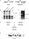
Fig. 1. The dox-repressible binary transgenic system. (A) The driver transgene contains the tek promoter driving the expression of the dox responsive transactivator, tTA. The responder transgene contains the tTA binding site (tetos) upstream of the tek cDNA. The regions corresponding to the RNase protection probe and the RT–PCR oligos are depicted. (B) RNase protection assay from total RNA prepared from E8.5 embryos harvested from matings between hemizygous driver and responder transgenic lines which are wildtype for the tek locus. The 300 bp transgene-specific message and the 200 bp endogenous tek transcript are indicated (tek). (C) RT–PCR analysis on untreated E8.5 embryos or E9.5 embryos that have been treated with dox for 1 day (8.5–9.5) using oligos specific for the transgene or for β-actin demonstrates the presence of a 140 bp transgene-specific fragment in untreated embryos alone. (D) Schematic of the genotypes of the mice used in these studies. Mice heterozygous for the tekΔsp allele and homozygous for either of the transgenes were crossed. The genotype of the desired double transgenic tekΔsp-rescued offspring is indicated. (E) Southern blot analysis of DNA extracted from E12.5 embryos obtained from the mating outlined above. The presence of the mutant and wildtype alleles is indicated. An asterisk above the lane indicates the presence of a homozygous-tekΔsp embryo. All embryos were hemizygous for each transgene as determined by PCR (data not shown).
None
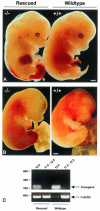
Fig. 2. E13.5 and E14.5 rescued embryos present with signs of vascular hemorrhage and anemia. (A) Homozygous tekΔsp double transgenic (rescued) embryos (–/–) obtained on E13.5 are anemic and the livers are reduced in size when compared to double transgenic wildtype (+/+) littermates. Extensive hemorrhaging in the tail and lower limbs of rescued embryos can also be observed. (B) Similar defects are found in rescued embryos obtained on E14.5. (C) RT–PCR analysis on untreated (12.5) or dox-treated (11.5–12.5) E12.5 wildtype or rescued embryos with oligos specific for the transgene or for β-actin demonstrates repression of tek-responder transgene expression with overnight dox treatment. Scale bars: (A), 450 µm; (B), 625 µm.
None
Fig. 3. Cardiac development in double transgenic rescued and wildtype embryos. H & E stained sections of the heart region of E13.5 embryos indicate extensive ventricular trabeculation in rescued embryos (A) comparable to that seen in wildtype littermates (B). Heart sections prepared from an independent litter stained with hematoxylin alone demonstrate that the general architecture of the heart in rescued embryos (C) is indistinguishable from normal littermates (D). Arrowheads indicate loose endothelium in both rescued and wildtype embryos. Scale bars: (A–D), 600 µm
None
Fig. 4. Histological analysis of tekΔsp rescued embryos. Thin sections prepared from rescued E13.5 embryos or yolk sacs were stained for the expression of vWF or SMA. The yolk sac vasculature of E13.5 rescued embryos had sparse EC lining (A) when compared with wildtype littermates (C), although both yolk sacs were positive for SMA (B and D). Similarly, a number of vessels in the embryo proper of double transgenic tekΔsp-null mice show intact endothelium surrounded by SMCs (E and F) while others display reduced EC lining despite retaining many SMCs (GL). Arrowheads indicate vWF-positive ECs. Scale bars: (A–D), 1.7 µm; (E–L), 15 µm.
None
Fig. 5. Extensive programmed cell death in hemorrhagic regions of rescued embryos. Sagittal sections through the lower part of the neural tube taken from either normal (B) or rescued (A) E12.5 littermates which were treated for a single day with dox illustrate the differences in the integrity of the leptomeninges. H & E staining reveals that in the normal embryo, the leptomeninges (L) are strongly adhered to the neural tube (NT) whereas in the rescued littermate this region is not well adhered and extravasated blood (Bl) can be observed. Near adjacent sections to those presented in (A) and (B) were analyzed for the presence of apoptotic cells by the TUNEL procedure. Very few TUNEL-positive cells were detected in wildtype embryos (D) whereas numerous apoptotic cells were identified in sections from rescued embryos (C). Scale bars: (A–D), 21 µm.
None
Fig. 6. EC-specific apoptosis in cells that do not express Tek. vWF or Tek (both red) were used to perform double staining with TUNEL (green) on near adjacent cryosections of E10.5, E12.5 or dox-treated E12.5 wildtype and rescued embryos. TUNEL-positive ECs were not observed in E10.5 embryos (AD) and the image in (G) represents an isolated case of overlap between vWF and TUNEL staining in E12.5 embryos (EH). In contrast, numerous TUNEL-positive cells could be observed in blood vessels of dox-treated E12.5 rescued embryos and these cells stained positive for vWF (I) but not Tek (K). In wildtype littermates, few TUNEL-positive cells could be detected although vWF (J) and Tek (L)-expressing cells could be readily observed lining blood vessels. Arrowheads indicate double-positive (yellow) cells. Scale bars: 20 µm.

References

    1. Davis S. et al. (1996) Isolation of angiopoietin-1, a ligand for the TIE2 receptor, by secretion-trap expression cloning. Cell, 87, 1161–1169. - PubMed
    1. Dumont D.J., Yamaguchi, T.P., Conlon, R.A., Rossant, J. and Breitman, M.L. (1992) tek, a novel tyrosine kinase gene located on mouse chromosome 4, is expressed in endothelial cells and their presumptive precursors. Oncogene, 7, 1471–1480. - PubMed
    1. Dumont D.J., Gradwohl, G., Fong, G.H., Puri, M.C., Gertsenstein, M., Auerbach, A. and Breitman, M.L. (1994) Dominant-negative and targeted null mutations in the endothelial receptor tyrosine kinase, tek, reveal a critical role in vasculogenesis of the embryo. Genes Dev., 8, 1897–1909. - PubMed
    1. Dumont D.J., Fong, G.H., Puri, M.C., Gradwohl, G., Alitalo, K. and Breitman, M.L. (1995) Vascularization of the mouse embryo: a study of flk-1, tek, tie, and vascular endothelial growth factor expression during development. Dev. Dyn., 203, 80–92. - PubMed
    1. Folkman J. and D’Amore, P.A. (1996) Blood vessel formation: what is its molecular basis? Cell, 87, 1153–1155. - PubMed

Publication types

MeSH terms