

Utility of inoculum counting (Walshe and English criteria) in clinical diagnosis of onychomycosis caused by nondermatophytic filamentous fungi
- PMID: 11376044
- PMCID: PMC88098
- DOI: 10.1128/JCM.39.6.2115-2121.2001
Utility of inoculum counting (Walshe and English criteria) in clinical diagnosis of onychomycosis caused by nondermatophytic filamentous fungi
Abstract
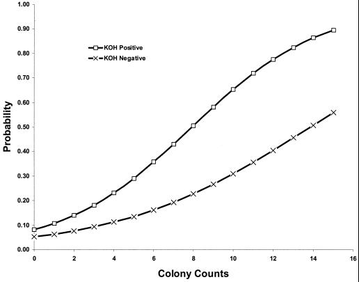
Opportunistic onychomycosis caused by nondermatophytic molds may differ in treatment from tinea unguium. Confirmed diagnosis of opportunistic onychomycosis classically requires more than one laboratory analysis to show consistency of fungal outgrowth. Walshe and English in 1966 proposed to extract sufficient diagnostic information from a single patient consultation by counting the number of nail fragments positive for inoculum of the suspected fungus. Twenty fragments were plated per patient, and each case in which five or more fragments grew the same mold was considered an infection by that mold, provided that compatible filaments were also seen invading the nail tissue by direct microscopy. This widely used and often recommended method has never been validated. Therefore, the validity of substituting any technique based on inoculum counting for conventional follow-up study in the diagnosis of opportunistic onychomycosis was investigated. Sampling of 473 patients was performed repeatedly. Nail specimens were examined by direct microscopy, and 15 pieces were plated on standard growth media. After 3 weeks, outgrowing dermatophytes were recorded, and pieces growing any nondermatophyte mold were counted. Patients returned on two to eight additional occasions over a 1- to 3-year period for similar examinations. Onychomycosis was etiologically classified based on long-term study. Opportunistic onychomycosis was definitively established for 86 patients. Counts of nondermatophyte molds in initial examinations were analyzed to determine if they successfully predicted both true cases of opportunistic onychomycosis and cases of insignificant mold contamination. There was a strong positive statistical association between mold colony counts and true opportunistic onychomycosis. Logistic regression analysis, however, determined that even the highest counts predicted true cases of opportunistic onychomycosis only 89.7% of the time. The counting criterion suggested by Walshe and English was correct only 23.2% of the time. Acremonium infections were especially likely to be correctly predicted by inoculum counting. Inoculum counting could be used to indicate a need for repeat studies in cases of false-negative results from laboratory direct microscopy. Inoculum counting cannot serve as a valid substitute for follow-up study in the diagnosis of opportunistic onychomycosis. It may, nonetheless, provide useful information both to the physician and to the laboratory, and it may be especially valuable when the patient does not present for follow-up sampling.
Figures
Similar articles
-
Onychomycosis caused by nondermatophytic molds: clinical features and response to treatment of 59 cases.J Am Acad Dermatol. 2000 Feb;42(2 Pt 1):217-24. doi: 10.1016/S0190-9622(00)90129-4. J Am Acad Dermatol. 2000. PMID: 10642676
-
Onychomycosis: a critical study of techniques and criteria for confirming the etiologic significance of nondermatophytes.Med Mycol. 2005 Feb;43(1):39-59. doi: 10.1080/13693780410001712043. Med Mycol. 2005. PMID: 15712607
-
Mycology of nail disorders.J Am Acad Dermatol. 1994 Sep;31(3 Pt 2):S68-74. doi: 10.1016/s0190-9622(08)81272-8. J Am Acad Dermatol. 1994. PMID: 8077512 Review.
-
Other fungi causing onychomycosis.Clin Dermatol. 2010 Mar 4;28(2):160-3. doi: 10.1016/j.clindermatol.2009.12.009. Clin Dermatol. 2010. PMID: 20347658 Review.
-
Non-dermatophytic molds as agents of onychomycosis in Izmir, Turkey - a prospective study.Mycopathologia. 2005 Sep;160(2):125-8. doi: 10.1007/s11046-005-6872-z. Mycopathologia. 2005. PMID: 16170607
Cited by
-
Prevalence, Etiology, and Risk Factors of Tinea Pedis and Tinea Unguium in Tunisia.Can J Infect Dis Med Microbiol. 2017;2017:6835725. doi: 10.1155/2017/6835725. Epub 2017 Aug 9. Can J Infect Dis Med Microbiol. 2017. PMID: 28852411 Free PMC article.
-
Identification of infectious agents in onychomycoses by PCR-terminal restriction fragment length polymorphism.J Clin Microbiol. 2012 Mar;50(3):553-61. doi: 10.1128/JCM.05164-11. Epub 2011 Dec 14. J Clin Microbiol. 2012. PMID: 22170903 Free PMC article.
-
Epidemiology of onychomycosis and paronychia in the area of ANCONA (ITALY) over a period of 5 years.Mycopathologia. 2004 Oct;158(3):271-4. doi: 10.1007/s11046-004-9615-7. Mycopathologia. 2004. PMID: 15645167
-
Molecular identification and susceptibility of Trichosporon species isolated from clinical specimens in Qatar: isolation of Trichosporon dohaense Taj-Aldeen, Meis & Boekhout sp. nov.J Clin Microbiol. 2009 Jun;47(6):1791-9. doi: 10.1128/JCM.02222-08. Epub 2009 Mar 25. J Clin Microbiol. 2009. PMID: 19321719 Free PMC article.
-
High Diversity of Fusarium Species in Onychomycosis: Clinical Presentations, Molecular Identification, and Antifungal Susceptibility.J Fungi (Basel). 2023 Apr 30;9(5):534. doi: 10.3390/jof9050534. J Fungi (Basel). 2023. PMID: 37233245 Free PMC article.
References
-
- Achten G, Wanet-Rouard J. Onychomycoses in the laboratory. Mykosen. 1978;23(Suppl. 1):125–127. - PubMed
-
- Arrese J E, Piérard-Franchimont C, Greimers R, Piérard G E. Fungi in onychomycosis. A study by immunohistochemistry and dual flow cytometry. J Eur Acad Dermatol Venereol. 1995;4:123–130.
-
- Clayton Y M. Clinical and mycological diagnostic aspects of onychomycoses and dermatomycoses. Clin Exp Dermatol. 1992;17:37–40. - PubMed
-
- Clayton Y M. Relevance of broad-spectrum and fungicidal activity of antifungals in the treatment of dermatomycoses. Br J Dermatol. 1994;130(Suppl. 43):7–8. - PubMed
MeSH terms
LinkOut - more resources
Full Text Sources
