

Human babesiosis in Japan: isolation of Babesia microti-like parasites from an asymptomatic transfusion donor and from a rodent from an area where babesiosis is endemic
- PMID: 11376054
- PMCID: PMC88108
- DOI: 10.1128/JCM.39.6.2178-2183.2001
Human babesiosis in Japan: isolation of Babesia microti-like parasites from an asymptomatic transfusion donor and from a rodent from an area where babesiosis is endemic
Abstract
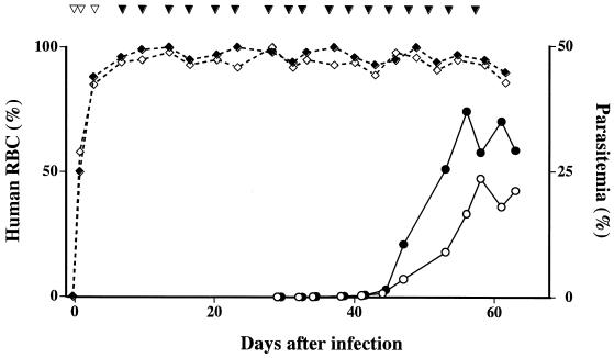
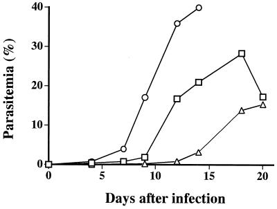
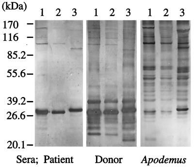
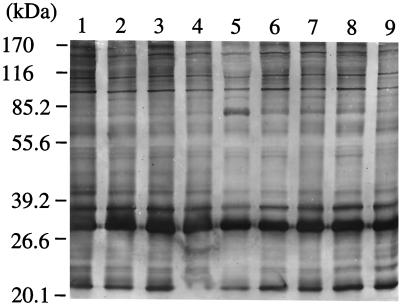
To determine the source of infection for the Japanese index case of human babesiosis, we analyzed blood samples from an asymptomatic individual whose blood had been transfused into the patient. In addition, we surveyed rodents collected from near the donor's residence. Examination by microscopy and PCR failed to detect the parasite in the donor's blood obtained 8 months after the donation of the blood that was transfused. However, we were able to isolate Babesia parasites by inoculating the blood sample into SCID mice whose circulating red blood cells (RBCs) had been replaced with human RBCs. A Babesia parasite capable of propagating in human RBCs was also isolated from a field mouse (Apodemus speciosus) captured near the donor's residential area. Follow-up surveys over a 1-year period revealed that the donor continued to be asymptomatic but had consistently high immunoglobulin G (IgG) titers in serum and low levels of parasitemia which were microscopically undetectable yet which were repeatedly demonstrable by inoculation into animals. The index case patient's sera contained high titers of IgM and, subsequently, rising titers of IgG antibodies, both of which gradually diminished with the disappearance of the parasitemia. Analysis of the parasite's rRNA gene (rDNA) sequence and immunodominant antigens revealed the similarity between donor and patient isolates. The rodent isolate also had an rDNA sequence that was identical to that of the human isolates but that differed slightly from that of the human isolates by Western blot analysis. We conclude that the index case patient acquired infection by transfusion from a donor who became infected in Japan, that parasitemia in an asymptomatic carrier can persist for more than a year, and that A. speciosus serves as a reservoir of an agent of human babesiosis in Japan.
Figures
Similar articles
-
Human babesiosis in Japan: epizootiologic survey of rodent reservoir and isolation of new type of Babesia microti-like parasite.J Clin Microbiol. 2001 Dec;39(12):4316-22. doi: 10.1128/JCM.39.12.4316-4322.2001. J Clin Microbiol. 2001. PMID: 11724838 Free PMC article.
-
Transfusion-acquired, autochthonous human babesiosis in Japan: isolation of Babesia microti-like parasites with hu-RBC-SCID mice.J Clin Microbiol. 2000 Dec;38(12):4511-6. doi: 10.1128/JCM.38.12.4511-4516.2000. J Clin Microbiol. 2000. PMID: 11101588 Free PMC article.
-
Demonstrable parasitemia among Connecticut blood donors with antibodies to Babesia microti.Transfusion. 2005 Nov;45(11):1804-10. doi: 10.1111/j.1537-2995.2005.00609.x. Transfusion. 2005. PMID: 16271108
-
[Babesosis--difficulty of diagnosis].Wiad Parazytol. 2001;47(3):527-33. Wiad Parazytol. 2001. PMID: 16894770 Review. Polish.
-
Investigation of transfusion transmission of a WA1-type babesial parasite to a premature infant in California.Transfusion. 2002 Nov;42(11):1482-7. doi: 10.1046/j.1537-2995.2002.00245.x. Transfusion. 2002. PMID: 12421222 Review.
Cited by
-
Human babesiosis: recent advances and future challenges.Curr Opin Hematol. 2020 Nov;27(6):399-405. doi: 10.1097/MOH.0000000000000606. Curr Opin Hematol. 2020. PMID: 32889826 Free PMC article. Review.
-
First case of human babesiosis in Korea: detection and characterization of a novel type of Babesia sp. (KO1) similar to ovine babesia.J Clin Microbiol. 2007 Jun;45(6):2084-7. doi: 10.1128/JCM.01334-06. Epub 2007 Mar 28. J Clin Microbiol. 2007. PMID: 17392446 Free PMC article.
-
Piroplasmosis in wildlife: Babesia and Theileria affecting free-ranging ungulates and carnivores in the Italian Alps.Parasit Vectors. 2014 Feb 17;7:70. doi: 10.1186/1756-3305-7-70. Parasit Vectors. 2014. PMID: 24533742 Free PMC article.
-
Preventing Transfusion-Transmitted Babesiosis.Pathogens. 2021 Sep 13;10(9):1176. doi: 10.3390/pathogens10091176. Pathogens. 2021. PMID: 34578209 Free PMC article. Review.
-
Cultivation of Babesia and Babesia-like blood parasites: agents of an emerging zoonotic disease.Clin Microbiol Rev. 2002 Jul;15(3):365-73. doi: 10.1128/CMR.15.3.365-373.2002. Clin Microbiol Rev. 2002. PMID: 12097245 Free PMC article. Review.
References
-
- Arai S, Tsuji M, Kim S-J, Nakada K, Kirisawa R, Ohta M, Ishihara C. Antigenic and genetic diversities of Babesia ovata in persistently infected cattle. J Vet Med Sci. 1998;60:1321–1327. - PubMed
-
- Brandt F, Healy G R, Welch M. Human babesiosis: the isolation of Babesia microti in golden hamsters. J Parasitol. 1977;63:934–937. - PubMed
-
- Fay F H, Rausch R L. Parasitic organisms in the blood of arvicoline rodents in Alaska. J Parasitol. 1969;55:1258–1265.
-
- Gorenflot A, Moubri K, Precigout E, Carcy B, Schetters T P M. Human babesiosis. Ann Trop Med Parasitol. 1998;92:489–501. - PubMed
Publication types
MeSH terms
Substances
LinkOut - more resources
Full Text Sources
Medical
Molecular Biology Databases
