

Phaeohyphomycotic cyst caused by Colletotrichum crassipes
- PMID: 11376082
- PMCID: PMC88136
- DOI: 10.1128/JCM.39.6.2321-2324.2001
Phaeohyphomycotic cyst caused by Colletotrichum crassipes
Abstract
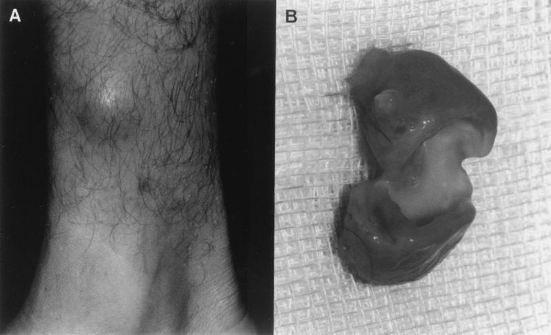
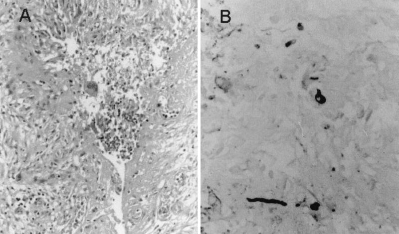
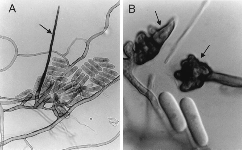
A case of phaeohyphomycosis is reported in a male renal transplant recipient with a nodular lesion in the right leg who was treated with immunosuppressing drugs. The lesion consisted of a purulent cyst with thick walls. The cyst was excised surgically, and the patient did not receive any antifungal therapy. One year later he remains well. Histological study of the lesion showed a granulomatous reaction of epithelioid and multinucleate giant cells, with a central area of necrosis and pus. Fontana-Masson staining demonstrated the presence of pigmented hyphal elements. The fungus Colletotrichum crassipes was grown in different cultures from the cyst. The in vitro inhibitory activities of eight antifungal drugs against the isolate were tested. Clotrimazole and UR-9825 were the most active drugs. This case represents the first known reported infection caused by this rare species.
Figures
Similar articles
-
[Phaeohyphomycosis caused by Colletotrichum gloeosporioides and Alternaría infectoria in renal transplant recipient].Rev Chilena Infectol. 2014 Aug;31(4):468-72. doi: 10.4067/S0716-10182014000400014. Rev Chilena Infectol. 2014. PMID: 25327202 Spanish.
-
Subcutaneous phaeohyphomycotic cyst caused by Pyrenochaeta romeroi.Med Mycol. 2010 Aug;48(5):763-8. doi: 10.3109/13693780903440383. Med Mycol. 2010. PMID: 20648971
-
Subcutaneous phaeohyphomycotic cysts caused by Exophiala jeanselmei in a lung transplant patient.Dermatol Surg. 2001 Apr;27(4):343-6. doi: 10.1046/j.1524-4725.2001.00308.x. Dermatol Surg. 2001. PMID: 11298703
-
[Phaeomycotic cyst caused by Phaeoacremonium parasiticum].Nihon Ishinkin Gakkai Zasshi. 2000;41(2):89-95. doi: 10.3314/jjmm.41.89. Nihon Ishinkin Gakkai Zasshi. 2000. PMID: 10777819 Review. Japanese.
-
Phaeohyphomycosis caused by Coniothyrium.Cutis. 2004 Feb;73(2):127-30. Cutis. 2004. PMID: 15027518 Review.
Cited by
-
Colletotrichum gloeosporioides sensu lato causing deep soft tissue mycosis following a penetrating injury.Med Mycol Case Rep. 2013 Feb 9;2:40-3. doi: 10.1016/j.mmcr.2013.01.003. eCollection 2013 Feb 9. Med Mycol Case Rep. 2013. PMID: 24432213 Free PMC article.
-
Angioinvasive, cutaneous infection due to Colletotrichum siamense in a stem cell transplant recipient: Report and review of prior cases.Transpl Infect Dis. 2019 Oct;21(5):e13153. doi: 10.1111/tid.13153. Epub 2019 Aug 13. Transpl Infect Dis. 2019. PMID: 31357231 Free PMC article. Review.
-
Non-traumatic keratitis due to Colletotrichum truncatum.JMM Case Rep. 2016 Aug 30;3(4):e005047. doi: 10.1099/jmmcr.0.005047. eCollection 2016 Aug. JMM Case Rep. 2016. PMID: 28348770 Free PMC article.
-
Molecular and morphological identification of Colletotrichum species of clinical interest.J Clin Microbiol. 2004 Jun;42(6):2450-4. doi: 10.1128/JCM.42.6.2450-2454.2004. J Clin Microbiol. 2004. PMID: 15184418 Free PMC article.
-
Colletotrichum truncatum: an unusual pathogen causing mycotic keratitis and endophthalmitis.J Clin Microbiol. 2011 Aug;49(8):2894-8. doi: 10.1128/JCM.00151-11. Epub 2011 Jun 8. J Clin Microbiol. 2011. PMID: 21653772 Free PMC article.
References
-
- Ajello L. Hyalohyphomycosis and phaeohyphomycosis: two global disease entities of public health importance. Eur J Epidemiol. 1986;2:243–251. - PubMed
-
- Bailey J A, Jegger M J, editors. Colletotrichum: biology, pathology and control. Wallingford, U.K: CAB International; 1992.
-
- Baxter A P, van der Westhuizen G C A, Eicker A. Morphology and taxonomy of South African isolates of Colletotrichum. S Af J Bot. 1983;2:259–289.
-
- De Hoog G S, Guarro J, Gené J, Figueras M J. Atlas of clinical fungi. 2nd ed. Utrecht, The Netherlands: Centraalbureau voor Schimmelcultures; 2000.
Publication types
MeSH terms
Substances
LinkOut - more resources
Full Text Sources
