. 2001 Jun 1;29(11):2292-302.
doi: 10.1093/nar/29.11.2292.

Multiple features contribute to efficient constitutive splicing of an unusually large exon

Affiliations

Multiple features contribute to efficient constitutive splicing of an unusually large exon

S R Bruce et al. Nucleic Acids Res. .

Abstract

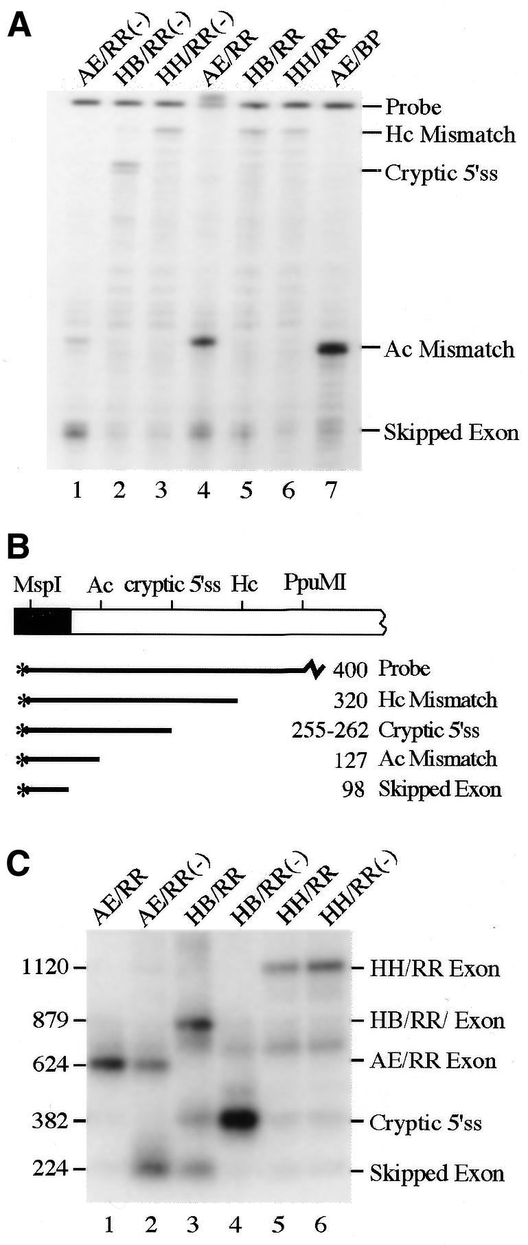
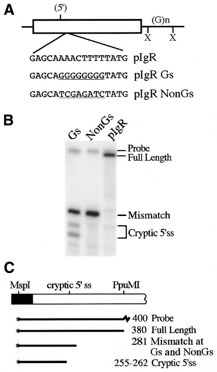
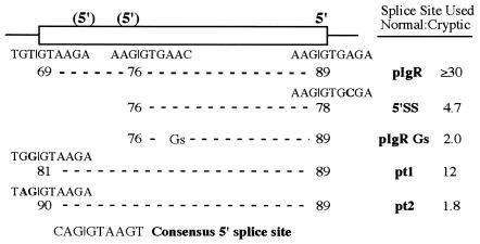
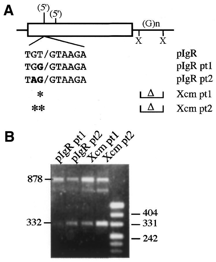
Vertebrate internal exons are usually between 50 and 400 nt long; exons outside this size range may require additional exonic and/or intronic sequences to be spliced into the mature mRNA. The mouse polymeric immunoglobulin receptor gene has a 654 nt exon that is efficiently spliced into the mRNA. We have examined this exon to identify features that contribute to its efficient splicing despite its large size; a large constitutive exon has not been studied previously. We found that a strong 5' splice site is necessary for this exon to be spliced intact, but the splice sites alone were not sufficient to efficiently splice a large exon. At least two exonic sequences and one evolutionarily conserved intronic sequence also contribute to recognition of this exon. However, these elements have redundant activities as they could only be detected in conjunction with other mutations that reduced splicing efficiency. Several mutations activated cryptic 5' splice sites that created smaller exons. Thus, the balance between use of these potential sites and the authentic 5' splice site must be modulated by sequences that repress or enhance use of these sites, respectively. Also, sequences that enhance cryptic splice site use must be absent from this large exon.


Figures

Figure 1
Figure 1
Chimeric Dd–pIgR construct. (A) The 654 bp pIgR exon 4 with 262 bp of upstream intron and 420 bp of downstream intron was placed into the KpnI (K) site of the Dd gene as an EcoRI (R)–HindIII (H) fragment. The open box represents the pIgR exon, the black boxes are Dd exons and thin lines are introns. (B) Several deletions between restriction sites within pIgR exon 4 and the downstream intron were constructed as shown. The 5′ splice site sequence at the end of exon 4 is shown; the A at the +4 position was mutated to C in the construct 5′SS. The lengths of the deletions are indicated; (5′) indicates the cryptic 5′ splice site identified in Figure 2; * indicates locations of 7–12 nt long purine-rich sequences; X indicates locations of 7–9 nt long A/C-rich sequences.
Figure 2
Figure 2
RT–PCR analysis of the Dd–pIgR construct and its derivatives. (A) RNA from HepG2 cells transiently transfected with the construct shown above each lane were analyzed by RT–PCR. M, marker lane; sizes, in bp, of some of the bands are shown. (B) Diagram of Dd–pIgR splicing reactions. The primers used in the RT–PCR analysis, Dd Ex3 and Dd Ex4, are indicated by the arrows. The 878 bp RT–PCR product is from RNA containing the complete pIgR exon 4, the 224 bp product is from RNA that has omitted the exon (see Dd control in lane 1) and any products migrating between 224 and 878 bp are from RNA that had spliced exon 4 using cryptic splice sites. RT–PCR products from complete exon 4 inclusion from the exon deletion constructs are 878 bp minus the size of the deletion. The cryptic splice reaction activated by the 5′SS mutation is shown on the diagram of the gene. The structure of the 382 bp RT–PCR product, as determined by DNA sequence analysis, is also shown.
Figure 3
Figure 3
S1 nuclease quantitation of Dd–pIgR spliced products. (A) S1 nuclease protection assays of RNA from HepG2 cells transfected with the constructs shown above each lane. Protected bands are identified on the right. (B) S1 nuclease reactions were quantitated on a phosphorimager and graphed as the ratio of full-length to cryptically spliced RNA. These numbers represent at least two independent transfections, each analyzed two or more times. (C) Diagram of the S1 nuclease probe. RNA that has spliced the full-length pIgR exon into Dd protects the probe to the PpuMI site and cryptically spliced RNA protects the probe to the cryptic 5′ splice site; the expected sizes of each product are indicated. Multiple protected bands are observed with the cryptic 5′ splice site in 5′SS and Xcm 5′SS RNA due to fortuitous partial homology between the probe and sequences in Dd exon 4 to which the cryptic splice site is joined; the fragments were combined for quantitation. There is a single cryptic splice band in Bsu36I since it is not spliced directly to Dd exon 4.
Figure 4
Figure 4
pIgR exon 4 replacement constructs. The EcoRI–HindIII pIgR fragment is shown with the restriction sites used to delete internal exon sequence: Ac, AccI; Hc, HincII; Bsu, Bsu36I; EcN, EcoNI. pIgR exon sequence was replaced with cDNA fragments from the constant region of the immunoglobulin M gene (Cµ). Shown at the bottom of the figure is the Cµ region of the gene and cDNA fragment cloned into pIgR; Cµ exons are filled boxes and the hatched box denotes the µs-specific portion of exon Cµ4. The restriction sites are: B, BamHI; R, RsaI; P, PstI. The size of each deletion and replacement fragment and the final chimeric exon are indicated; construct names are shown, with (–) indicating fragment insertion in the antisense orientation.
Figure 5
Figure 5
Expression from exon replacement constructs. (A) S1 nuclease protection assays of RNA from HepG2 cells transfected with the constructs shown above each lane. The protected fragments are identified on the right. (B) Diagram of the probe used in this assay. With the AE/RR and AE/BP constructs the S1 probe will only be protected to the AccI site, the site of fragment insertion; the HH/RR and HB/RR constructs will protect the probe to the HincII site. Skipped exon represents those RNAs in which the chimeric pIgR exon is excluded from the Dd gene. (C) Southern blot analysis of RT–PCR reactions from the RNA in (A) using the Dd exon primers (Fig. 2). The sizes in bp of the RT–PCR products are shown on the left; identities of the products are shown on the right. The blot was probed with a cDNA fragment that spans the Dd exon 3–pIgR splice junction.
Figure 6
Figure 6
A purine-rich sequence within pIgR intron 4 is conserved between the mouse and human genes. Purine-rich sequences just downstream from the 5′ splice junction in the mouse and human pIgR genes are underlined; additional sequence homology extends beyond this stretch of purines and is underlined with a dotted line. Upper case letters are the 3′-end of exon 4, lower case letters are intron sequence. The XcmI sites that were used in the Dd–Xcm construct are shown. The 144 nt of human sequence that is available is shown compared to the same sequence of the mouse intron. The sequence we obtained for the mouse intron matches the recently published sequence, GenBank accession no. AB001489 (30). The sequences shown here were submitted to GenBank, accession nos AF261083 (human) and AF261084 (mouse).
Figure 7
Figure 7
G-rich sequences activate the cryptic 5′ splice site. (A) Sequences beginning 27 nt downstream of the cryptic 5′ splice site were mutated to create a run of eight G residues or changed to a non-G-rich sequence, shown underlined below the wild-type sequence in this region. (B) S1 nuclease analysis of RNA from HepG2 cells transfected with the constructs in (A). Protected bands are identified on the right. (C) Diagram of the S1 nuclease probe and protected fragments; the intact spliced IgR Gs and pIgR NonGs exons protect the probe only to the site of these mutations.
Figure 8
Figure 8
Relative use of 5′ splice sites in pIgR constructs compared to their match to consensus scores. The frequency with which each nucleotide occurs at each position of a 5′ splice site has been determined and used to calculate a match to consensus score for individual 5′ splice sites (see for example ref. 31). The scores for the natural pIgR exon 4 5′ splice site, the upstream cryptic 5′ splice sites and the 5′SS, pt1 and pt2 mutations are shown below each splice site. The consensus 5′ splice site sequence is shown below. Also included is relative use of the 5′ splice sites in each construct, taken from the data in Figures 3 and 9 and Table 2.
Figure 9
Figure 9
Cryptic 5′ splice site enhancement mutations. (A) A potential cryptic 5′ splice site 50 nt upstream from the identified cryptic site was mutated at one or two nucleotides to more closely resemble the consensus 5′ splice site sequence; the mutations are indicated in bold. The point mutations were analyzed on their own or in combination with the intronic XcmI deletion. (B) RT–PCR analysis of the constructs shown in (A). Activation of the new cryptic splice site is indicated by the 332 bp RT–PCR product; full-length pIgR exon 4 inclusion is also seen as the 878 bp product. The ∼800 bp band likely represents a hybrid product between the full-length product and the cryptic 5′ splice site product as seen before (32).
Figure 10
Figure 10
Summary of sequences in and surrounding pIgR exon 4 shown to contribute to the intact splicing of this large exon. The regions identified by each experiment are denoted by the hatched ovals; the experiments are identified on the right. Symbols and restriction sites are as identified in Figures 1 and 4. * indicates locations of the 7–12 nt long purine-rich sequences and X indicates locations of 7–9 nt long A/C-rich sequences that could potentially contribute to exon recognition.

Similar articles

Cited by

References

    1. Hawkins J.D. (1988) A survey of intron and exon lengths. Nucleic Acids Res., 16, 9893–9908. - PMC - PubMed
    1. Berget S.M. (1995) Exon recognition in vertebrate splicing. J. Biol. Chem., 270, 2411–2414. - PubMed
    1. Cooper T.A. and Ordahl,C.P. (1989) Nucleotide substitutions within the cardiac troponin T alternative exon disrupt pre-mRNA alternative splicing. Nucleic Acids Res., 17, 7905–7921. - PMC - PubMed
    1. Dominski Z. and Kole,R. (1991) Selection of splice sites in pre-mRNAs with short internal exons. Mol. Cell. Biol., 11, 6075–6083. - PMC - PubMed
    1. Tian H. and Kole,R. (1995) Selection of novel exon recognition elements from a pool of random sequences. Mol. Cell. Biol., 15, 6291–6298. - PMC - PubMed

Publication types

MeSH terms

Associated data