. 2001 May 15;11(10):793-7.
doi: 10.1016/s0960-9822(01)00232-9.

Fibroblast growth factor homologous factors are intracellular signaling proteins

Affiliations

Fibroblast growth factor homologous factors are intracellular signaling proteins

J Schoorlemmer et al. Curr Biol. .

Abstract

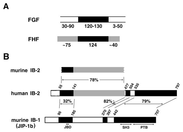
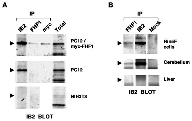
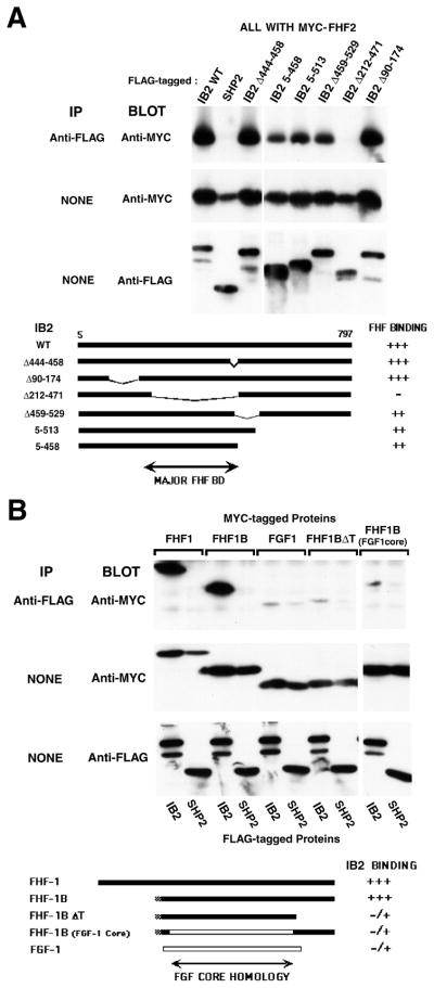
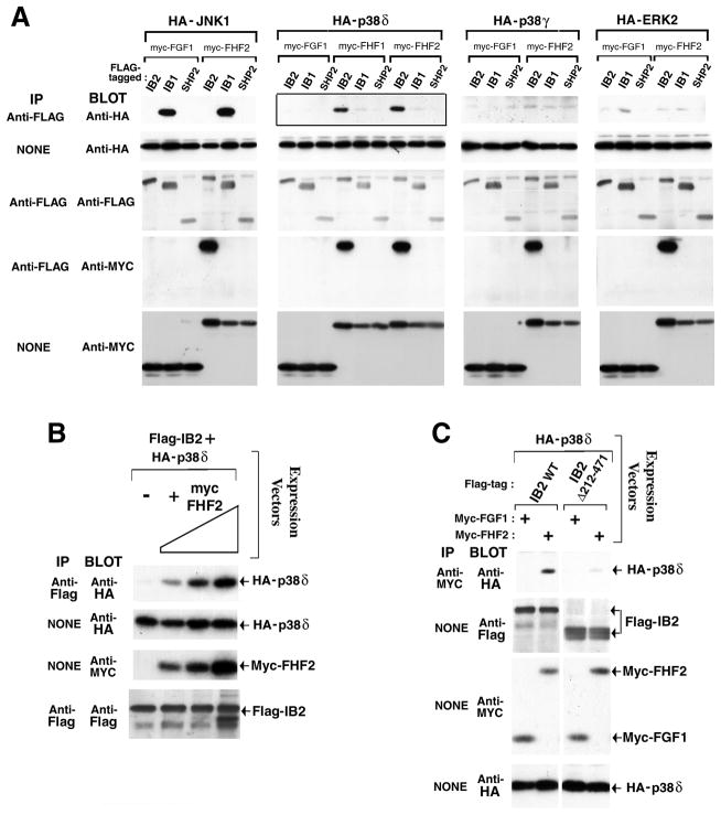
Fibroblast growth factors (FGFs) mediate cell growth, differentiation, migration, and morphogenesis by binding to the extracellular domain of cell surface receptors, triggering receptor tyrosine phosphorylation and signal transduction [1-5]. FGF homologous factors (FHFs) were discovered within vertebrate DNA sequence databases by virtue of their sequence similarity to FGFs [3, 6, 7], but the mechanism of FHF action has not been reported. We show here that FHF-1 is associated with the MAP kinase (MAPK) scaffold protein Islet-Brain-2 (IB2) [8] in the brain and in specific cell lines. FHF/IB2 interaction is highly specific, as FHFs do not bind to the related scaffold protein IB1(JIP-1b) [9, 10], nor can FGF-1 bind to IB2. We further show that FHFs enable IB2 to recruit a specific MAPK in transfected cells, and our data suggest that the scaffolds IB1 and IB2 have different MAPK specificities. Hence, FHFs are intracellular components of a tissue-specific protein kinase signaling module.


Figures

Figure 1
Figure 1
FHF/FGF and IB2/IB1 sequence similarities. A, Relatedness of FHF and FGF proteins. The core homology region shared by FGFs and FHFs is denoted in black. Additional regions of sequence similarity among FHFs are in grey. The lengths (in residues) of cores and flanking regions are indicated. B, Alignment of IB2 with IB1(JIP-1b). Human IB2 is aligned with the cloned segment of murine IB2 and with IB1. Three regions of IB2/IB1 similarity are blackened, and the percent amino acid sequence identities indicated. Grey denotes additional sequence similarity between human and murine IB2. PTB, SH3, and JNK-binding (JBD) domains of IB1 are indicated.
Figure 2
Figure 2
FHF-1/IB2 complexes in cell lines and brain. A, Complexes in FHF-transfected PC12 cells. Lysates from NIH3T3 cells (IB2 RNAFHF-1 RNA), PC12 cells (IB2 RNA+FHF-1 RNA) and a stably transfected clonal derivative expressing myc-tagged FHF-1 were immunoprecipitated with antibodies against IB2, FHF-1, or myc-tag, and immunoprecipitates or total lysates were electrophoresed and immunoblot probed with anti-IB2. Anti-FHF-1 or anti-myc coprecipitated IB2 only from FHF1-transfected PC12 cells. Arrowheads mark position of IB2. B, Native FHF-1/IB2 complexes. Lysates from RIN5F insulinoma cells (IB2 RNA+FHF-1 RNA+), rat cerebellum (IB2 RNA+FHF-1 RNA+), and liver (IB2 RNAFHF-1 RNA) were immunoprecipitated with antibodies to IB2, FHF-1, or an unrelated antigen (SNT-1, designated Mock) and immunoblotted for IB2, as in A. Anti-FHF-1 coprecipitated IB2 from RIN5F cells and cerebellum.
Figure 3
Figure 3
Sequence requirements for FHF/IB2 interaction. A, Major FHF-binding domain on IB2 is unrelated to IB1. Total lysates or M2 anti-flag immunoprecipitates from transfected 293T cells expressing flag-tagged wild-type IB2, mutant IB2 derivatives, or SHP-2 together with myc-FHF-2 were electrophoresed, and blots probed with 9E10 anti-myc and rabbit anti-flag antibodies. Schematic depicts alignment of mutant IB2 proteins and relative magnitude of interactions with FHF-2. B, FGF core homology region and C-terminus of FHF-1 are required for IB2 interaction. Total lysates or M2 anti-flag immunoprecipitates from transfected 293T cells expressing flag-tagged IB2 or SHP-2 and myc-tagged FGF-1, FHF-1, FHF-1B, FHF-1BFGF1core, or FHF-1BΔT were electrophoresed, and blots probed with anti-myc and anti-flag antibodies. Schematic shows different FHF and FGF proteins employed and relative magnitude of interaction with IB2. White bar, FGF-1 derived sequence; black bar, FHF-1 derived sequence.
Figure 4
Figure 4
FHFs promote IB2 interaction with p38δ MAPK. A, FHF-dependent IB2/p38δ interaction. Lysates from transfected 293T cells expressing flag-tagged IB2, IB1, or SHP-2, myc-tagged FGF-1, FHF-1, or FHF-2, and HA-tagged MAPKs (p38δ, p38γ, ERK-2, or JNK-1) were assayed for scaffold-associated kinases and scaffold-associated FHFs by anti-flag immunoprecipitation and anti-HA or anti-myc immunoblot, respectively. Protein expression was assayed by direct immunoblots with appropriate antibodies. FHF-dependent IB2/p38δ interaction (solid-boxed panel) is highlighted. B, IB2/p38δ interaction is FHF-2 dose dependent. Expression plasmids for flag-IB2 (0.12 μg), HA-p38δ (0.02 μg), and myc-FHF-2 (0, 0.12, 0.35, or 1.2 μg) were cotransfected into 293T cells, and IB2/p38δ complexes were detected by anti-flag immunoprecipitation and anti-HA immuoblot. IB2, p38δ, and FHF expression levels were also assayed on immunoblots. C, Heterotrimeric FHF/IB2/p38δ complexes. Anti-myc immunoprecipitates from transfected 293T cells expressing HA-p38δ, myc-tagged FGF-1 or FHF-2, and flag-tagged IB2 or IB2Δ212–471 were immunoblot probed with anti-HA to detect wild-type IB2-dependent FHF/p38δ interaction. Protein expression levels were assayed on anti-flag, anti-myc, and anti-HA immunoblots.

References

    1. Basilico C, Moscatelli D. The FGF family of growth factors and oncogenes. Adv Cancer Res. 1992;59:115–65. - PubMed
    1. Goldfarb M. Functions of FGFs in vertebrate development. Cytokine Growth Factor Rev. 1996;7:311–325. - PubMed
    1. Coulier F, Pontarotti P, Roubin R, Hartung H, Goldfarb M, Birnbaum D. Of worms and men: an evolutionary perspective on the fibroblast growth factor (FGF) and FGF receptor families. J Mol Evol. 1997;44:43–56. - PubMed
    1. Bellot F, Crumley G, Kaplow JM, Schlessinger J, Jaye M, Dionne CA. Ligand-induced transphosphorylation between different FGF receptors. Embo J. 1991;10:2849–54. - PMC - PubMed
    1. Ornitz DM, Xu J, Colvin JS, McEwen DG, MacArthur CA, Coulier F, et al. Receptor specificity of the fibroblast growth factor family. J Biol Chem. 1996;271:15292–7. - PubMed

Publication types

MeSH terms

Substances