. 2001 May 28;153(5):957-70.
doi: 10.1083/jcb.153.5.957.

COOH-terminal truncations promote proteasome-dependent degradation of mature cystic fibrosis transmembrane conductance regulator from post-Golgi compartments

Affiliations

COOH-terminal truncations promote proteasome-dependent degradation of mature cystic fibrosis transmembrane conductance regulator from post-Golgi compartments

M Benharouga et al. J Cell Biol. .

Abstract

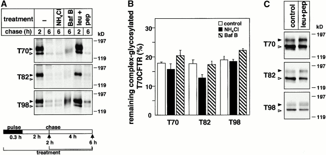
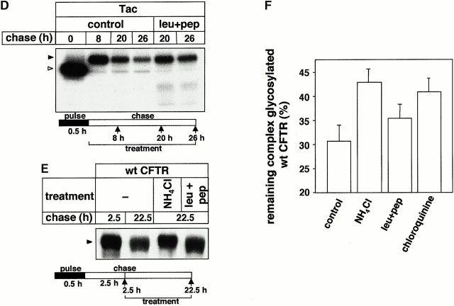
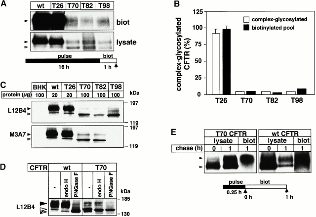
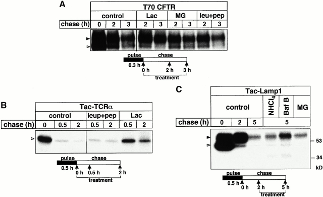
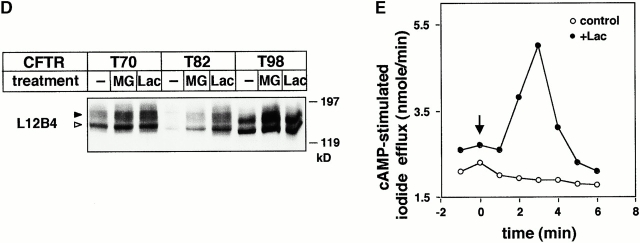
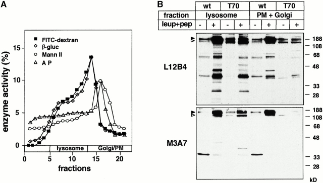
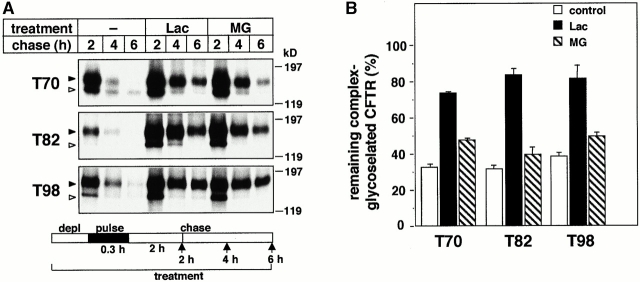
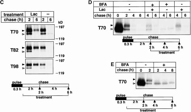
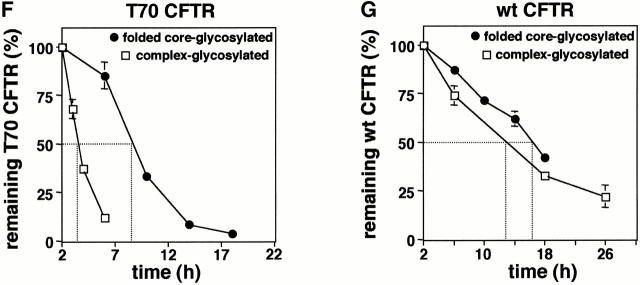
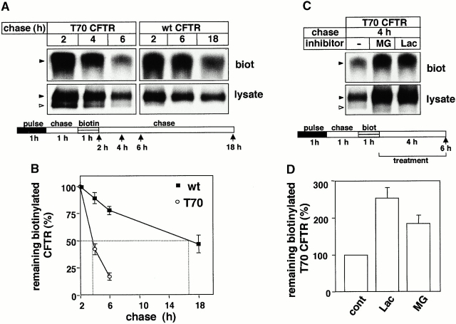
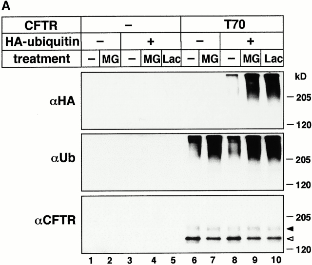
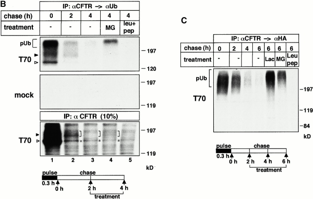
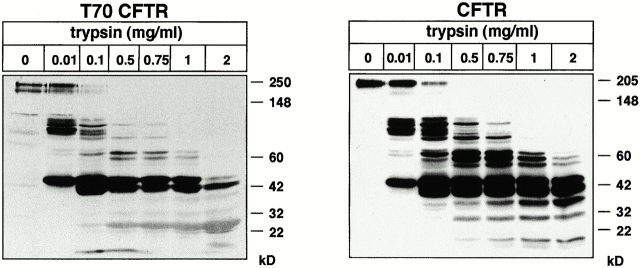
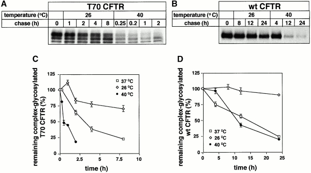
Impaired biosynthetic processing of the cystic fibrosis (CF) transmembrane conductance regulator (CFTR), a cAMP-regulated chloride channel, constitutes the most common cause of CF. Recently, we have identified a distinct category of mutation, caused by premature stop codons and frameshift mutations, which manifests in diminished expression of COOH-terminally truncated CFTR at the cell surface. Although the biosynthetic processing and plasma membrane targeting of truncated CFTRs are preserved, the turnover of the complex-glycosylated mutant is sixfold faster than its wild-type (wt) counterpart. Destabilization of the truncated CFTR coincides with its enhanced susceptibility to proteasome-dependent degradation from post-Golgi compartments globally, and the plasma membrane specifically, determined by pulse-chase analysis in conjunction with cell surface biotinylation. Proteolytic cleavage of the full-length complex-glycosylated wt and degradation intermediates derived from both T70 and wt CFTR requires endolysosomal proteases. The enhanced protease sensitivity in vitro and the decreased thermostability of the complex-glycosylated T70 CFTR in vivo suggest that structural destabilization may account for the increased proteasome susceptibility and the short residence time at the cell surface. These in turn are responsible, at least in part, for the phenotypic manifestation of CF. We propose that the proteasome-ubiquitin pathway may be involved in the peripheral quality control of other, partially unfolded membrane proteins as well.


Figures

Figure 2
Figure 2
The role of lysosomal proteases in the metabolism of truncated and wt CFTR. (A) After the pulse labeling of T70, T82, and T98 CFTR expressors, BHK cells were chased for 2 h to ensure the conversion of core-glycosylated form to complex-glycosylated CFTR at 37°C. Subsequent chase was performed in the presence of NH4Cl (15 mM). Bafilomycin B (Baf B; 2 μM) and leupeptin plus pepstatin A (leu+pep; 50 μg/ml) were present during the entire pulse–chase. CFTR was immunoprecipitated and visualized by fluorography. (B) The radioactivity remaining in the complex-glycosylated T70, T82, and T98 CFTR after the chase was quantified by PhosphorImage analysis in experiments shown in A and expressed as the percentage of the initial amount present after 2 h of chase. Data represent means ± SEM, n = 3. (C) The effect of chathepsin inhibitors on the expression level of truncated CFTR. BHK transfectants were incubated with or without leupeptin and pepstatin A (50 μg/ml) for 4 h. Equal amounts of protein were probed by immunoblotting as described in the legend to Fig. 1 C. (D) The metabolic stability of Tac in BHK cells. The turnover of Tac was determined in the presence or absence of leupeptin and pepstatin A (50 μg/ml). Metabolically labeled cells were chased for the indicated time, and Tac was isolated by immunoprecipitation and visualized by fluorography. (E and F) Metabolic stability of the complex-glycosylated wt CFTR was determined in stably transfected CHO cells in the presence of NH4Cl (15 mM), chloroquin (200 μM), or leupeptin plus pepstatin A (leu+pep; 50 μg/ml) as described for A and B. Statistical analysis was performed on samples subjected to a 20-h chase (F). Data are means ± SEM, n = 3.
Figure 2
Figure 2
The role of lysosomal proteases in the metabolism of truncated and wt CFTR. (A) After the pulse labeling of T70, T82, and T98 CFTR expressors, BHK cells were chased for 2 h to ensure the conversion of core-glycosylated form to complex-glycosylated CFTR at 37°C. Subsequent chase was performed in the presence of NH4Cl (15 mM). Bafilomycin B (Baf B; 2 μM) and leupeptin plus pepstatin A (leu+pep; 50 μg/ml) were present during the entire pulse–chase. CFTR was immunoprecipitated and visualized by fluorography. (B) The radioactivity remaining in the complex-glycosylated T70, T82, and T98 CFTR after the chase was quantified by PhosphorImage analysis in experiments shown in A and expressed as the percentage of the initial amount present after 2 h of chase. Data represent means ± SEM, n = 3. (C) The effect of chathepsin inhibitors on the expression level of truncated CFTR. BHK transfectants were incubated with or without leupeptin and pepstatin A (50 μg/ml) for 4 h. Equal amounts of protein were probed by immunoblotting as described in the legend to Fig. 1 C. (D) The metabolic stability of Tac in BHK cells. The turnover of Tac was determined in the presence or absence of leupeptin and pepstatin A (50 μg/ml). Metabolically labeled cells were chased for the indicated time, and Tac was isolated by immunoprecipitation and visualized by fluorography. (E and F) Metabolic stability of the complex-glycosylated wt CFTR was determined in stably transfected CHO cells in the presence of NH4Cl (15 mM), chloroquin (200 μM), or leupeptin plus pepstatin A (leu+pep; 50 μg/ml) as described for A and B. Statistical analysis was performed on samples subjected to a 20-h chase (F). Data are means ± SEM, n = 3.
Figure 1
Figure 1
Steady state expression and cell surface targeting of wt and truncated CFTR. (A) Expression level of biotinylated CFTR. An identical number of BHK-21 cells expressing wt, T26, T70, T82, or T98 CFTR were metabolically labeled overnight with [35S]methionine and [35S]cysteine. Plasma membrane proteins biotinylated with 1 mg/ml sulfo-NHS-SS-biotin for 60 min at 37°C were isolated by immunoprecipitation with L12B4 and M3A7 anti-CFTR Abs and then on Streptavidin-Sepharose (top). 10% of the immunoprecipitate was directly loaded (bottom). To avoid clonal variations, this, and the experiments shown in C, were carried out on a mixture of clones. The sulfo-NHS-SS-biotin remains membrane impermeant during the labeling, indicated by the lack of biotinylated core-glycosylated forms. Complex- and core-glycosylated CFTR are indicated by black and white arrowheads, respectively. (B) The expression level of wt and truncated CFTR at the cell surface (biotinylated) and at post-ER compartments (complex glycosylated). The radioactivity of biotinylated CFTR was measured by PhosphorImage analysis in experiments as described in the legend to A. The level of the complex-glycosylated CFTR was determined by densitometry of immunoblots (C). Data represent means ± SEM (n = 3), and are expressed as the percentage of the wt, designated as 100%. (C) Expression of wt, T26, T70, T82, and T98 CFTR in BHK-21 cells. CFTR expression in cell lysate was assessed by immunoblotting using the L12B4 and M3A7 anti-CFTR Abs. BHK lane contains a lysate of the parental cells. (D) Glycosidase sensitivity of complex- and core-glycosylated wt and T70 CFTR. Cell lysates were incubated in the presence or absence of endoglycosidase H (endo H) or peptide-N-glycosidase F (PNGase F) for 3 h at 37°C. Polypeptides were separated by SDS-PAGE and probed by the L12B4 anti-CFTR Ab. Complex-, core-, and deglycosylated CFTR are indicated by black, white, and hatched arrowheads, respectively. (E) Targeting efficiency of newly synthesized CFTR to the cell surface. After the pulse labeling of wt or T70 CFTR for 20 min, plasma membrane insertion of the channels was determined by biotinylation during a 1-h chase at 37°C using freshly dissolved sulfo-NHS-SS-biotin (1 mg/ml) every 15 min. Biotinylated CFTR was precipitated with L12B4 and M3A7 Abs and then isolated on Streptavidin-Sepharose (biot). Labeled CFTR was visualized by fluorography. To monitor the pulse labeling of total CFTR pools, 10% of the precipitate was loaded directly (lysate).
Figure 5
Figure 5
Functional rescue of the T70 CFTR by lactacystin is exerted by selective inhibition of proteasomes. (A) Protease inhibitors have no effect on the disposal and maturation efficiency of the core-glycosylated T70 CFTR. Pulse-labeled (20 min) BHK transfectants were chased in the presence of lactacystin (10 μM), MG 132 (10 μM), or leupeptin and pepstatin A (50 μg/ml) as indicated. T70 CFTR was visualized by immunoprecipitation and fluorography. PhosphorImage analysis showed that none of the drug had significantly delayed the degradation of the core-glycosylated form (not shown). (B) Lactacystin (10 μM), but not leupeptin and pepstatin A (50 μg/ml), impedes the degradation of the core-glycosylated Tac-TCRα. Stable transfectants were pulse labeled for 15 min and chased in the presence of the indicated drugs. Fluorography was performed as described in the legend to Fig. 2 D. (C) The degradation of the Tac-Lamp1 chimera is sensitive to the inhibition of the vacuolar H+-ATPase with bafilomycin B (Baf B). BHK cells stably expressing Tac-Lamp1 were labeled (30 min) and chased for 2 h. After the addition of NH4Cl (10 mM), bafilomycin B (2 μM), or MG132 (10 μM) the cells were chased for an additional 3 h and Tac-Lamp1 was isolated by immunoprecipitation. (D) The effect of lactacystin and MG132 on the steady state expression of mutant CFTR. Equal amounts of whole cell lysate, obtained from cells exposed to lactacystin (Lac; 10 μM) and MG132 (10 μM) for 3 h, were separated by SDS-PAGE and immunoblotted with the L12B4 anti-CFTR Ab. Complex- and core-glycosylated CFTR are indicated by black and white arrowheads, respectively. (E) Functional rescue of T70 CFTR by lactacystin. The cAMP-activated halide conductance of T70 CFTR–expressing BHK cells was measured by the iodide efflux assay in the absence (control) or presence of lactacystin treatment (+Lac; 10 μM for 3 h). Data points are averages of triplicate determinations and show the difference of iodide release in the presence and absence of the protein kinase A agonist cocktail (10 μM forskolin, 0.5 mM dibutyryl-cAMP, and 0.2 mM IBMX), added as indicated.
Figure 5
Figure 5
Functional rescue of the T70 CFTR by lactacystin is exerted by selective inhibition of proteasomes. (A) Protease inhibitors have no effect on the disposal and maturation efficiency of the core-glycosylated T70 CFTR. Pulse-labeled (20 min) BHK transfectants were chased in the presence of lactacystin (10 μM), MG 132 (10 μM), or leupeptin and pepstatin A (50 μg/ml) as indicated. T70 CFTR was visualized by immunoprecipitation and fluorography. PhosphorImage analysis showed that none of the drug had significantly delayed the degradation of the core-glycosylated form (not shown). (B) Lactacystin (10 μM), but not leupeptin and pepstatin A (50 μg/ml), impedes the degradation of the core-glycosylated Tac-TCRα. Stable transfectants were pulse labeled for 15 min and chased in the presence of the indicated drugs. Fluorography was performed as described in the legend to Fig. 2 D. (C) The degradation of the Tac-Lamp1 chimera is sensitive to the inhibition of the vacuolar H+-ATPase with bafilomycin B (Baf B). BHK cells stably expressing Tac-Lamp1 were labeled (30 min) and chased for 2 h. After the addition of NH4Cl (10 mM), bafilomycin B (2 μM), or MG132 (10 μM) the cells were chased for an additional 3 h and Tac-Lamp1 was isolated by immunoprecipitation. (D) The effect of lactacystin and MG132 on the steady state expression of mutant CFTR. Equal amounts of whole cell lysate, obtained from cells exposed to lactacystin (Lac; 10 μM) and MG132 (10 μM) for 3 h, were separated by SDS-PAGE and immunoblotted with the L12B4 anti-CFTR Ab. Complex- and core-glycosylated CFTR are indicated by black and white arrowheads, respectively. (E) Functional rescue of T70 CFTR by lactacystin. The cAMP-activated halide conductance of T70 CFTR–expressing BHK cells was measured by the iodide efflux assay in the absence (control) or presence of lactacystin treatment (+Lac; 10 μM for 3 h). Data points are averages of triplicate determinations and show the difference of iodide release in the presence and absence of the protein kinase A agonist cocktail (10 μM forskolin, 0.5 mM dibutyryl-cAMP, and 0.2 mM IBMX), added as indicated.
Figure 3
Figure 3
The effect of leupeptin and pepstatin A on the metabolism of wt and T70 CFTR in lysosomes. (A) Separation of subcellular organelles on Percoll density gradient. Postnuclear supernatants of wt- and T70 CFTR–expressing BHK cells were fractionated on a 25% Percoll density gradient as described in Materials and Methods. Organellar distribution was established by the activity of specific marker enzymes (plasma membrane, alkaline phosphatase [A P]; Golgi, mannosidase II [Mann II]; and lysosomes, β-glucuronidase [β-gluc]) as described previously (Lukacs et al. 1997). Enrichment of lysosomes in the high density fractions was confirmed by the accumulation of fluorescein-dextran (70 kD, 1.5 mg/ml) as well after an overnight labeling and a 3-h chase. (B) BHK cells expressing wt or T70 CFTR were treated with leupeptin and pepstatin A overnight (50 μg/ml) and for 4 h (100 μg/ml), respectively. Microsomes were isolated on Percoll density gradient, and fractions 5–13 and 15–18, comprising the majority of lysosomes and plasma membrane, Golgi regions, and endosomes, respectively, were pooled. Equal amounts (50 μg) of protein were loaded and probed with NBD1 (L12B4) and NBD2 (M3A7) specific anti-CFTR Abs using ECL.
Figure 4
Figure 4
Biochemical rescue of the complex-glycosylated form of truncated CFTR. (A and B) The complex-glycosylated truncated CFTR is stabilized by proteasome inhibitors. BHK cells expressing T70, T82, and T98 CFTR were metabolically labeled and chased in the presence of lactacystin (Lac; 10 μM) or MG132 (MG; 10 μM). The stability of the complex-glycosylated form was monitored by immunoprecipitation and fluorography. The radioactivity remaining in the complex-glycosylated CFTR was measured by PhosphorImage analysis and expressed as the percentage of the amount after a 2-h chase (B). Data are means ± SEM, n = 3–4. (C and D) To rule out that lactacystin (10 μM) delays the degradation of the complex-glycosylated form by promoting the biosynthesis or the conversion of the core-glycosylated form, cells were treated with lactacystin after the pulse labeling (C) or after the conversion of the core- to complex-glycosylated T70 (D). This protocol provided similar inhibition of the complex-glycosylated turnover as reported for A. When indicated, BFA (5 μg/ml) was included to prevent the conversion of the residual core-glycosylated form (D and E). (E–G) Comparison of the turnover rate of folded wt and T70 CFTR in the ER and post-Golgi compartments. After the pulse labeling of BHK transfectants, the fully mature core-glycosylated T70 and wt CFTR were accumulated in the ER by BFA (5 μg/ml) during a 2-h chase. The disappearance of the folded core-glycosylated form was monitored during a subsequent 15-h chase in the presence of BFA and was expressed as the percentage of the amount detected after a 2-h chase (F and G). The stability of the complex-glycosylated T70 and wt CFTR, representing post-Golgi pools, was determined as described for A.
Figure 4
Figure 4
Biochemical rescue of the complex-glycosylated form of truncated CFTR. (A and B) The complex-glycosylated truncated CFTR is stabilized by proteasome inhibitors. BHK cells expressing T70, T82, and T98 CFTR were metabolically labeled and chased in the presence of lactacystin (Lac; 10 μM) or MG132 (MG; 10 μM). The stability of the complex-glycosylated form was monitored by immunoprecipitation and fluorography. The radioactivity remaining in the complex-glycosylated CFTR was measured by PhosphorImage analysis and expressed as the percentage of the amount after a 2-h chase (B). Data are means ± SEM, n = 3–4. (C and D) To rule out that lactacystin (10 μM) delays the degradation of the complex-glycosylated form by promoting the biosynthesis or the conversion of the core-glycosylated form, cells were treated with lactacystin after the pulse labeling (C) or after the conversion of the core- to complex-glycosylated T70 (D). This protocol provided similar inhibition of the complex-glycosylated turnover as reported for A. When indicated, BFA (5 μg/ml) was included to prevent the conversion of the residual core-glycosylated form (D and E). (E–G) Comparison of the turnover rate of folded wt and T70 CFTR in the ER and post-Golgi compartments. After the pulse labeling of BHK transfectants, the fully mature core-glycosylated T70 and wt CFTR were accumulated in the ER by BFA (5 μg/ml) during a 2-h chase. The disappearance of the folded core-glycosylated form was monitored during a subsequent 15-h chase in the presence of BFA and was expressed as the percentage of the amount detected after a 2-h chase (F and G). The stability of the complex-glycosylated T70 and wt CFTR, representing post-Golgi pools, was determined as described for A.
Figure 6
Figure 6
Residence time of cell surface biotinylated wt and T70 CFTR. (A) After metabolic labeling of the complex-glycosylated wt or T70 CFTR, cell surface resident proteins were biotinylated for 45 min and chased for the indicated time in complete medium. Biotinylated CFTR was immunoprecipitated by L12B4 and M3A7 anti-CFTR Abs and then isolated on Streptavidin-Sepharose (top). For comparison, 10% of the immunoprecipitate was loaded for fluorography (bottom). (B) Turnover rates of biotinylated wt and T70 CFTR. Radioactivity incorporated into the biotinylated CFTR was measured with PhosphorImage analysis and expressed as the percentage of the initial label. Data represent means ± SEM, n = 3. (C and D) Proteasome inhibitors delay the degradation of the biotinylated T70 CFTR. Radioactivity remaining in the biotinylated T70 CFTR was measured in pulse–chase experiments after 4 h of chase in the absence or presence of lactacystin (Lac; 10 μM) or MG132 (MG; 10 μM) by PhosphorImage analysis. Data are expressed as a percentage of the amount remaining for the control T70 CFTR and represent means ± SEM, n = 3.
Figure 7
Figure 7
Proteasome inhibitors induce the accumulation of the polyubiquitinated T70 CFTR. (A) COS-1 cells transiently expressing HA-Ub, T70 CFTR, or a combination of these, were treated with MG132 (MG; 10 μM) or lactacystin (Lac; 10 μM) for 3 h at 37°C. Equal amounts of cellular proteins were immunoprecipitated with L12B4 and M3A7 anti-CFTR Abs and immunoblotted with mouse monoclonal anti-HA (top) or antiubiquitin (middle) Ab. Expression of T70 CFTR was verified by probing 10% of the cell lysate with L12B4 anti-CFTR Ab (bottom). (B) Detection of polyubiquitinated complex-glycosylated T70 CFTR. After the pulse labeling (20 min) of T70 CFTR–expressing (T70) or nontransfected (mock) BHK cells, the degradation of the core-glycosylated T70 CFTR was ensured during a 2-h chase. An additional 2-h chase was performed in the presence of MG132 (MG; 10 μM) or leupeptin and pepstatin A (leu+pep; 50 μg/ml) (lanes 4 and 5). CFTR–ubiquitin conjugates were isolated with sequential immunoprecipitation (IP) using L12B4 and M3A7 Abs first and then antiubiquitin Ab (top and middle) and visualized by fluorography. The processing of the pulse-labeled T70 CFTR pool was monitored by visualizing 10% of the CFTR-immunoprecipitate (bottom). (C) Detection of polyubiquitinated complex-glycosylated T70 CFTR in COS-1 cells. The experimental protocols, described for B, were performed on transiently transfected COS-1 cells expressing T70 CFTR and HA-Ub. Incubation with protease inhibitors was extended to 4 h as indicated and lactacystin (Lac) was used at 20 μM. The second immunoprecipitation was done with anti-HA Abs.
Figure 7
Figure 7
Proteasome inhibitors induce the accumulation of the polyubiquitinated T70 CFTR. (A) COS-1 cells transiently expressing HA-Ub, T70 CFTR, or a combination of these, were treated with MG132 (MG; 10 μM) or lactacystin (Lac; 10 μM) for 3 h at 37°C. Equal amounts of cellular proteins were immunoprecipitated with L12B4 and M3A7 anti-CFTR Abs and immunoblotted with mouse monoclonal anti-HA (top) or antiubiquitin (middle) Ab. Expression of T70 CFTR was verified by probing 10% of the cell lysate with L12B4 anti-CFTR Ab (bottom). (B) Detection of polyubiquitinated complex-glycosylated T70 CFTR. After the pulse labeling (20 min) of T70 CFTR–expressing (T70) or nontransfected (mock) BHK cells, the degradation of the core-glycosylated T70 CFTR was ensured during a 2-h chase. An additional 2-h chase was performed in the presence of MG132 (MG; 10 μM) or leupeptin and pepstatin A (leu+pep; 50 μg/ml) (lanes 4 and 5). CFTR–ubiquitin conjugates were isolated with sequential immunoprecipitation (IP) using L12B4 and M3A7 Abs first and then antiubiquitin Ab (top and middle) and visualized by fluorography. The processing of the pulse-labeled T70 CFTR pool was monitored by visualizing 10% of the CFTR-immunoprecipitate (bottom). (C) Detection of polyubiquitinated complex-glycosylated T70 CFTR in COS-1 cells. The experimental protocols, described for B, were performed on transiently transfected COS-1 cells expressing T70 CFTR and HA-Ub. Incubation with protease inhibitors was extended to 4 h as indicated and lactacystin (Lac) was used at 20 μM. The second immunoprecipitation was done with anti-HA Abs.
Figure 8
Figure 8
Protease susceptibility of native wt and T70 CFTR. After treating the cells with cyclohexamide (100 μg/ml) for 2.5 h at 37°C to deplete the core-glycosylated forms, microsomes were isolated with differential centrifugation from BHK-21 cells stably expressing wt or T70 CFTR. Microsomes were subjected to limited proteolysis at the indicated concentrations of trypsin for 15 min at 4°C. The proteolytic digestion pattern was probed by L12B4 anti-CFTR Ab and ECL.
Figure 9
Figure 9
Thermostability of the complex-glycosylated wt and T70 CFTR in vivo. (A and B) Temperature-dependent turnover of the complex-glycosylated T70 and wt CFTR. Metabolic stability of the pulse-labeled complex-glycosylated T70 and wt CFTR was monitored as described in the legend to Fig. 4. Chase was conducted at 26°C, 37°C, or 40°C. (C and D) The disappearance kinetics of complex-glycosylated T70 and wt CFTR were determined by PhosphorImage analysis in experiments shown in A and B. Data are means ± SEM, n = 3.

References

    1. Amigorena S., Webster P., Drake J., Newcomb J., Cresswell P., Mellman I. Invariant chain cleavage and peptide loading in major histocompatibility complex class II vesicles. J. Exp. Med. 1995;181:1729–1741. - PMC - PubMed
    1. Aridor M., Balch W.E. Integration of endoplasmic reticulum signaling in health and disease. Nat. Med. 1999;5:745–751. - PubMed
    1. Armstrong J., Patel S., Riddle P. Lysosomal sorting mutants of coronavirus E1 protein, a Golgi membane protein. J. Cell Sci. 1990;95:191–197. - PubMed
    1. Baumeister W., Walz J., Zuhi F., Seemuller E. The proteasomeparadigm of a self-compartmentalizing protease. Cell. 1998;92:367–380. - PubMed
    1. Bonifacino J.S., Weismann A.M. Ubiquitin and the control of protein fate in the secretory and endocytic pathway. Ann. Rev. Cell Dev. Biol. 1998;14:19–57. - PMC - PubMed

Publication types

MeSH terms