. 2001 May 28;153(5):999-1010.
doi: 10.1083/jcb.153.5.999.

Cathepsin B acts as a dominant execution protease in tumor cell apoptosis induced by tumor necrosis factor

Affiliations

Cathepsin B acts as a dominant execution protease in tumor cell apoptosis induced by tumor necrosis factor

L Foghsgaard et al. J Cell Biol. .

Abstract

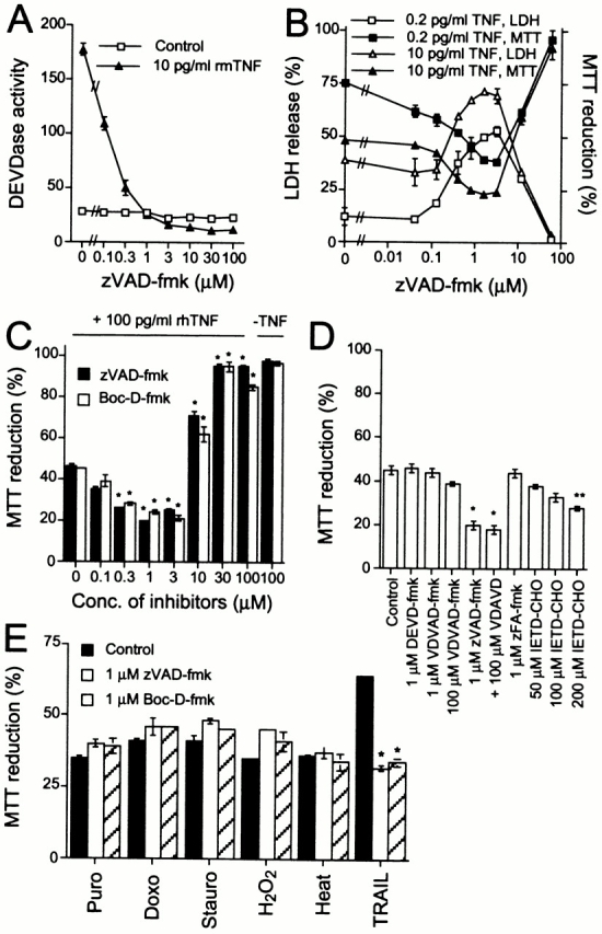
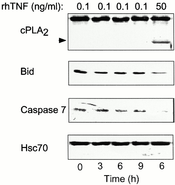
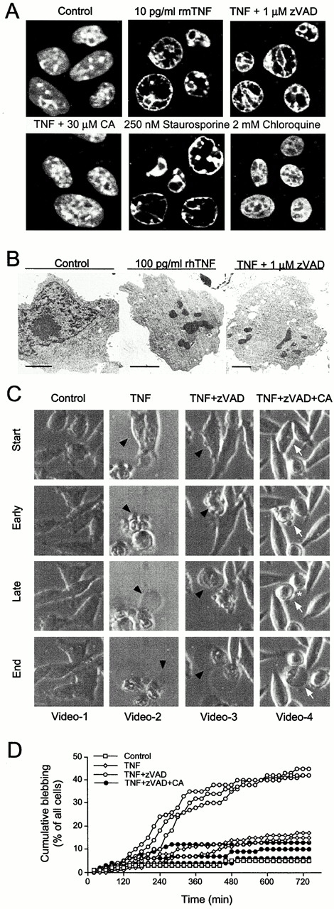
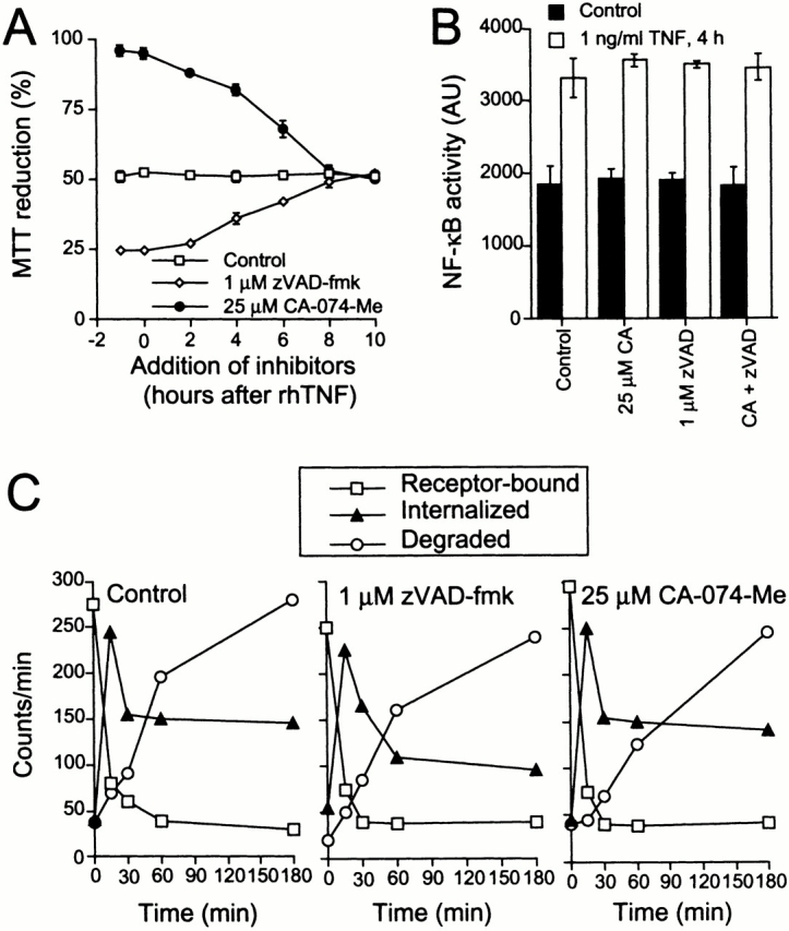
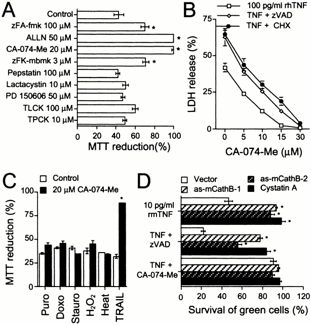
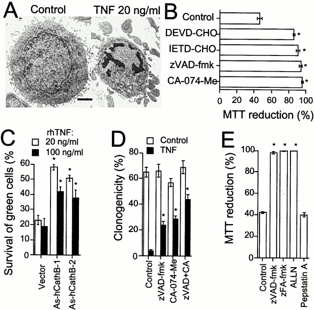
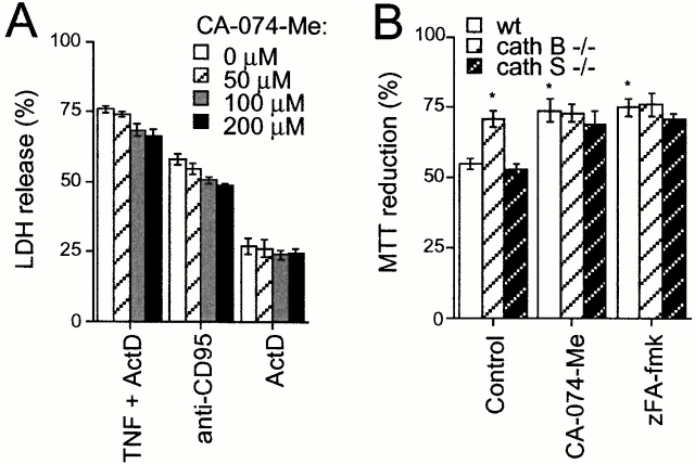
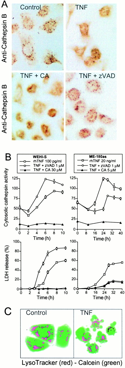
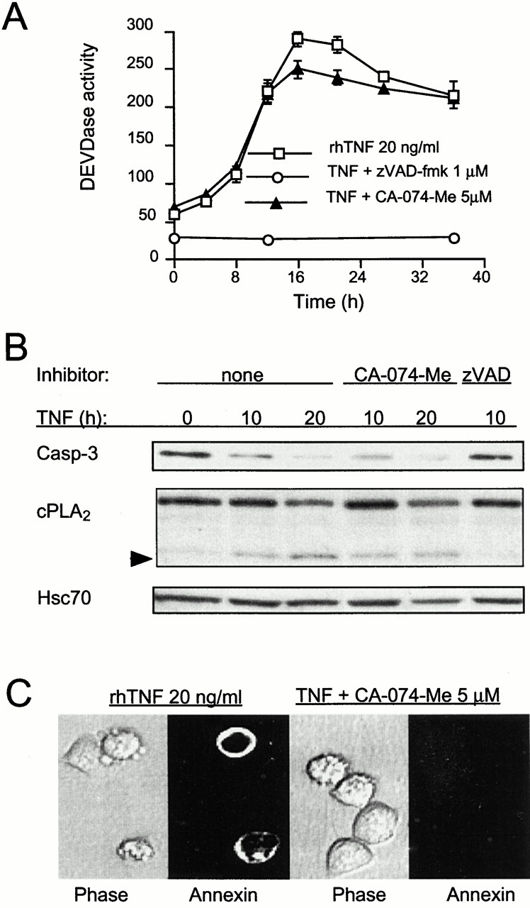
Death receptors can trigger cell demise dependent or independent of caspases. In WEHI-S fibrosarcoma cells, tumor necrosis factor (TNF) induced an increase in cytosolic cathepsin B activity followed by death with apoptotic features. Surprisingly, this process was enhanced by low, but effectively inhibiting, concentrations of pan-caspase inhibitors. Contrary to caspase inhibitors, a panel of pharmacological cathepsin B inhibitors, the endogenous cathepsin inhibitor cystatin A as well as antisense-mediated depletion of cathepsin B rescued WEHI-S cells from apoptosis triggered by TNF or TNF-related apoptosis-inducing ligand. Thus, cathepsin B can take over the role of the dominant execution protease in death receptor-induced apoptosis. The conservation of this alternative execution pathway was further examined in other tumor cell lines. Here, cathepsin B acted as an essential downstream mediator of TNF-triggered and caspase-initiated apoptosis cascade, whereas apoptosis of primary cells was only minimally dependent on cathepsin B. These data imply that cathepsin B, which is commonly overexpressed in human primary tumors, may have two opposing roles in malignancy, reducing it by its proapoptotic features and enhancing it by its known facilitation of invasion.


Figures

Figure 1
Figure 1
Inhibition of caspase activation sensitizes WEHI-S cells to death receptor–mediated apoptosis. (A) The DEVDase activity in lysates of cells treated as indicated for 4 h was quantitated by measurement of the cleavage of DEVD-AFC. The enzyme activity is expressed as arbitrary units. (B) Cells were incubated with the indicated concentrations of zVAD-fmk for 1 h before a 10-h treatment with rmTNF. The viability of the cells was measured by LDH-release and MTT assays. (C) Cells were treated for 1 h with indicated concentrations of pan-caspase inhibitors before an 11-h treatment with or without rhTNF. The survival of the cells was measured by the MTT assay. (D) WEHI-S cells were pretreated with indicated protease inhibitors for 1 h before the addition of 100 pg/ml of rhTNF. The survival of the cells was analyzed 12 h later by the MTT assay. (E) Cells were treated with caspase inhibitors for 1 h before a 22-h treatment with 4 μg/ml of puromycin (Puro) or doxorubicin (Doxo), 100 ng/ml of staurosporine (Stauro), or 50 μM H2O2, a 16-h treatment with 75 ng/ml of Flag-TRAIL plus 2 μg/ml of anti-Flag antibody (TRAIL), or a 45-min treatment at 43.5°C followed by 18 h at 37°C (Heat). The survival of the cells was measured by the MTT assay. *p-value < 0.001, **p-value < 0.01; as compared with the control. All values represent means of triplicate determinations ± SD.
Figure 3
Figure 3
Absence of significant caspase-mediated intracellular proteolysis in WEHI-S cells exposed to low but lethal concentration of TNF. Equal amounts of protein from WEHI-S cells treated for indicated times with 0.1 or 50 ng/ml of rhTNF were analyzed for cleavage of caspase-7 or caspase substrates cPLA2 and Bid by immunoblot analysis. The arrowhead indicates a caspase cleavage product of cPLA2 (Wissing et al. 1997). Hsc70 serves as a loading control.
Figure 2
Figure 2
TNF-treated WEHI-S cells display apoptotic morphology in the absence of caspase activity. (A) Chromatin of WEHI-S cells was imaged by confocal microscopy. Cells treated with rmTNF or TNF plus zVAD-fmk are shown at the initiation of chromatin condensation. Cotreatment with 30 μM CA-074-Me inhibited chromatin condensation. For comparison, chromatin of untreated control cells is shown. Chloroquine- and staurosporine-exposed cells are shown as control for necrosis and apoptosis, respectively. (B) WEHI-S cells were treated for 6 h as indicated, fixed in situ, and prepared for transmission electron microscopy. Condensed and fragmented chromatin lumps, nuclear fragmentation, and loss of cell surface microvilli were induced by TNF in the presence or absence of zVAD-fmk. Bars, 2 μm. (C) WEHI-S cells were exposed to 5 pg/ml rmTNF or indicated combinations of 0.2 pg/ml rmTNF, 1 μM zVAD-fmk, and CA-074-Me (30 μM) and recorded by time-lapse microscopy (also see supplementary videos available at http://www.jcb.org/cgi/content/full/153/5/999/DC1). Note the continuous membrane movements in control cells. Two different types of blebbing in TNF–treated cells are marked as early (highly dynamic, multifocal, apoptotic blebbing) and late (formation of a single large bleb on one side of the cell) events. In necrotic death, only the latter occurs (data not shown). Blebbing characteristics were not significantly modified by zVAD-fmk, and quantitative analysis showed an average blebbing time of 21 ± 2 min (n = 43) for TNF and 18 ± 2 min (n = 37) for TNF plus zVAD-fmk. The very few cells that died in the presence of CA-074-Me showed a distinct blebbing behavior with average blebbing time of 50 ± 9 min (white arrows). During that time cells recovered from blebbing and looked normal (*) for 13 ± 5 min (n = 20), before the final lethal bleb formed. (D) WEHI-S cells were exposed to indicated combinations of rmTNF (0.2 pg/ml), zVAD-fmk (1 μM), and CA-074-Me (30 μM) and recorded by time-lapse microscopy. For quantitative analysis of bleb formation, every cell (40–50) in the field was included, and the time between the addition of TNF and the start of the blebbing was measured. Data for each experiment is presented as cumulative frequency of cells that have initiated blebbing at a given time point.
Figure 4
Figure 4
Inhibition of caspase or cathepsin B activities does not affect TNF-induced NF-κB activation or binding, internalization, or degradation of TNF. (A) WEHI-S cells were treated with 100 pg/ml rhTNF for 11 h and protease inhibitors as indicated. The survival of the cells was measured by the MTT assay. (B) WEHI-S cells transfected with p3K-INF-LUC and pEBS7-β-Gal were left untreated (control) or treated with 1 ng/ml of rhTNF for 4 h. 1 μM zVAD-fmk, 25 μM CA-074-Me, or their combination was added to the cells 1 h before the addition of TNF. NF-κB activity is expressed as arbitrary units of luciferase activity relative to β-galactosidase activity. (A and B) The values represent means of triplicate determinations ± SD, and the experiments were repeated twice with similar results. (C) Subconfluent WEHI-S cells were treated with indicated protease inhibitors for 1 h before a 90-min incubation with 1 nM 125I-labeled TNF on ice. After careful washing, prewarmed complete medium containing the indicated protease inhibitors was added, and the cells were placed at 37°C (0 min). Receptor-bound, internalized, and degraded TNF at indicated time points are shown. The values represent means of triplicate determinations. The experiment was repeated once with similar results.
Figure 5
Figure 5
Inhibition of cathepsin B activity or expression protects WEHI-S cells from death receptor–induced apoptosis. (A) WEHI-S cells were pretreated with indicated protease inhibitors for 1 h before the addition of 100 pg/ml rhTNF. The survival of the cells was analyzed 12 h later by the MTT assay. (B) WEHI-S cells were treated for 11 h with indicated concentrations of CA-074-Me and rhTNF in the presence or absence of 1 μM zVAD-fmk or 10 μM cycloheximide (CHX). The cytotoxicity was measured by the LDH release assay. (C) The survival of WEHI-S cells pretreated with CA-074-Me for 1 h before the treatment with cytotoxic agents, as indicated in the legend for Fig. 1 E (except for TRAIL which was used at 150 ng/ml) was measured by the MTT assay. (D) WEHI-S cells were transfected with indicated plasmids plus pEGFP-N1 and treated 22 h later with rmTNF alone or with 1 μM zVAD-fmk or 20 μM CA-074-Me. The survival of green cells was determined 12 h later. The values represent means of triplicate determinations (A–C) or means of 10 (D) randomly chosen fields of ≥100 cells ± SD. *p-value < 0.01, as compared with control cells (A and C) or to similarly treated vector-transfected cells (D).
Figure 6
Figure 6
Cathepsin B is a key mediator in TNF-induced caspase-dependent apoptosis of tumor cells. (A) ME-180as cells were treated as indicated for 24 h and analyzed by transmission electron microscopy. Nuclear and chromatin condensation as well as formation of apoptotic bodies was induced by TNF treatment. Bar, 2 μm. (B) The survival of ME-180as cells pretreated with 200 μM DEVD-CHO or IETD-CHO, 1 μM zVAD-fmk, or 5 μM CA-074-Me for 1 h before a 48-h treatment with 20 ng/ml of rhTNF was analyzed by the MTT assay. (C) ME-180as cells were transfected with indicated plasmids plus pEGFP-N1 and treated 48 h later as indicated. The survival of the green cells was analyzed after 48-h treatment. (D) After 3 d of treatment with indicated combinations of 100 ng/ml rhTNF, 5 μM zVAD-fmk, and 5 μM CA-074-Me in serum-free medium, ME-180as cells were replated on cloning plates in medium containing 6% FCS, and the clonogenic potential was determined 7 d later by counting viable colonies. (E) The survival of MCF-7 breast cancer cells pretreated with 1 μM zVAD-fmk, 100 μM zFA-fmk, or 12.5 μM ALLN for 1 h before a 32-h treatment with 10 ng/ml of rhTNF was analyzed by the MTT assay. The values represent means of triplicate determinations (B and E) or means of 10 randomly chosen fields of ≥100 cells (C and D) ± SD. *p-value < 0.01, as compared with control cells (B, D, and E) or similarly treated vector-transfected cells (C).
Figure 7
Figure 7
Cathepsin B plays a minor role in death receptor–induced apoptosis of primary cells. (A) The survival of hepatocytes pretreated with the indicated concentrations of CA-074-Me for 1 h before the addition of 200 nM actinomycin D plus 28 ng/ml of rmTNF or 100 ng/ml of anti-CD95 antibody (Jo-2) was analyzed after 22-h treatment by the LDH release assay. (B) MEF (passage 4) originating from wild-type, cathepsin B–, or cathepsin S–deficient mice were left untreated or treated with 50 μM CA-074-Me or 100 μM zFA-fmk for 1 h before an 18-h treatment with 100 ng/ml of rhTNF plus 10 μM cycloheximide. The survival was analyzed by the MTT assay. The values represent means of triplicate determinations ± SD. *p-value < 0.01, as compared with control cells.
Figure 8
Figure 8
TNF induces the appearance of active cysteine cathepsins in the cytosol. (A) ME-180as cells were left untreated (control) or treated with 20 ng/ml rhTNF, TNF plus 5 μM CA-074-Me, or TNF plus 1 μM zVAD-fmk and stained with anti–cathepsin B after 16-h treatment. (B) WEHI-S or ME-180as cells were treated as indicated and analyzed for cytosolic cysteine cathepsin activity within intact cells and for released LDH activity. The values represent means of a triplicate determination ± SD. (C) ME-180as cells were left untreated (control) or treated with 20 ng/ml rhTNF for 30 h and stained with LysoTracker and calcein-AM.
Figure 9
Figure 9
TNF induces cathepsin B–independent activation of caspases and cathepsin B–dependent translocation of PS in ME-180as cells. (A and B) Equal amount of protein from cells treated for indicated times with 20 ng/ml of rhTNF in the presence or absence of 1 μM zVAD-fmk (zVAD) or 5 μM CA-074-Me (CA) was analyzed for caspase-3–like activity by DEVDase assay (DEVDase activity is expressed as arbitrary units and values represent means of triplicate determinations ± SD) or immunoblot analysis of caspase-3 or its substrate cPLA2. The arrowhead indicates a cleavage product of cPLA2. Hsc70 serves as a loading control. (C) ME-180as cells treated as indicated for 30 h were analyzed for PS translocation by staining with FITC–annexin V. Note that in the presence of CA-074-Me, TNF induces cellular shrinkage and rounding in the absence of PS translocation.

References

    1. Ashkenazi A., Dixit V.M. Apoptosis control by death and decoy receptors. Curr. Opin. Cell Biol. 1999;11:255–260. - PubMed
    1. Brunk U.T., Dalen H., Roberg K., Hellquist H.B. Photo-oxidative disruption of lysosomal membranes causes apoptosis of cultured human fibroblasts. Free Radic. Biol. Med. 1997;23:616–626. - PubMed
    1. Chautan M., Chazal G., Cecconi F., Gruss P., Golstein P. Interdigital cell death can occur through a necrotic and caspase-independent pathway. Curr. Biol. 1999;9:967–970. - PubMed
    1. Deiss L.P., Galinka H., Berissi H., Cohen O., Kimchi A. Catepsin D protease mediates programmed cell death induced by interferon-γ, Fas/APO-1, and TNF-α. EMBO J. 1996;15:3861–3870. - PMC - PubMed
    1. Deussing J., Roth W., Saftig P., Peters C., Ploegh H.L., Villadangos J.A. Cathepsins B and D are dispensable for major histocompatibility complex class II-mediated antigen presentation. Proc. Natl. Acad. Sci. USA. 1998;95:4516–4521. - PMC - PubMed

Publication types

MeSH terms