

doi: 10.1083/jcb.153.5.1133.
Circulating skeletal stem cells
Affiliations
- PMID: 11381097
- PMCID: PMC2174322
- DOI: 10.1083/jcb.153.5.1133
Item in Clipboard
Circulating skeletal stem cells
J Cell Biol.
.
Abstract
We report the isolation of adherent, clonogenic, fibroblast-like cells with osteogenic and adipogenic potential from the blood of four mammalian species. These cells phenotypically resemble but are distinguishable from skeletal stem cells found in bone marrow (stromal stem cells, "mesenchymal stem cells"). The osteogenic potential of the blood-borne cells was proven by an in vivo transplantation assay in which either polyclonal or single colony-derived strains were transplanted into the subcutis of immunocompromised mice, and the donor origin of the fully differentiated bone cells was proven using species-specific probes. This is the first definitive proof of the existence of circulating skeletal stem cells in mammals.
Figures
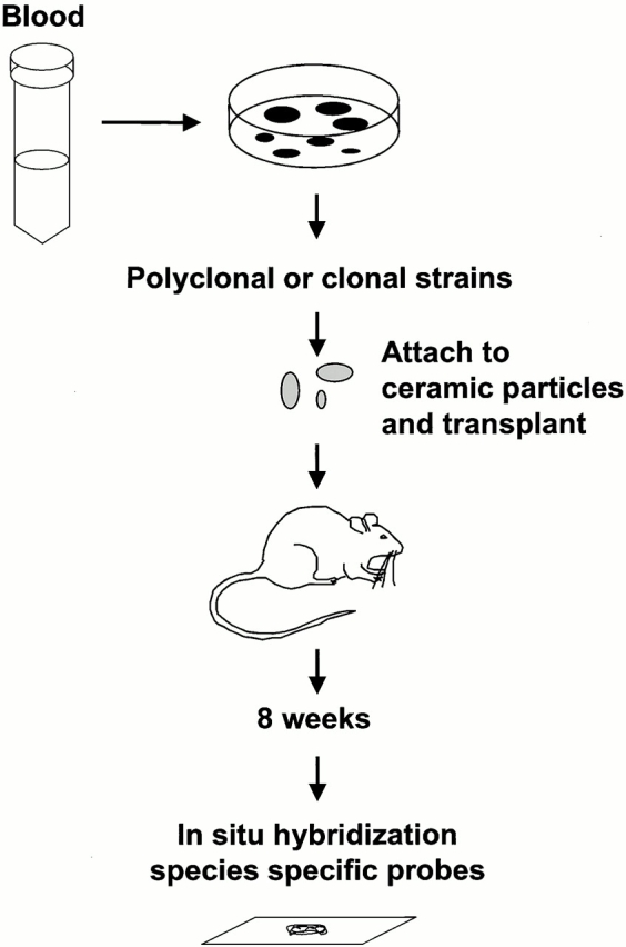
Identification of skeletal stem cells in peripheral blood. Blood obtained from mice, rabbits, guinea pigs, and humans was plated in vitro. In all four species, adherent clonogenic colonies were formed. After ex vivo expansion, they were attached to ceramic particles and transplanted in the subcutis of immunocompromised mice. After 8 wk, the transplants were examined for bone formation. The donor origin of bone in guinea pig and human transplants was determined using species-specific repetitive DNA sequences.
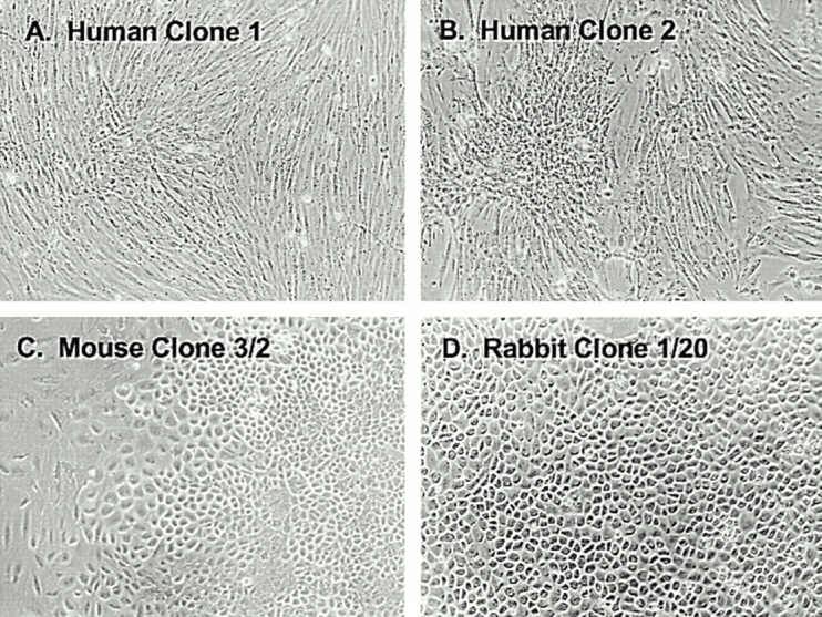
Morphological features of blood-derived adherent clonogenic cells. The majority of clones that developed from all animal species, including the two human colonies (A and B) consisted of cells with a fibroblastic morphology. However, between 5 and 13% of the colonies, depending on the species, had a distinctly polygonal morphology in mouse (C), rabbit (D), and guinea pig (not shown).
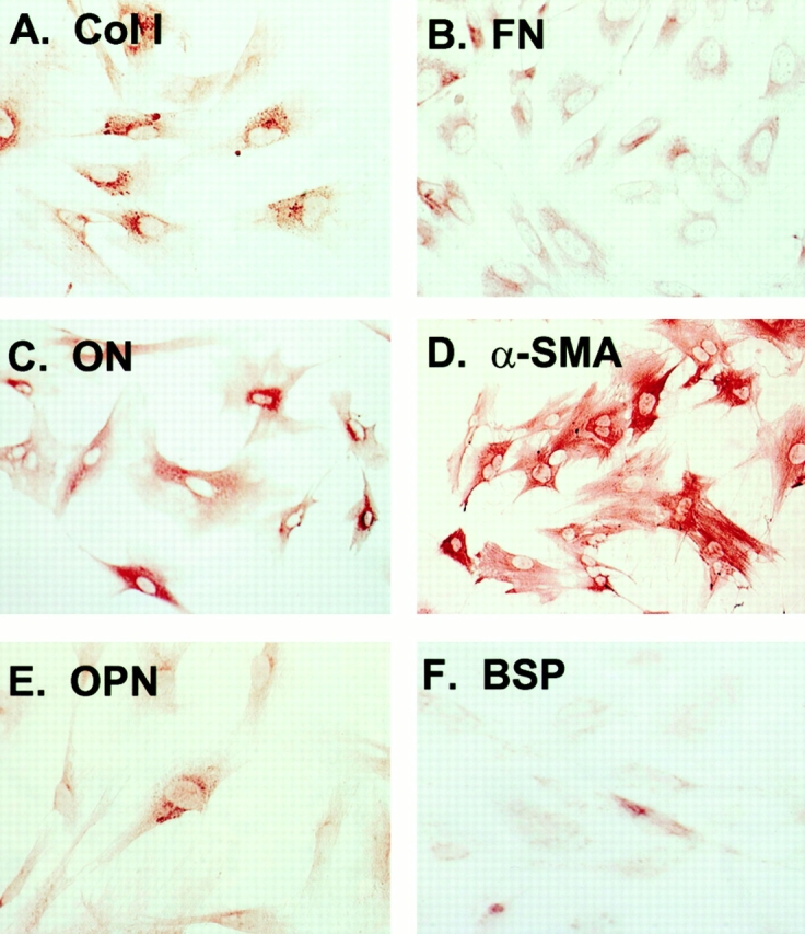
Representative immunophenotype of blood-derived adherent cells. Although two morphologically distinct types of cells were detected in three of the four animal species, their immunophenotype was identical, and this did not vary significantly from one species to another. Their similarity to marrow stromal cells is demonstrated by their expression of (A) type I collagen, (B) fibronectin, (C) osteonectin, and (D) α-smooth muscle actin, and variable expression of (E) osteopontin and (F) bone sialoprotein.
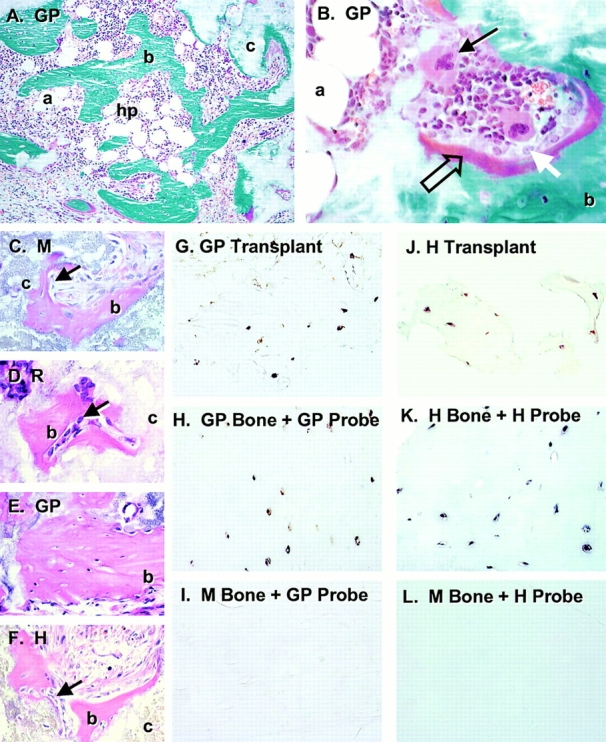
Bone formation by in vivo transplantation of blood-derived adherent cells. Strains of blood-derived adherent cells from guinea pig (GP, in A, B, and E), mouse (M, in C), rabbit (R, in D), and human (H, in F) were attached to ceramic particles (c) and transplanted into the subcutis of immunocompromised mice. After 8 wk, the transplants were harvested and sections were stained with Goldner's to distinguish between bone (b, green in A and B) and osteoid (open arrow, red in B). In all species, bone was deposited on the surfaces of the ceramic particles by mature osteoblasts (white arrow in B, arrows in C, D, and F). In transplants in which there was a significant amount of bone formed, a fully functional hematopoietic marrow (hp) was also established (A and B), complete with megakaryocytes (arrow in B) and adipocytes (a). Sections of paraffin-embedded transplants generated by guinea pig (G) and human (J) blood-derived adherent cells were used for in situ hybridization using a guinea pig–specific sequence (G, H, and I) and a human-specific alu sequence (J, K, and L) as probes. Osteocytes completely embedded in bone were easily identifiable in both guinea pig and human transplants, indicating the donor nature of the cells that formed the bone. Specificity of the probes was demonstrated by the positive reaction of the guinea pig probe with guinea pig bone (H) and negative reaction with mouse bone (I), and by positive reaction of the human probe with human bone (K) and negative reaction with mouse bone (L).
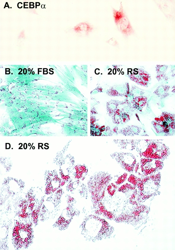
Adipocytic conversion of human blood-derived adherent cells in vitro. Immunohistochemistry of human blood-derived adherent cells revealed expression of the adipocytic transcription factor, CEBPα (A). The ability of blood-derived cells to form adipocyte-like cells was further investigated by assessing lipid accumulation as demonstrated by Oil Red O staining after culture with rabbit serum. As has been seen in marrow stromal cells and in most blood-derived cell types, small lipid-containing structures are present in cultures grown in 20% FBS (B). In cultures grown in 20% rabbit serum, there is a dramatic increase in the number and size of lipid vacuoles (C), which begin to coalesce in vitro (D).
References
-
- Beresford J.N., Bennett J.H., Devlin C., Leboy P.S., Owen M.E. Evidence for an inverse relationship between the differentiation of adipocytic and osteogenic cells in rat marrow stromal cell cultures. J. Cell Sci. 1992;102:341–351. - PubMed
-
- Bianco P., Cossu G. Uno, nessuno e centomilasearching for the identity of mesodermal progenitors. Exp. Cell Res. 1999;251:257–263. - PubMed
-
- Bianco P., Costantini M., Dearden L.C., Bonucci E. Alkaline phosphatase positive precursors of adipocytes in the human bone marrow. Br. J. Haematol. 1988;68:401–403. - PubMed
-
- Bianco P., Riminucci M. The bone marrow stroma in vivoontogeny, structure, cellular composition and changes in disease. In: Beresford J.N., Owen M., editors. Marrow Stromal Cell Cultures. Cambridge University Press; Cambridge, UK: 1998. pp. 10–25.
-
- Cossu G., Tajbakhsh S., Buckingham M. How is myogenesis initiated in the embryo? Trends Genet. 1996;12:218–223. - PubMed
Publication types
MeSH terms
Grants and funding
LinkOut - more resources
Full Text Sources
Other Literature Sources
Medical
