. 2001 Jun;65(2):261-87 ; second page, table of contents.
doi: 10.1128/MMBR.65.2.261-287.2001.

Phi29 family of phages

Affiliations

Phi29 family of phages

W J Meijer et al. Microbiol Mol Biol Rev. 2001 Jun.

Abstract

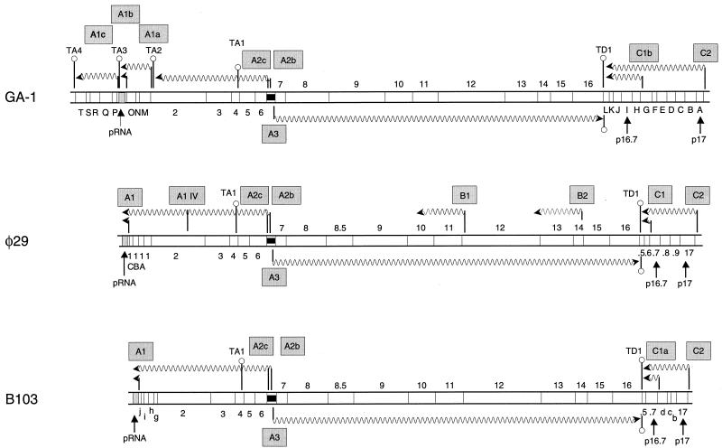
Continuous research spanning more than three decades has made the Bacillus bacteriophage phi29 a paradigm for several molecular mechanisms of general biological processes, such as DNA replication, regulation of transcription, phage morphogenesis, and phage DNA packaging. The genome of bacteriophage phi29 consists of a linear double-stranded DNA (dsDNA), which has a terminal protein (TP) covalently linked to its 5' ends. Initiation of DNA replication, carried out by a protein-primed mechanism, has been studied in detail and is considered to be a model system for the protein-primed DNA replication that is also used by most other linear genomes with a TP linked to their DNA ends, such as other phages, linear plasmids, and adenoviruses. In addition to a continuing progress in unraveling the initiation of DNA replication mechanism and the role of various proteins involved in this process, major advances have been made during the last few years, especially in our understanding of transcription regulation, the head-tail connector protein, and DNA packaging. Recent progress in all these topics is reviewed. In addition to phi29, the genomes of several other Bacillus phages consist of a linear dsDNA with a TP molecule attached to their 5' ends. These phi29-like phages can be divided into three groups. The first group includes, in addition to phi29, phages PZA, phi15, and BS32. The second group comprises B103, Nf, and M2Y, and the third group contains GA-1 as its sole member. Whereas the DNA sequences of the complete genomes of phi29 (group I) and B103 (group II) are known, only parts of the genome of GA-1 (group III) were sequenced. We have determined the complete DNA sequence of the GA-1 genome, which allowed analysis of differences and homologies between the three groups of phi29-like phages, which is included in this review.


Figures

FIG. 1
FIG. 1
Schematic representation of a φ29 phage particle. The various proteins are indicated.
FIG. 2
FIG. 2
Genetic and transcriptional maps of GA-1 (group III, 21,129 bp), φ29 (group I, 19,285 bp), and B103 (group II, 18,630 bp). The maps are aligned according to the A2b, A2c, A3 promoter region. The direction of transcription and lengths of the transcripts are indicated by wavy arrows. The transcripts of late- and early-expressed operons and the late and early promoters (boxed) are shown below and above the map, respectively. The positions of the various genes and ORFs are indicated between the two DNA strands. Genes are indicated by numbers, ORFs are indicated by letters (capital for GA-1 and φ29, and lowercase for B103). The positions of genes 17 and 16.7 that are conserved in all three phage genomes, located in the right early operon, are indicated. The positions of φ29 ORFs 16.9, 16.8, 16.6, and 16.5 and B103 ORF 16.5, located at the right side of their genomes, are indicated by the numbers .9, .8, .6, and .5. Transcriptional terminators are indicated by hairpin structures. A light grey box indicates the DNA region encoding the pRNA, and a black box indicates the region spanning the early A2b, A2c, and late A3 promoters.
FIG. 3
FIG. 3
Alignment of the deduced transcriptional regulatory protein sequences encoded by φ29 (p4phi29), B103 (p4B103), and GA-1 (p4GA-1). Black and grey boxes enclose residues that are conserved in all three or two of the three sequences, respectively. The following amino acids were considered conservative: L, I, V, A, and M; F, Y, and W; K and R; D and E; Q and N; and S and T. The position up to which the N-terminal part of the respective gene 4 can be translated, considering the mRNA length terminated at the transcriptional terminator TA1, is indicated by a vertical arrow.
FIG. 4
FIG. 4
Schematic representation of the mechanism of φ29 protein-primed DNA replication. Primer and parental TP are shown in black and grey, respectively. p6, double-stranded DNA binding protein; p5, single-stranded DNA binding protein (SSB). Only the left DNA end has been drawn, except for type I and type II molecules, where both DNA ends are shown. See the text for details. Adapted with permission from reference .
FIG. 5
FIG. 5
Structural and functional map of the DNA polymerase. The N-terminal domain, required for proofreading and strand displacement, and the C-terminal initiation and polymerization domain are indicated by white and black rectangular boxes, respectively (32). The ExoI, ExoII, and ExoIII motifs, as well as the motifs of the C-terminal domain, are indicated: motif 1 (also called motif A), motif 2a (also called motif B), motif 2b, motif 3 (also called motif C), and motif 4. In addition, the position of the YxG(G/A) motif, important for the coordination between 3′-5′ exonuclease and 5′-3′ polymerization, is indicated by CT (cross talk). All these motifs are conserved in B-type proofreading-proficient DNA polymerases. The amino acid sequences of each of these various motifs present in the DNA polymerases of φ29, B103 and GA-1, together with each consensus sequence, are indicated below the map. The three Asp residues that form a metal binding triad required for catalysis at the polymerization active site are indicated by an asterisk. Finally, the position of the TPR-1 and TPR-2 motifs, characteristic of DNA polymerases involved in protein-primed DNA replication, are indicated. See the text for further details.
FIG. 6
FIG. 6
Comparison of the deduced p16.7 protein sequences encoded by φ29 (group I), B103 (group II), and GA-1 (group III). Amino acid residues are given on the right. Black and grey boxes enclose residues that are conserved in all three or in two of the three sequences, respectively. The following amino acids were considered conservative: L, I, V, M, and A; F, Y, and W; K and R; D and E; Q and N; and S and T. The transmembrane, coiled-coil, and putative DNA binding domain are indicated above the protein sequence. Note that the p16.7 sequences of φ29 and B103 have a high level of homology throughout their entire protein sequences but that only the putative DNA binding domain is conserved in all three p16.7 protein sequences. An alignment of the deduced p16.7 protein sequences from φ29 (group I) and B103 (group II), together with those of the group I phages φ15, PZA, and BS32, has been published previously (135). The percent identities between the deduced p16.7 protein sequence of φ29 and those of φ15, PZA, and BS32 are 91, 89, and 87%, respectively.
FIG. 7
FIG. 7
Alignment of the deduced holin (p14) sequences of φ29 (p14phi29; group I), B103 (p14B103; group II) and GA-1 (p14GA-1; group III). Amino acid residues are given on the right. Black and grey boxes enclose residues that are conserved in all three or in two of the three sequences, respectively. The following amino acids were considered conservative: L, I, V, M, and A; F, Y, and W; K and R; D and E; Q and N; and S and T. Note that the holin sequence of GA-1 lacks the dual start motif.

Similar articles

Cited by

References

    1. Abril A M, Marco S, Carrascosa J L, Salas M, Hermoso J M. Oligomeric structures of the phage φ29 histone-like protein p6. J Mol Biol. 1999;292:581–588. - PubMed
    1. Abril A M, Salas M, Andreu J M, Hermoso J M, Rivas G. Phage φ29 protein p6 is in a monomer-dimer equilibrium that shifts to higher association states at the millimolar concentrations found in vivo. Biochemistry. 1997;36:11901–11908. - PubMed
    1. Abril A M, Salas M, Hermoso J M. Identification of residues within two regions involved in self-association of viral histone-like protein p6 from phage φ29. J Biol Chem. 2000;275:26404–26410. - PubMed
    1. Ackermann H-W. Tailed bacteriophages: the order Caudovirales. Adv Virus Res. 1998;51:135–201. - PMC - PubMed
    1. Anderson D L, Bodley J W. Role of RNA in bacteriophage φ29 DNA packaging. J Struct Biol. 1990;104:70–74. - PubMed

Publication types