. 2001 Jun 5;98(12):6593-8.
doi: 10.1073/pnas.041608698. Epub 2001 May 29.

Identification of GIT1/Cat-1 as a substrate molecule of protein tyrosine phosphatase zeta /beta by the yeast substrate-trapping system

Affiliations

Identification of GIT1/Cat-1 as a substrate molecule of protein tyrosine phosphatase zeta /beta by the yeast substrate-trapping system

H Kawachi et al. Proc Natl Acad Sci U S A. .

Abstract

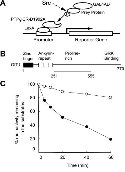
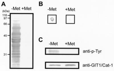
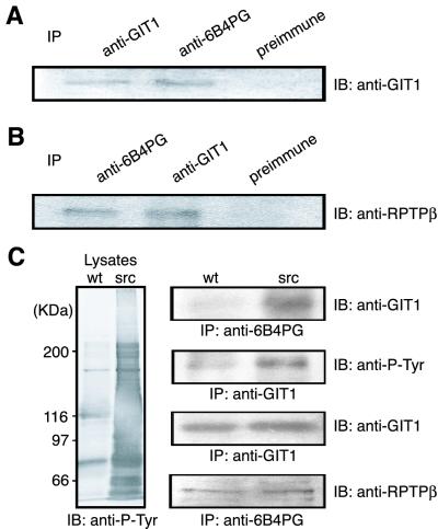
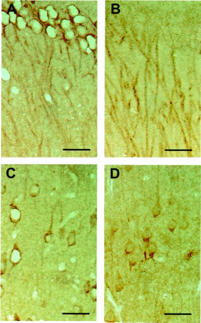
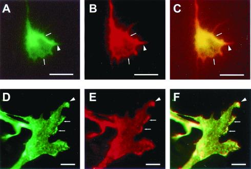
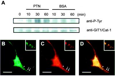
We used a genetic method, the yeast substrate-trapping system, to identify substrates for protein tyrosine phosphatases zeta (PTPzeta/RPTPbeta). This method is based on the yeast two-hybrid system, with two essential modifications: conditional expression of protein tyrosine kinase v-src (active src) to tyrosine-phosphorylate the prey proteins and screening by using a substrate-trap mutant of PTPzeta (PTPzeta-D1902A) as bait. By using this system, several substrate candidates for PTPzeta were isolated. Among them, GIT1/Cat-1 (G protein-coupled receptor kinase-interactor 1/Cool-associated, tyrosine-phosphorylated 1) was examined further. GIT1/Cat-1 bound to PTPzeta-D1902A dependent on the substrate tyrosine phosphorylation. Tyrosine-phosphorylated GIT1/Cat-1 was dephosphorylated by PTPzeta in vitro. Immunoprecipitation experiments indicated that PTPzeta-D1902A and GIT1/Cat-1 form a stable complex also in mammalian cells. Immunohistochemical analyses revealed that PTPzeta and GIT1/Cat-1 were colocalized in the processes of pyramidal cells in the hippocampus and neocortex in rat brain. Subcellular colocalization was further verified in the growth cones of mossy fibers from pontine explants and in the ruffling membranes and processes of B103 neuroblastoma cells. Moreover, pleiotrophin, a ligand for PTPzeta, increased tyrosine phosphorylation of GIT1/Cat-1 in B103 cells. All these results indicate that GIT1/Cat-1 is a substrate molecule of PTPzeta.


Figures

Figure 1
Figure 1
Schematic representations of the yeast substrate-trapping system and GIT1/Cat-1 protein isolated by this system. (A) In the presence of 1 mM methionine when v-src is not induced, only the standard two-hybrid bindings occur (not shown). In the absence of methionine, prey proteins (substrates for PTPζ) could be phosphorylated by the induced v-src, and trapped by the bait consisting of the whole intracellular domain with an Asp-1902 → Ala mutation of PTPζ (PTPζICR-D1902A). This complex formation leads to activation of transcription of the reporter genes, HIS3 and LacZ. We performed screenings in two steps and selected the colonies that showed an increase in blue color development on expression of v-src (see Materials and Methods). (B) GIT1/Cat-1 contains a zinc-finger region, three ankyrin-repeat regions, a potential Src-homology 3 (SH3) binding site and a G protein-coupled receptor kinase (GRK) binding site. The region encoded by clone 10–1 is shown by the solid line underneath. (C) GIT1/Cat-1 (amino acid residues 251–555; ●) and RCM lysozyme (○) were tyrosine phosphorylated by p60c-src and then incubated with GST-PTPζICR for various periods. GST-PTPζICR efficiently dephosphorylated GIT1/Cat-1.
Figure 2
Figure 2
Substrate identification with the yeast substrate-trapping system. (A) Yeast proteins were highly tyrosine-phosphorylated only when v-src was induced in the absence of methionine (−Met), whereas almost no tyrosine-phosphorylation was observed in the presence of methionine (+Met). Yeast lysates (70 μg) were analyzed by Western blotting with antiphosphotyrosine antibody. (B) β-Galactosidase filter-lift assay for clone 10–1. A filter placed on the plate lacking leucine, tryptophan, and methionine (Left) and lacking leucine and tryptophan (Right) was streaked directly with clone 10–1 and pBridgeLexA/ζICR-D1902A/v-src cotransformants, incubated at 30°C for 2 days, and developed for 3 h for color detection. (C) The prey consisting of the GAL4 activation domain fused to GIT1/Cat-1 was actually tyrosine-phosphorylated by v-src induction (−Met). Yeast cell lysates (750 μg) were immunoprecipitated with anti-GIT1/Cat-1 antiserum and analyzed by Western blotting with antiphosphotyrosine antibody (Upper) and anti-GIT1/Cat-1 antiserum (Lower).
Figure 3
Figure 3
Coimmunoprecipitation of PTPζ and GIT1/Cat-1. (A and B) NIH 3T3 cells were cotransfected with GIT1/Cat-1 and PTPζ-D1902A expression plasmids. Following the transfection, cells were plated on fibronectin. The cell lysates were immunoprecipitated with the appropriate antibodies, and the precipitated proteins were detected with anti-GIT1/Cat-1 (A) and anti-RPTPβ (B) antibodies. (C) Native 3Y1 (wt) and v-src-transformed 3Y1 (src) cells were cotransfected with GIT1/Cat-1 and PTPζ-D1902A expression plasmids. The cell lysates were analyzed by Western blotting using antiphosphotyrosine antibody (Left). The cell lysates were subjected to immunoprecipitation (Right) with anti-6B4 PG (first blot) or anti-GIT1/Cat-1 (second blot) antibodies. Western blotting indicated that GIT1/Cat-1 is highly tyrosine-phosphorylated (second blot) and is present in complex with PTPζ-D1902A (first blot) in the v-src-transformed 3Y1 cells. Both cells expressed comparable amounts of GIT1/Cat-1 and PTPζ-D1902A (third and fourth blots, respectively).
Figure 4
Figure 4
Immunohistochemical localization of GIT1/Cat-1 and PTPζ in the rat brain. Rat brain serial sections were stained with anti-GIT1/Cat-1 (A and C) and anti-RPTPβ (B and D) antibodies, respectively. (A and B) Hippocampal CA1 region. The upper portion is the pyramidal cell layer. (C and D) Layer V of the frontal cortex. Layer IV is above. (Scale bars, 50 μm.)
Figure 5
Figure 5
Immunohistochemical localization of GIT1/Cat-1 and PTPζ in the growth cones and B103 neuroblastoma cells. The mossy fibers extending from pontine explants were doubly immunostained with anti-GIT1/Cat-1 (A) and anti-6B4 PG (B) antibodies. A combined image is shown in C. The colocalization of both proteins was evident at the peripheral regions (arrows) and filopodial processes (arrowhead) of the growth cones. B103 cells were doubly immunostained with anti-GIT1/Cat-1 (D) and anti-6B4 PG (E) antibodies. A combined image is shown in F. GIT1/Cat-1 and PTPζ were colocalized in the processes (arrowhead) and the ruffling membranes (arrows) of the cells. [Scale bars, 5 μm (AC) and 10 μm (DF).]
Figure 6
Figure 6
PTN stimulates tyrosine phosphorylation of GIT1/Cat-1 in the B103 cells. (A) Time course of the tyrosine phosphorylation of GIT1/Cat-1 in response to polystyrene beads coated with PTN (20 μg/ml) or BSA (20 μg/ml). B103 cell lysates (800 μg) were immunoprecipitated with anti-GIT1/Cat-1 antiserum and analyzed by Western blotting with antiphosphotyrosine antibody (Upper) and anti-GIT1/Cat-1 antibody (Lower). PTN-coated beads induced accumulations of GIT1/Cat-1 (B) and PTPζ (C) around the beads after 30 min of incubation (arrows and arrowheads). The combined image (D) shows that the two proteins were coaggregated around the beads. (Insets) A magnified view of the beads marked by arrows. A similar accumulation was also observed around BSA-coated beads (data not shown). [Scale bars, 20 μm (BD).]

Similar articles

Cited by

References

    1. Kavin M, Walton K M, Dixon J E. Annu Rev Biochem. 1993;62:101–120. - PubMed
    1. Sun H, Tonks N K. Trends Biol Sci. 1994;19:480–485. - PubMed
    1. Keegan K, Cooper J A. Oncogene. 1996;12:1537–1544. - PubMed
    1. Mousseau D D, Banville D, L'Abbe D, Bouchard P, Shen S-H. J Biol Chem. 2000;275:4467–4474. - PubMed
    1. Flint A J, Tiganis T, Barford D, Tonks N K. Proc Natl Acad Sci USA. 1997;94:1680–1685. - PMC - PubMed

Publication types

LinkOut - more resources