. 2001 Jun 5;98(12):6771-6.
doi: 10.1073/pnas.121101598. Epub 2001 May 29.

Immune-type receptor genes in zebrafish share genetic and functional properties with genes encoded by the mammalian leukocyte receptor cluster

Affiliations

Immune-type receptor genes in zebrafish share genetic and functional properties with genes encoded by the mammalian leukocyte receptor cluster

J A Yoder et al. Proc Natl Acad Sci U S A. .

Abstract

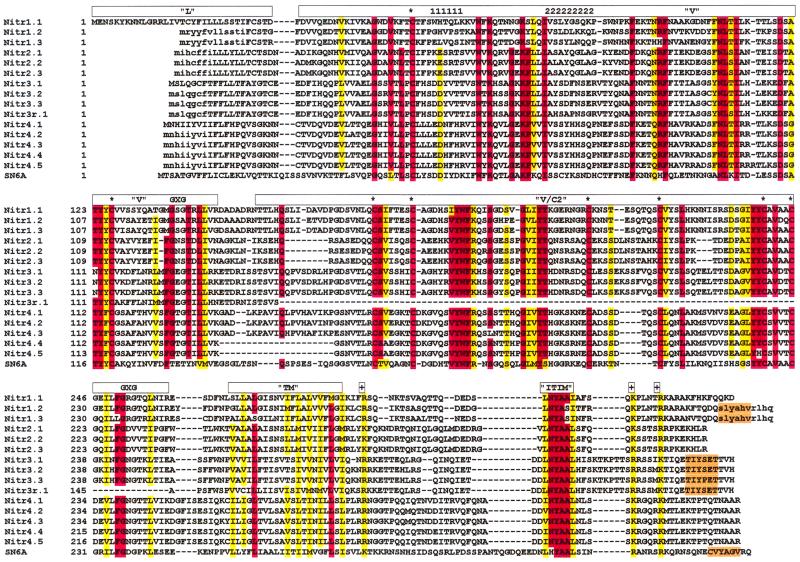
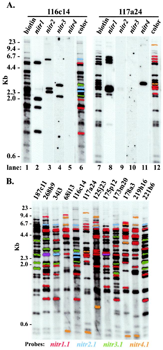
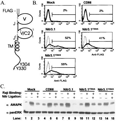
An extensive, highly diversified multigene family of novel immune-type receptor (nitr) genes has been defined in Danio rerio (zebrafish). The genes are predicted to encode type I transmembrane glycoproteins consisting of extracellular variable (V) and V-like C2 (V/C2) domains, a transmembrane region and a cytoplasmic tail. All of the genes examined encode immunoreceptor tyrosine-based inhibition motifs in the cytoplasmic tail. Radiation hybrid panel mapping and analysis of a deletion mutant line (b240) indicate that a minimum of approximately 40 nitr genes are contiguous in the genome and span approximately 0.6 Mb near the top of zebrafish linkage group 7. One flanking region of the nitr gene complex shares conserved synteny with a region of mouse chromosome 7, which shares conserved synteny with human 19q13.3-q13.4 that encodes the leukocyte receptor cluster. Antibody-induced crosslinking of Nitrs that have been introduced into a human natural killer cell line inhibits the phosphorylation of mitogen-activated protein kinase that is triggered by natural killer-sensitive tumor target cells. Nitrs likely represent intermediates in the evolution of the leukocyte receptor cluster.


Figures

Figure 1
Figure 1
Sequence identity patterns in Nitrs. clustalw alignment of predicted translation products from zebrafish nitr cDNAs and the representative pufferfish cDNA, SN6A. Leader (L), V, V/C2, transmembrane (TM), and ITIM sequences are indicated above the alignment. A second putative ITIM in Nitr1, Nitr3, and SN6A is highlighted in orange. Identical residues are enclosed in red. Residues that are in similar functional groups (defined in ref. 10) are boxed in yellow. Conserved cysteines are indicated with an asterisk. The glycine bulges of the J domains (GXG) are indicated. Conserved charged residues are indicated (+). CDR1 and CDR2 are indicated by “1111111” and “222222222”, respectively. Residues shown in lowercase letters are predicted from genomic sequence. Note that Nitr4 transcripts encode long (Nitr4.1, Nitr4.2, and Nitr4.3) or short (Nitr4.4 and Nitr4.5) forms, which differ by 19 amino acids between the V and V/C2 exons. The nucleotide identity between members within each nitr subfamily ranges from 80% to 99%.
Figure 2
Figure 2
Characterization of nitr-containing P1 artificial chromosomes. (A) Zebrafish nitr PAC clones 116c14 and 117a24 were digested with HindIII, 5′ end-labeled with biotin, electrophoresed, and transferred to a nylon membrane. Restriction fragments were identified by a streptavidin–enzyme-linked chemiluminescent assay (lanes 1 and 7). The membrane was stripped and hybridized sequentially with probes complementing the four different subfamilies of nitr genes (lanes 2–5 and 8–11). The fragments that hybridized to each probe are digitally color-coded on the original image (lanes 6 and 12); color-coding can be discerned from coding in B. (B) Similar analyses were performed for the 12 PACs that exhibit the greatest number of banding differences and are presumed to correspond to different regions of the extended nitr gene complex. Purple bands reflect hybridization with both nitr1 and nitr3 probes. Color-coding corresponding to different nitr subfamilies is indicated at the bottom of B and also corresponds to lanes 6 and 12 in A.
Figure 3
Figure 3
Characterization of the nitr genomic region. (A) Intronic STSs from nitr-encoding PACs were mapped to LG7 with the T51 RH panel (14) and the LN54 RH panel (15). The regions of shared synteny between LG7 and the human and mouse genomes are shown. Mapping positions of genes shown have been published (25) except for tip30, which was identified for this study. (B) PCR analysis of nitr STSs in wild type (wt) and the b240 deficiency strain zebrafish (Δ). The positive control is noggin (nog3) (50), which is retained, and the negative control is sonic hedgehog (shh), which is deleted in b240 (A. Fritz, personal communication). Zebrafish strain b240 has lost a large region of LG7 and does not encode sequences from PACs 116c14, 117a24, 175p12, 173m20, and 187c11, strongly supporting the hypothesis that all of the nitr genes are located on LG7. (C) The nitr gene complex on LG7 is flanked by the men1 and tip30 genes in both RH panels. Exon 10 of the men1 gene has been mapped to one end of the nitr gene complex in both RH panels. Exon 2 of the men1 gene has been mapped 33 centiRays distal to the nitr genes on the T51 RH panel. A nitr-encoding PAC was identified that also encodes the 3′ end of the men1 gene placing the nitr gene complex within 120 kb of men1. The figure shown is not to scale and merges position data from both RH panels. Corresponding regions of the mouse and human genomes are shown below the line. (D) Multiple genes from the Tip30/Dbx region on mouse chromosome 7 are shown. Corresponding regions of the human genome are shown below the line. Note that this region of mouse chromosome 7 shares conserved synteny with human 11p15 and 19q13.
Figure 4
Figure 4
Inhibition of MAPK activation in NK cells. (A) Predicted protein structure of recombinant Nitr3.1; tyrosines 304 and 330 are indicated as Y304 and Y330, respectively. (B) Human NK92 cells, mock-infected or infected with vaccinia virus carrying CD56, FLAG-tagged Nitr3.1, FLAG-tagged mutant Nitr3.1Y304A, or FLAG-tagged Nitr3.1Y304S, were analyzed for surface expression of the receptors by flow cytometry. (C) The same NK92 cells were then incubated with Raji tumor cells before lysis and Western analysis for active MAPK. The membrane was stripped and probed for panERK to verify equal loading; triplicate determinations verified the findings.

Similar articles

Cited by

References

    1. Medzhitov R, Janeway C A J. Cell. 1997;91:295–298. - PubMed
    1. Wende H, Colonna M, Ziegler A, Volz A. Mamm Genome. 1999;10:154–160. - PubMed
    1. Wilson M J, Torkar M, Haude A, Milne S, Jones T, Sheer D, Beck S, Trowsdale J. Proc Natl Acad Sci USA. 2000;97:4778–4783. . (First Published April 18, 2000, 10.1073/pnas.080588597) - PMC - PubMed
    1. Kubagawa H, Cooper M D, Chen C C, Ho L H, Alley T L, Hurez V, Tun T, Uehara T, Shimada T, Burrows P D. Curr Top Microbiol Immunol. 1999;244:137–149. - PubMed
    1. Moretta L, Biassoni R, Bottino C, Mingari M C, Moretta A. Immunol Today. 2000;21:420–422. - PubMed

Publication types

Substances

LinkOut - more resources