. 2001 Jun 5;98(12):6923-8.
doi: 10.1073/pnas.121119298. Epub 2001 May 29.

Hyperphosphorylation induces self-assembly of tau into tangles of paired helical filaments/straight filaments

Affiliations

Hyperphosphorylation induces self-assembly of tau into tangles of paired helical filaments/straight filaments

A Alonso et al. Proc Natl Acad Sci U S A. .

Abstract

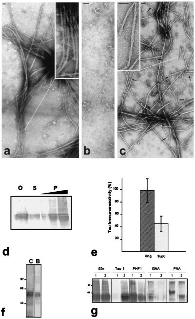
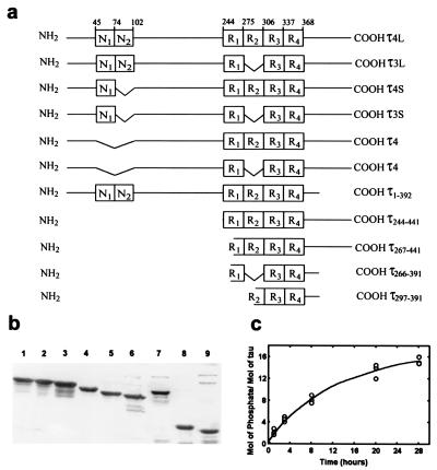
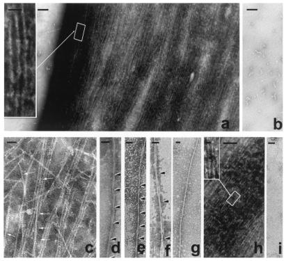
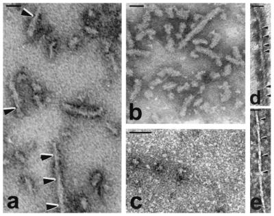
The microtubule-associated protein tau is a family of six isoforms that becomes abnormally hyperphosphorylated and accumulates in the form of paired helical filaments (PHF) in the brains of patients with Alzheimer's disease (AD) and patients with several other tauopathies. Here, we show that the abnormally hyperphosphorylated tau from AD brain cytosol (AD P-tau) self-aggregates into PHF-like structures on incubation at pH 6.9 under reducing conditions at 35 degrees C during 90 min. In vitro dephosphorylation, but not deglycosylation, of AD P-tau inhibits its self-association into PHF. Furthermore, hyperphosphorylation induces self-assembly of each of the six tau isoforms into tangles of PHF and straight filaments, and the microtubule binding domains/repeats region in the absence of the rest of the molecule can also self-assemble into PHF. Thus, it appears that tau self-assembles by association of the microtubule binding domains/repeats and that the abnormal hyperphosphorylation promotes the self-assembly of tau into tangles of PHF and straight filaments by neutralizing the inhibitory basic charges of the flanking regions.


Figures

Figure 1
Figure 1
In vitro polymerization of AD P-τ into tangles of PHF/SF and the effects of dephosphorylation and deglycosylation. AD P-τ, 0.4 mg/ml, without treatment (a), dephosphorylated by AP (b), or deglycosylated by endoglycosidase F/N-glycosidase F (c), was incubated for 90 min, and the products of the assembly were examined by NSEM. Dephosphorylation, but not deglycosylation, completely abolished AD P-τ polymerization. Bar represents 50 nm. (Insets) PHF at higher magnifications. Arrows label examples of 10–15-nm (straight) and 4-nm (arrowhead) filaments. (d) AD P-τ, 0.4 mg/ml, was incubated as above to induce assembly, and the aggregated protein was separated from the nonaggregated protein by centrifugation at 35°C and 100,000 × g for 15 min. The pellet (P) was resuspended to its original volume, and equivalent samples of the original mixture (O), the supernatant (S), and the pellet (1, 2, and 4×) were analyzed by Western blots by using Tau-1 antibody and dephosphorylation of the proteins on the blot with AP. (e) The amount (mean ± SD of 4 values) of AD P-τ present in the original and the supernatant fractions was quantitated by scanning the immunoblots. (f) SDS/PAGE (10% gel) of AD P-τ and blot of a lane from the same gel developed with Tau-1 antibody after dephosphorylation. One strip (8 μg of protein/lane) was stained with Coomassie blue (C), and another strip (2 μg of protein/lane) was developed with Tau-1 antibody after dephosphorylation of the proteins on the membrane (B). (g) For in vitro dephosphorylation and deglycosylation of AD P-τ, aliquots of AD P-τ were treated with (2) or without (1) the addition of AP to dephosphorylate (panels labeled 92e, Tau-1, and PHF1) or endoglycosidase F/N-glycosidase F to deglycosylate (panels labeled GNA and PNA) the proteins as described. The immunoblots were developed with 92e (dilution 1/5,000) to detect the total amount of τ, Tau-1 (1/50,000) to detect dephosphorylated τ, and PHF1 (1/250) to detect phosphorylated τ. The increase in Tau-1 staining and decrease in PHF1 staining show dephosphorylation of AD P-τ. The immunoblots were developed with lectin GNA or PNA to detect glycosylation. Decrease in the staining with the lectins shows deglycosylation of AD P-τ by the glycosidase.
Figure 2
Figure 2
Schematic of human τ isoforms and τ fragments, SDS/PAGE, and phosphorylation. (a) The amino acid residue numbers are according to τ441, (τ4L). (b) Coomassie blue-stained patterns of the SDS/PAGE (10% gel) of human τ isoforms and fragments shown in a, 4 μg/lane, except constructs τ266–391 and τ297–391, which were shown previously (36). Lanes 1, τ4L; 2, τ3L; 3, τ4S; 4, τ3S; 5, τ4; 6, τ3; 7, τ4L1–392; 8, τ4L244–441; and 9, τ4L267–441. (c) Time course of incorporation of [32P]. τ was hyperphosphorylated as described under Materials and Methods. Similar incorporation was obtained with any of the six τ isoforms. The curve shows phosphorylation of τ4L.
Figure 3
Figure 3
The formation of tangles of PHF-like and SF from in vitro hyperphosphorylated τ. τ, 0.5 mg/ml, was incubated with rat brain extract in the presence of ATP to induce hyperphosphorylation of τ (a) or incubated with nonhydrolyzable ATP, AMP–PNP, as a control (b). τ hyperphosphorylation (≈12–15 mol of phosphate per mol of protein) induced its self-polymerization into straight and PHF-like (Inset) filaments (a), irrespective of the τ isoform used for the phosphorylation assay. The magnification bars in a and b represent 500 nm and, in the Inset, 50 nm. Intertwining 4-nm filaments generate small PHF-like structures (arrows) as follows: τ4 (c); PHF, τ3L (d); τ protofilaments forming PHF, τ4S (e); τ protofilaments forming bigger PHF, τ4S (f); ≈15-nm straight filament, τ3 (g); mixture of all six τ isoforms hyperphosphorylated (h), and Inset shows a PHF from the tangle formed; and control sample incubated without ATP, τ4S (i). Bars represent 40 nm in c–g and i, 200 nm in h, and 25 nm in Inset.
Figure 4
Figure 4
Self-assembly of whole τ and of microtubule binding region. Polymerization of τ4L, 0.04 mg/ml (a), and the association of τ4L with normal brain τ (b) is shown; similar PHF with fuzzy coat were obtained when τ4L was coassembled with τ3L, τ3S, τ3, τ4S, or τ4 (not shown). Acid and heat treatment of τ4L (0.04 mg/ml) inhibited the self-assembly of τ (c). τ constructs τ266–391 and τ297–391 containing the microtubule binding region (0.5 mg/ml) were able to polymerize into short PHF-like filaments, with twists every ≈40 nm (d) or every ≈80 nm (e); similar but several twists longer than those formed from τ4L. Bar represents 50 nm.

Similar articles

Cited by

References

    1. Finch C, Tanzi R E. Science. 1997;278:407–411. - PubMed
    1. Tomlinson B E, Blessed G, Roth M J. Neurol Sci. 1970;11:205–242. - PubMed
    1. Arigada P A, Growdon J H, Hedley-White E T, Hyman B T. Neurology. 1992;42:631–639. - PubMed
    1. Grundke-Iqbal I, Iqbal K, Quinlan M, Tung Y-C, Zaidi M S, Wisniewski H M. J Biol Chem. 1986;261:6084–6089. - PubMed
    1. Grundke-Iqbal I, Iqbal K, Tung Y-C, Quinlan M, Wisniewski H M, Binder L I. Proc Natl Acad Sci USA. 1986;83:4913–4917. - PMC - PubMed

Publication types